Murat Durdu
142 Turk J Dermatol 2017;11:142-3Başkent University Faculty of Medicine, Adana Dr. Turgut Noyan Application and Research Center, Clinic of Dermatology, Adana, Turkey
©Copyright 2017 by Turkish Society of Dermatology Turkish Journal of Dermatology published
by Galenos Publishing House.
Murat Durdu, Başkent University Faculty of Medicine, Adana Dr. Turgut Noyan Application and Research Center, Clinic of Dermatology, Adana, Turkey Phone: +90 322 327 27 27 E-mail: [email protected] ORCID ID: orcid.org/0000-0003-1247-3932 Correspondence/ Yazışma Adresi:
Pearls in Dermatology / Dermatolojik İnciler
Apoptotic Bodies due to Erythema Multiforme:
Cytological, Histopathological and
Immunofluorescence Findings
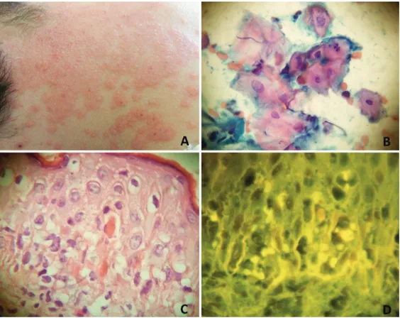
A 12-year-old male patient was admitted with alopecic patches on the scalp. In his history, he had been diagnosed with alopecia areata for two years. Despite topical steroid creams and intralesional steroid injections, he continued to develop new alopecic patches. Topical immunotherapy with diphencyprone (DPCP) was planned due to severe (>50% involvement) alopecia areata lesions. Sensitization with 2% DPCP solution was applied to the occipital region. Two days later, vesicular and bullous lesions on the erythematous base developed on the sensitization site. One week later, erythematous target-like papules and plaques localized on the occipital region and back of the neck (Figure 1a). There were vesicles in the mid of some papules. Skin scraping smear from the vesicular lesions were taken and the smears were stained with the Papanicolaou (PAP) stain. Cytological examination showed apoptotic bodies characterized by karyolysis, karyorrhexis, and pyknosis in the keratinocytes (Figure 1b). Histopathological examination of the punch biopsy specimens revealed epidermal apoptotic cells (Figure 1c). Hematoxylin and eosin (H&E) stained specimens were evaluated using immunofluorescence microscopy and autofluorescence was obtained from
Figure 1. Clinical, cytological, histopathological, and immunofluorescence microscopic findings of a case with erythema multiforme. a) Erythema multiforme-induced target-like lesions on the sensitization site with DPCP, b) Cytology examination showed apoptotic bodies, c) Histopathological examination revealed abundant apoptotic keratinocytes in the epidermis, d) In hematoxylin and eosin stained slides, apoptotic bodies showed autofluorescence under an immunofluorescence microscope (b, PAP x1000; c, H&E x1000, DIF x1000)
the epidermal apoptotic bodies (Figure 1d). The patient was suspected of DPCP-related erythema multiforme. Target-like lesions completely resolved with topical steroid creams.
Pearls;
Clinical: In addition to some infectious diseases, increased
apoptosis in the keratinocytes is seen in the course of a number of inflammatory processes. Erythema multiforme is one of these conditions which induce apoptosis. In the majority of cases, the most common causes are infections and medications. Not only systemic agents but also topical drugs such as DPCP can induce erythema multiforme (1).
Cytological: Cytological findings of apoptosis are karyolysis,
karyorrhexis, and pyknosis. These nuclear changes can be easily detected through PAP stain (2).
Histopathological: In patients with erythema multiforme,
apoptotic cells can be found in the basal layers, papillary dermis, upper spinous layer, and occasionally stratum corneum (3). In addition, autofluorescence can be detected, when H&E stained slides are examined under a fluorescent microscope.
References
1. Oh CW, Han KD, Kim TH. Bullous erythema multiforme following topical diphenylcyclopropenone application. Contact Dermatitis 1998;38:220-1. 2. Durdu M, Baba M, Seçkin D. The value of Tzanck smear test in diagnosis of
erosive, vesicular, bullous, and pustular skin lesions. J Am Acad Dermatol 2008;59:958-64.
3. Joshi R. Interface dermatitis. Indian J Dermatol Venereol Leprol 2013;79: 349-59.
143