KADIR HAS UNIVERSITY
GRADUATE SCHOOL OF SCIENCE AND ENGINEERING
IN SILICO SCREENING OF TANGIBLE-POTENTIAL INHIBITOR
OF METHIONINE AMINOPEPTIDASE 2 FOR THE TREATMENT
OF CANCER
GRADUATE THESIS
By
JACKSON WEAKO
IN SILICO SCREENING OF TANGIBLE-POTENTIAL INHIBITOR
OF METHIONINE AMINOPEPTIDASE 2 FOR THE TREATMENT
OF CANCER
By
Jackson Weako
Submitted to the Graduate School of Science and Engineering in partial fulfillment of the requirements for the degree of
Master of Science in
COMPUTATIONAL BIOLOGY AND BIOINFORMATICS
KADIR HAS UNIVERSITY
i
ABSTRACT
Methionine Aminopeptidases (MetAPs) are divalent-cofactor dependent enzymes that are responsible for cleaving the initiator Methionine from the newly synthesized polypeptides. These metalloproteases are classified into two distinct isoforms- MetAP1 and MetAP2. The MetAP2 isoform is upregulated in many cancerous cells. A selective inhibition of MetAP2 is an effective means of suppressing vascularization and limiting both the size and metastasis of solid tumors in a model organism. A selective and potent inhibitor of MetAP2 is the natural product – fumagillin. Fumagillin and its semi-synthetic analogs have been shown as promising candidates in various clinical trials for treating cancer and in rent time for treating obesity. However, their further developments have received a great setback due to their poor pharmacokinetic properties and neurotoxicities in clinical studies. Here, in an effort to find potential inhibitors of MetAP2, In-silico Screening and Molecular Modelling were applied to generate a new class of inhibitors. The OTAVA’s Chemical Library was screened and ten best compounds were selected based on their structural, physicochemical properties and inhibitory potentials against MetAP2. PyRx, AutoDock 4.2, and Accelrys (BIOVIA Discovery Studio version 2016) were deployed to obtain these potential inhibitors. Utilizing OTAVA’s Chemical Library, 130 potential drug candidates were selected based on a threshold of -9.0Kcal/mole using PyRx. In order to re-evaluate and validate these 130 inhibitors, AutoDock 4.2 was employed to dock these selected candidates. A total of 71 potential candidates were selected based on AutoDock result. Further analysis of their inhibition constants and Gibbs free energies led to the ten best potential candidates. Accelrys (BIOVIA Discovery Studio version 2016) was used to identify the positions of these candidates in the active site of MetAP2. Discovery Studio’s ADMET protocols was used to determine the pharmacokinetics properties of these candidates. It is anticipated herein that, these candidates will serve as a new class of inhibitors and/or lead compounds for MetAP2.
Keywords: Key Words: Methionine Aminopeptidases, In Silico Screening, Virtual Screening, Tangible Compounds, Docking, Inhibition Constant (Ki), Gibbs free energy (ΔG)
ii
ÖZET
Metiyonin Aminopeptidazlar (MetAP’ler), metiyoninleri yeni sentezlenmiş polipeptitlerin başından kesmekle sorumlu, divalent kofaktörlere bağlı çalışan enzimlerdir. Bu metalloproteaz enzimi iki gruba ayrılır: MatAP1 ve MetAP2. MetAP2 izoformunun seviyesinin pek çok kanser türünde arttığı gözlemlenmiştir. Model organizmada yapılan çalışmalar, MetAP2’nin seçimli olarak engellenmesinin damar oluşumunu baskıladığını, katı tümörlerde tümör boyunu ve metastazı sınırlandırdığını göstermiştir. Met2AP’nin seçimli ve kuvvetli bir inhibitörü, doğal bir ürün olan fumagillindir. Fumagillin ve yarı sentetik analogları, kanser tedavisi klinik deneylerinde ve muhtemelen obezite tedavisinde umut vaat eden adaylardır. Fakat bunların klinik çalışmalarda daha da geliştirilmesinin önünde farmakokinetik ve nörotoksik özelliklerin bilinmiyor olması engel oluşturmaktadır. Bu çalışmada, MatAP2’nin potansiyel inhibitörlerini bulmak ve yeni bir sınıf inhibitörler yaratmak amacı ile, in silico tarama ve moleküler modelleme uygulandı. OTAVA’nın kimyasal kütüphanesi tarandı ve yapısal, fizikokimyasal özellikleri ve MET2AP baskılama potansiyelleri açısından en iyi on bileşik seçildi. Bu potansiyel inhibitörleri bulmak için PyRx, AutoDock 4.2, ve Accelrys (BIOVIA Discovery Studio versiyon 2016) kullanıldı. OTAVA kimyasal kütüphanesini kullanarak PyRx aracı ile -9.0 Kcal/mol eşiğini aşan 130 ilaç adayı seçildi. Bu 130 aday, tekrar değerlendirmek ve teyit etmek için AutoDock 4.2 kullanılarak hedefe yuvalandı. AutoDock sonuçlarına göre toplamda 71 potansiyel aday seçildi. İnhibisyon sabitleri ve Gibbs serbest enerjileri ile yapılan daha kapsamlı bir analiz en iyi on adayı ortaya çıkardı. Accelrys (BIOVIA Discovery Studio versiyon 2016) ise bu adayların MetAP2 enzimi aktif yüzeyinde yuvalandığı yeri bulmaya yardımcı oldu. Discovery Studio’nun ADMET protokolü adayların farmakokinetik özelliklerini açığa çıkarmak için kullanıldı. Bulunan bu adaylar MetAP2 enzimi için yeni bir inhibitör sınıfı olacaktır veya yeni bileşiklerin bulunmasına öncülük edecektir.
Anahtar kelimeler: Metiyonin Aminopeptidaz, in silico tarama, sanal tarama, somut bileşikler, yuvalama, inhibisyon sabiti (Ki), Gibbs serbest enerji (∆G)
AP PE
iii
Acknowledgements
First of all, I would like to express my heartfelt and sincere appreciation to my academic advisor – Prof. Dr. Kemal Yelekçi for the privilege he proffered me to work under his supervision. His patience, commitment, diligence, continual support and moral guidance provided were excellent. Working with him in such a scientific arena was memorable.
Besides my academic advisor, I extend my gratitude to the Scientific and Technological Research Council of Turkey (TÜBITAK) for funding my master program through the 2235- Graduate Scholarship for the Least Developed Countries. My warmest thanks and appreciations to my parents who have provided unwavering support and moral guidance throughout my educational sojourn. They have instilled in me a desire to explore new things and to excel among the professionals.
Moreover, I also extend my profound thanks to Mr. Christian B. Nicol, Dean of the College of natural Science, Cuttington University, Mr. J. Kelvin Fallah, Human Resource Director, and Dr. Henrique F. Tokpa erstwhile President of Cuttington University, for their initial cooperation and financial support.
Finally, hats off to my colleagues and staff of the Bioinformatics and Genetics Department of Kadir University for the knowledge imparted onto us. You served as a moral role model and exhibited patience as you were vibrant in executing your duties with commitment, fairness, and diligence.
AP PE
iv
Dedication
v Table of Content Abstract……….. ….i Özet………...ii Acknowledgement…..………...…..iii Dedication………...iv Table of Content………...v-vi List of figures ….……….…...vii
List of Tables………...viii
Chapter 1: Objective ……….1-2 Chapter 2: Introduction ………...3-22 2.1 The Structure of Methionine Aminopeptidase 2……….6-7 2.2 The Divalent center of Methionine Aminopeptidase 2………...7-8 2.3 The binding cavity of Methionine Aminopeptidase 2………8
2.4 The nature of the Methionine Aminopeptidases reaction mechanism…8-9 2.5 .The major function of Methionine Aminopeptidase2 in biological process………...10
2.6. Clinical Significance of Methionine Aminopeptidase 2 Enzyme…..10-11 2.7. Natural products as source of Lead Compound………...11
2.8 Drug Design and In Silico Approach………..12-14 2.9. Stages of Modern rational Drug Discovery and Development…………15
2.10 In Silico ADMET properties Prediction of a drug molecule……….16-17 2.11. Virtual Screening in Drug Development………..18-19 2.12. Docking process in Drug Design………..19-21 2.13. AutoDock- a Tool for Docking in Drug Design………...21-22 Chapter 3: Material and Method ………...23-30 3.1. Preparation of the receptor’s structure for docking purpose………24
3.2. Preparation of the Receptor’s native ligands for re-docking as test case………....24-28 3.3. Screening the OTAVA’s Chemical Library ………...29
vi
Chapter 4: Result and Discussion ……….31-58 Chapter 5: Conclusion ………...59-60 Definition of key words ………61-63 Reference: ……….64-69 .
vii
List of Figure
Figure 1: Structure of the Methionine Amino acid ………..3 Figure 2: Comparison of Methionine Aminopeptidases primary structure…...7 Figure 3: Reaction mechanism of Methionine Aminopeptidase in E. coli………….9 Figure 4: In Silico Approach during drug discovery process……….14 Figure 5: 3D structure of Spiroepoxytriazole binds into the active of MetAP2…….27 Figure 6: 2D diagram showing various interactions between MetAP2 and
Spiroepoxytriazole………..27 Figure 7: 3D Structure of fumagillin binds into the active of MetAP2…………...28 Figure 8: 2D diagram that depicts various interactions between MetAP2 and
fumagillin ………...28 Figure 9: Summary of the selection process………...33 Figure 10: 2D diagram showing various interactions between the receptor and
compound1………..55 Figure 11: 3D orientation of compound 1 in the binding cavity of MetAP2
enzyme………....55 Figure 12. Compound 1 binding in MetAP2’s active site ……….56 Figure 13: 2D diagram showing various interactions between the receptor and
compound 2……….57 Figure 14: 3D orientation of compound 2 in the binding cavity of MetAP2
enzyme………....57 Figure 15: Compound 2 binding in MetAP2’s active site………..58 Figure 16: The ADMET PSA 2D (Polar surface area) versus ADMET Alogp98 Plot………58
viii List of Table
Table 1: Native ligands Calculated inhibition and Experimental inhibition constants ……….26 Table 2: 71 selected inhibitors with their Ki values, ΔG values, run number and torsional angles………..34-50 Table 3: Ten candidates selected based on lowest ΔG values………51-53
1
Chapter 1
Objective
Methionine Aminopeptidases (MetAPs) are “metalloproteases” that are responsible for regulating post-translational processes such as the removal of initiator methionine from a newly synthesized polypeptide. There are two isoforms of these nascent proteins processing enzymes; namely, MetAP subtype1 and MetAP subtype2. Both of these isoforms are found in Eukaryotes such as human, while prokaryotes contains only subtype 1. The failure of MetAPs to remove the methionine residue from the nascent protein can lead to a protein product that is not functional or cannot produce an immune response [1][2] .
MetAP2 plays a critical role in the development of cancer and angiogenesis which have made it a molecular target for the anti-angiogenic compounds, the natural products fumagillin and its analogs [3]. These two isoforms of MetAPs have active sites that have highly structural and sequence similarity and have a very important function in living cells. For example, the specific inactivation of MetAPs enzymes in bacteria is lethal while in yeast, slow growth phenotype is observed when individual genes are impeded. All these MetAPs isozymes in human are overexpressed in cancer cells and inhibition of these enzymes is important and beneficial [4]. The fungal product- fumagillin and its derivatives are selective and potential inhibitors of MetAP2. They have shown great specificity toward MetAP2 enzymes and have been considered as promising candidates in clinical studies for treating cancer and recently for obesity. However, many attempts to use them as drug have failed in clinical trials due to their poor pharmacokinetic properties, neurotoxicities, and other related side effects. Even though, they have shown different activities, their inhibition mode in both subtype 1 and subtype 2 of MetAPs seems similar, that is, by covalently modifying the histidine residue in the active site of
2
the enzymes. Besides these natural products, no other compounds have displayed such highly selectivity and potency toward any of these protein processing enzymes both vivo and in-vitro[4][5].
Here, these problems are addressed via an Insilco Screening and Molecular Modelling by screening several thousand of chemical compounds via the OTAVA’s Chemicals Library to select potential inhibitors of MetAP2. The underlying principle of the screening process is that these compounds will be:
(i) Easily synthesizable and Possess chemical features to serve as a new class of inhibitors and/or lead compounds of MetAP-2
(ii) Be potent and selective with less side effect and an increase in better pharmacodynamic and pharmacokinetic properties
(iii) Be sterochemically less complex than fumagillin and obey Lipinski and Veber rules and (iv) Finally the ADMET values of candidates or compounds should pass the required properties. The goal of this study and research is to unveil a new class of inhibitors and/or lead compounds against MetAP2 not inspired by fumagillin. It is anticipated herein that, these inhibitors will be selective, potent with less side effect and an improved pharmacokinetic and pharmacodynamics properties than fumagillin and its derivatives for the treatment of cancer. In so doing, the researcher expects that these selected inhibitors will be important and beneficial for better understanding and treatment of various types of cancers in the near future.
3
Chapter 2
Introduction
Methionine (Met) is an essential-alpha (α) amino acid that has α-amino group (NH2),
α-carboxylic group (COOH), hydrogen (H) atom and S-methyl (CH3S) thioether side chain. Based on
the S-methyl thioether side chain, it is classified as a non-polar, aliphatic amino acid. Methionine is coded by the initiator codon (AUG) that serves as the first amino acid in the NH2
-terminal position of a nascent polypeptide, which signifies its significance in the biosynthesis of proteins. Methionine’s metabolism serves as a biosynthetic precursors of polyamines and S-adenosylmethionine – that donates methyl (CH3) group that is used in DNA methylation or
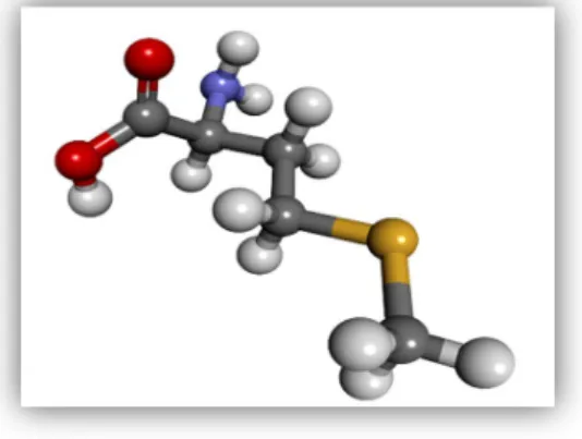
cellular reactions such as cell division [6][7]. In human, this amino acid plays a significant role in angiogenesis and supplementation benefit people suffering from diseases such as Parkinson, Asthma, drug withdrawal, Schizophrenia, Allergy or depression etc. On the contrary, over-consumption is directly related to the cancer growth. Restriction of methionine is a very essential technique for targeting cancer growth, especially in cancers that depend on methionine for survival and proliferation [8][9]. The structure of methionine is shown below.
Figure 1. Structure of methionine. Each element is colored as follow: Carbon (black), Hydrogen (white), Oxygen (red), Nitrogen (blue) and sulfur (deep yellow).
4
The “proteolytic system” plays a critical role in protein synthesis and degradation. Protein turnover, protein maturation, signal peptide processing, abnormal polypeptide degradation as well as inactivation of regulatory proteins are major processes that are enhanced by various proteases in the cell. One of the major processing events that nearly all synthesized nascent polypeptides undergo is the amino-terminal modification which can occur either co or post-translationally[1].
Protein synthesis is pivotal in all living cells and it begins with the initiator methionine. In both prokaryotes and eukaryotes, the translational process of a matured mRNA by the ribosome begins with initiator codon (AUG) which code for methionine[10]. Methionine Amino Peptidases (MetAPs) are polypeptides processing enzymes that are primarily responsible for the co-translational cleavage of the initiator methionine from newly synthesized proteins [5]. In transporting the newly synthesized polypeptide to its intracellular location, the methionine residue at the amino-terminal (NH2) position has to be cleaved[10]. These protein processing
enzymes (MetAP2) exhibit general specificity only to NH2-terminal methionine residue cleaving
no other natural amino acid residues. For instance, the protease deformylase does the removal of the formyl group in formylmethionine before the removal of the actual methionine residue can take place by the MetAPs enzymes. The penultimate amino acids are the driving force of specificity, in that, methionine removal takes place only when the penultimate amino acid residue is small and uncharged (G,A,S,C,T,P,V) and not when it is large (D,E,N,Q,K,H,R,L,I,M,F,Y,W). The cleavage of this methionine residue from the ribosomally newly synthesized polypeptide is cardinal for further amino-terminal modifications such acetylation by N-alpha-acetyltransferase and myristoylation of glycine by N-myristoyltransferase, NMT [1][10]. The retention of the NH2-terminal methionine residue can serve as hindrance for the
5
myristoylation of the N-terminal glycine residue. The polypeptide N-myristoylation is the co-translational attachment of 14 carbon saturated fatty acid via the amide bond to the NH2
-termimal glycine residue following the removal of the initiator methionine from the nascent protein. It can lead to weak and reversible protein-membrane and protein-protein interactions in the cells. N-myristoylproteins have variety of functions and intracellular destinations and are involved in several signal transductions cascades[11][12][13].
The post-translational modification of nascent proteins is carried out in about 60% to 70% of all newly translated polypeptides[3]. There are two isozymes of MetAPs; namely, subtype I (MetAP1) and subtype II (MetAp2). The insertion of a sixty amino acids residues within the catalytic domain of MetAP2 differs it from MetAp1. Both isoforms are all present in Eukaryotes while on the other hand, Prokaryotes contain only MetAP1 isoform. MetAP1 proteases are further divided into four sub-divisions or subtypes (Ia-Id). MetAP1 subtype (Ia) is present in all known prokaryotes, Actinobacteria including Mycobacterium contain subtype (Ic) while Ib and Id are found in Eukaryotes[4]. In human, MetAP2 gene is responsible for encoding MetAP2 enzymes. These enzymes play a superior role in tissue repair, protein synthesis and protein degradation by proteases in living cells. Moreover, MetAP2 is paramount for every cell due to its essential role play in angiogenesis which aid in disease progression especially in solid tumor cancers and rheumatoid arthritis[14]. Further still, it is presumed that MetAP2 (alternatively-eukaryotic initiation factor-2 associated protein, 67-KD P67) has dual factions in vivo: regulating the biosynthesis of protein through the interacting with the eukaryotic initiation factor (eIF-2) and processing of the amino-terminal of the nascent protein [15]
6
2.1 The Structure of Methionine Amino Peptidase-2
As previously mentioned, ribosome-based protein synthesis is initiated by the start codon (AUG) in Eukaryotes or formylmethionine in prokaryotes such Escherichia coli (E. coli) s- Biologists most favored model organism. There are different genes that encode MetAPs enzymes in different organisms, for example, in E. coli, the only well know MetAP-2 is a 29.33 Da, a monomeric enzyme which is coded by a gene containing 264 codons; while in Homo sapiens MetAP1 and MetAP2 code for 42 KDa enzyme and 67 KDa enzyme respectively. MetAP-2 shares approximately twenty-two percent sequence identity with MetAp1 and is highly conserved between Saccharomyces Cerevisiae and Humans[16][17]
Previous studies have shown that in Prokaryotes, a single MetAP gene has been identified and the removal is lethal, while in Archaea, only MetAP2 is found. In contrast to both Prokaryotes and Archaea, Eukaryotes have multiple genes that encode both type1 and type2 of Methionine enzymes. The two isoforms of MetAPs and are differentiated by the insertion of an alpha (α)-helical domain of approximated sixty amino acid residues in length inserted within the surface loop of the C-terminal of MetAP2 which is half of the molecule. Further still, these nascent protein processing enzymes (MetAPs) in eukaryotes have an additional NH2-terminal extension
which is absent in the Methionine Aminopeptidases found in prokaryotes. It has also been revealed that Eukaryotic MetAP1 has two putative Zinc finger motifs in this 12-KDa region, while Eukaryotic MetAP2 has a highly charged N-terminal with alternating poly acidic and poly basic structures in a similarly sized segment which is assumed to associate with organelles such as the ribosome. All MetAPs C-terminal catalytic domains are well conserved in both prokaryotes and Eukaryotes.
7
There are five conserved amino acid residues that are found within the catalytic domain that usually bind up to two divalent cations s or cofactors. For example, in a typical E .Coli , the five conserved amino acid residues in the lining of the active site or in the catalytic domain are Asp97, Asp108, His171, Glu204, and Glu235[1][18].
Figure 2. Comparison of MetAPs primary structures. Conserved metal binding amino acids residues within the C-terminal catalytic domain are shown (DDHEE) [18].
2.2 The Divalent Center of Methionine Aminopeptidase MetAP2
The nature of the active site metal ion or coenzyme of MetAP2 enzymes still remains a controversial and an unresolved issue at physiological condition. This is due to the fact that MetAP2 enzymes have shown activities in the presence of many cations; namely, Cobalt (Co2+), Manganese (Mn2+), Zinc (Zn2+) and Iron (Fe2+). However, most MetAP-2 have been crystallized both in the presence of Zn2+ or Co2+ [19]. From Spectroscopy and kinetics analyses, it has been unearthed that MetAP2 depends on two metals ions to be functional, while in other instances,
8
only a single metal ion is needed for enzymatic activity, and the second metal ion shows positive or negative enzymatic activity. It has been stated that, in low glutathione concentration the prefer cofactor for yeast MetAP1 protease is Zn2+ not Co2+ at a physiological condition [1]. Other studies also indicated that Fe2+ is the metal ion of E. coli at physiological pH [20]. Further investigation based on selective inhibitors of MetAPs bond with various dimetal cofactors on human MetAp-2 has implicated Mn2+ as the cofactor at physiological pH[1]. With these suggestions from various studies, the actual identity or identities of cofactors utilized by MetAP-2 in vivo remains an unclear an unresolved issue at physiological condition [18]
2.3 The Binding Cavity of Methionine Aminopeptidase 2 (MetAP-2)
MetAP2 enzyme has a conserved active site across many organisms. It has been revealed that the active site of MetAP2 has a structural motif characteristic of many metalloproteases such as ribonucleotide reductase, leucine aminopeptidase, Urease, etc, and as well as several phosphatases and phosphoesterases - that includes two bridging carboxylate ligands and a bridging water (H2O) or hydroxide (OH) [21][22].
2.4
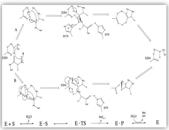
The nature of the MetAPs Reaction Mechanism
The exact reaction mechanism of MetAPs is not fully understood or established yet, but it is presumed that MetAP is involved in nucleophilic substitution reaction. In this so-called hydrolysis reaction, the H2O molecules or simply the hydroxide ligand acts as a nucleophile to
initiate the reaction. The Histidine (H) residues are among the well conserved residues of MetAP enzymes. For example, in the figure 3, H178 and H79 are well conserved in all MetAPs (type1 and type2) up to present pointing out their crucial role in catalysis. X-ray crystallography data
9
analysis unveils that HIS79 initiates the positioning of the initiator methionine residue into the binding cavity of the enzyme and protonate the newly exposed N-terminal amine. [23]. In E. Coli, Lower and colleagues have proposed that there are two main possible reaction mechanisms for MetAPs. The adopted two-fold reaction mechanism of MetAP enzyme is depicted below.
Figure 3. Reaction mechanism of Methionine Aminopeptidase in E. coli. In scheme A, a tetrahedral intermediate is being stabilized by Glu204 and a metal center, while in scheme B a tetrahedral intermediate is being stabilized by His178 residue and metal center[24].
10
2.5 The Major Function of MetAP-2 in Biological Processes
Although previous studies have indicated that MetAP-2 are “bifunctional” enzymes with a special pita-bread structure that are responsible for the cleavage of the N-terminal Methionine residues and protein’s synthesis regulation in-vitro, the actual functions of these enzymes go beyond this. For example, when MetAP2 enzyme activity is inhibited an arrest in cell growth is observed, thus implicating MetAP2 enzyme in endothelial cell proliferation. As a result of such correlation, MetAP2 is being considered as a potential target for inhibiting angiogenesis. Moreover, MetAP2 co-purifies and make interaction with the alpha (α) –subunit of the eukaryotic initiation factor two (eIf-2) -a heterotrimer that mediates the binding of tRNAiMet to
the ribosome, a finding suggested by other studies. Furthermore, MetAP2 provides protection for the elf-2α subunit from inhibitory phosphorylation by the elf-2α kinase and also interact with the protein kinase R [25].
2.6 Clinical significance of MetAP-2 Enzyme
It has been unveiled earlier that MetAP2 plays a critical role in angiogenesis. The Covalent binding of either the ovalicin or the fumagillin epoxide moiety to the conserved histidine residue in the binding cavity of the enzyme is believed to inhibit MetAP2 enzymatic activity leading to the inhibition of angiogenesis. The mechanism by which the inhibition of MetAP2 blocks angiogenesis is yet to be established. Additional studies and experiments that will validate the antiangiogenic activity that arises from inhibition of this enzyme are needed. Solid tumors progression, that is growth and metastasis heavily rely on new blood vessels formation and the natural product fumagillin and its allies such as (O-chloro-acetyl carbamoyl) fumagillol
(AGM-11
1470) commonly known as TNP-470, caplostatin as well as beloranib and ovalicin have shown potential anticancer activities in living cells. The authors suggest that fumagillin and TNP-470 both inhibit neovascularization via endothelia cell cycle arrest in the late G1 phase of the cell cycle. MetAP2 inhibition has effect on cells survival which have made it a potential target for antibacterial agents. Beloranib is also another potential inactivator of MetAP2 enzyme. They have shown significant efficacy in weight loss in obese subjects or patients by re-establishing equilibrium to how the human body metabolizes fat, a process that result in reducing body weight. It has failed in clinical trial due to its side effects or unwanted properties [2][25][26] [27].
2.7 Natural products as source of Lead Compound
Most natural products such as alkaloids or terpenoids are often good at crossing biological barriers and penetrating cells. These natural products have desirable pharmacokinetic properties making them essential for lead discovery in drug development. This is particularly important in areas such as antibiotic and anticancer therapies where natural products and their analogs constitute a meaningful percentage of drugs that are used for the treating diseases. Although they are an excellent source of lead compounds with regard to drug discovery and development, often times, there are needs to modify their structures in order to improve their pharmacological properties to be administered in a clinical trial. There are structural complexities of most natural products so semisynthetic approaches are used most often to optimize them. An example of a natural product that has undergone such approach is the fungal product or metabolite (fumagillin), that target MetAp-2 [5][28].
12
2.8 Drug Design and In Silico Approach
There are many life-threatening diseases, and the need for ideal drug development is prime in modern society. To design, develop and validate a new drug candidate to be marketed is quite an exhaustive process. In drug development process much or less than 75% of entirely process cost is consumed as a result of failures. Several years of intensive work would result in either success or failure. A rapid method of drug discovery process is significant to meet the challenges that are involved with the development of ideal drugs because the process of drug design, development, and marketing is tedious, expensive and very time-consuming.
High-throughput Experimental Screening has limitation such as accuracy, and it is cost intensive which hinders its application. As a result, the need to shift to an In Silico approach is worthwhile in drug development process. There are various methods used in In silico approach which includes but not limited to Virtual High-throughput screening, Homology Modelling, Molecular Docking, Comparative molecular field analysis (CoMFA), 3D pharmacophore mapping, quantitative structure activity relationship (QSAR).
Biological molecules such as proteins or nucleic acids are the actual target for drugs due to their signaling or metabolic pathways which are unique to disease processes. They are linked with disease progression by means of communication through protein-protein or protein –nucleic acids interactions that result into signaling propagation and increasing metabolic processes. By inhibiting or activating these processes with ideal drugs that have a greater competitive binding affinity as compared to their natural ligands is worth heeding in the drug development process. This is a two-fold process that can be achieved either by inhibiting the biomolecular interactions
13
between biomolecules or by activating biomolecules that are deregulated in some diseases, for example in cancer.
As it has been outlined earlier, to identify a good lead compound and to develop it into an effective drug is tedious and costly even if the target of interest is known. Drug development in recent times is being enhanced as result of the availability of Nuclear Magnetic Resonance (NMR) or X-ray structures or the 3D structures of biological molecules, docking tools as well as the computer aided drug design methods. Since drugs provide a lot of benefits for human, but their development process is expensive and time-consuming, the need to use an effective tool or technique is necessary to reduce the cost and time needed for development and marketing. An example of such technique is the In Silico drug designing technique that catalyzes the drug discovery process. This approach provides lots of information about binding energy, inhibition constant and predicts ADMET property. It does not maximize the utility alone so there is need to be coupled with an experimental method for proper validation.
In Silico drug designing process can be summarized into three major stages. In the first stage, a therapeutic target is firstly identified and then small heterogeneous molecules are built to be tested against the target. Stage two requires docking of selected hits to determine their binding specificity and affinity when docked into the binding sites of the targets. Finally, in the last stage, In Silico ADMET profiling studies are carried out to determine possible lead that could be modified into drug and then for use by human [29][30][31]. In Silico approach uses computational methods to model molecules. It is categorized into two, based on the knowledge of the substrate (ligands) and the protein-ligand interactions otherwise known as ligand-based and structure-based respectively. In this present work, structures-based In Silico method was
14
applied because the structure of the receptor was available. The receptor’s 3D structure of the receptor has been already determined through x-ray crystallography and deposited in the protein data bank (PDB) [28][29]. The figure below summarizes the three paramount steps that are involved in this approach
Figure 4 In Silico Approach during Drug Discovery process[29]
Target Selection and Optimization
Lead Discovery and Optimization
In Silico ADMET Profiling
Docking with Target and Lead Candidate
15
2.9 Stages of Modern Rational Drug Discovery and Development
In modern rational drug discovery and development process, there are several stages which include target identification, target validation, lead discovery and optimization, pre-clinical and clinical studies. The first stage in drug development pipeline is to identify a potential drug target. Most drug molecules target either proteins or nucleic acids (DNA or RNA)[28].The discovery of any potential drug for a particular disease, requires lots of researches, studies and projects about the disease in order to identify signaling cascades, and the gene encoding for the proteins that are involved. Critical pathways analyses are carried out along with genomic and proteomic approaches such as comparing the protein expression profile. This may take several years to establish the actual cause of the disease that may lead to an idea of how to treat that disease [29][30]. When a target has been identified and validated, the next step in the drug discovery process is to discover a lead compound for that target. A good lead compound for a target must have the ability to interact with the intended target to achieve a desirable effect, be amenable to synthetic modifications and finally possess some physical properties to enable it reach the target after being administered to a human patient. Natural ligand or substrate for the particular target of interest, natural product such as plant alkaloids, terpenoids and chemical compound libraries are good sources of lead compounds. Moreover, there are modern approaches for identifying possible lead compound which include but not limited to structure-based design, virtual High-throughput screening, literature, and patent-structure-based innovations.
16
2.10 In silico ADMET properties prediction of a drug molecule.
A potent drug should be less toxic and with a good pharmacokinetic properties which include: Absorption- the process by which a drug reaches the bloodstream from the site of administration; distribution- transporting of the drug molecule to its expected site of action in the body; metabolism- the conversion of the drug molecule into metabolites by actions of some specific proteases on the drug molecule, and finally be eliminated from the body a process referred to as excretion.
Nowadays, In silico screening tools play a critical role in the drug discovery process by predicting most relevant properties such as binding affinity, inhibition constant, pharmacokinetics, metabolism, and toxicity –all of which seems to be the cause of late failures in clinical studies in drug development. In order for a potential drug to reach the market, it must go through several stages, and these stages include preclinical development, clinical development, and regulatory approval. In the preclinical development, synthetic processes are carried out to enable the drug to be manufactured. This stage provides relevant information about the safety, efficacy, and affinity of the drug before beginning a clinical trial in human. Toxicity test of the drug candidate is carried out in model organism with permission from regulatory authority and afterward, a proof of experimental data is provided. The pre-clinical studies provide relevant information about the promising drug’s toxicity, pharmacodynamics, and pharmacokinetic properties, metabolism, organ sensitivity, as well as the starting dose. After this stage has been established, the clinical stage begins with the promising drug being tested in a human. The drug is administered to healthy volunteers to evaluate the safety, toxicity, and efficacy. This stage is subdivided into four (4) distinct phases namely phase (0, i, ii, iii and iv). Phase zero was
17
established by the Food and Drug Administration (FDA) in order to administer a single dose of the drug to about ten to fifteen healthy volunteers to get a preliminary human ADMET data. In phase i, the safety, tolerability, and both pharmacodynamics and pharmacokinetic properties are evaluated in about twenty to hundred healthy people and may last for about few months or a year and a half. Oftentimes, if it is a deadly disease, for example, Ebola, the drug may be administered to the actual diseased patients. Phase ii, on the other hand, assesses how effective the drug molecule is and determines its safety and side effects in few hundred diseased patients with duration ranging from one to three years. The next phase, phase (iii) is meant for a larger trial to be performed in thousands of patients and compared the drug with other marketed drugs. Phase iv is the final phase also known as the regulatory Approval for the market. In this phase, a New Drug Application (NDA) with a summary from clinical trials is submitted to the FDA for approval. Once the NDA is submitted, FDA then decides whether to approve the drug or not; if approved, then prescription for the drug is provided in order to enter the market to assess its real safety and tolerability[28][29].
Lead compounds can be obtained in few ways- de novo, natural products, biotechnology and High-throughput screening (HTS). Of all these approaches, the most popular used one is HTS which uses compound libraries to find lead compound for further modification. The driving force in this approach is a computer which provides means of testing and screening several thousands of compounds with the aim of getting promising lead compounds. These compounds are then docked to determine their inhibition constants (Ki) and binding energies using docking tools and their structure are investigated based on chemical groups and then linked to the target molecules[30]
18
2.11 Virtual Screening in Drug Development
In recent year, Pharmaceutical research has directed a great deal of attention on a computational screening of various databases to discover new inhibitors for intended targets. A famous method used by these pharmaceutical researchers is virtual screening, a method that uses a computer to discover new sets of inhibitor based on their biological structures or nature of the active site of the receptors. Screening is a term used to describe the practice of testing a large set of heterogeneous molecules for the activity in the model system which mimics the human disease [32]. Structure based In Silico approach can be viewed as a two-fold process, namely virtual screening and de novo design. Virtual screening also known as virtual throughput screening (VHTS) is a very important technique presently used to discover lead compounds in drug discovery process. In VHTS methods, there are two classes namely, Ligand-Centric and Receptor-centric virtual screenings. The former takes into consideration the comparative analysis of structural shape, chemical and pharmacophore similarities between compounds and known ligands, and also thorough knowledge of the selected active molecules. The later on the other hand, provides information about interaction of a given compound with a target receptor. In receptor-based virtual screening, the principal technique use is a molecular docking, that uses a computer to predict both the affinity and binding mode of a particular compound and a target receptor. There is a critical point of concern here, which is in cooperating a dynamic nature of the receptors, because in molecular docking algorithms, the receptors are kept rigid at a very low energy conformation, and only the ligands are considered to be flexible. In reality, however; receptors at similar energies can have different conformational states and their binding sites can undergo induce fit. With the availability of various number of small molecules and fast growing in corporate and public libraries of compounds, the importance of VHS has now
19
increased drastically in new drug development. Presently, there are various VHTS tools that available commercially, and with these tools new drug candidates can be tested, by determining complementarity between the receptor and its ligand (guest). These docking tools can predict the 3D binding mode (pose) of a docked ligand in the binding site of a receptor as well as binding energy. As a result of this, many poses or representations of the ligands are generated, and varied by conformations, positions and orientations in the receptor’s active site. The purpose of this process is to determine the best energetically favorable pose of the ligand in the receptor’s binding cavity. To ease the process of pose and affinity predictions of a ligand, efficient algorithms and scoring functions are developed and applied differently by various kind of docking programs [30][33] .
2.12 Docking Process in Drug Design
Docking is process that strives to find the best relation between two molecules: receptor traditionally a protein and a ligand usually a drug molecule. In other words, it is a computational method of structure based drug design intended for predicting receptor-ligand interactions, geometries, inhibition constants, and binding affinities. In recent time, automated receptor – ligand docking is an effective tool that has play a critical role in structure-based drug discovery. It predicts the translation, orientation, and conformation of a ligand relative to the receptor’s active site. There are two general methods of docking: rigid docking and flexible docking. In rigid docking, both the receptor and the ligand are treated as rigid objects while in flexible docking, only the ligand is considered as an interlocked object and the receptor as a rigid body. The most commonly used method is flexible docking simply because ligands are flexible
20
molecules. Alternative methods to these two methods are soft docking and partially flexible protein docking – all of which seems to reflect the flexibility of the protein’s side chains [32][34]. Docking process has been, and continue to play an important role in structure-based drug design because of its ability to identify lead compounds by means of energy minimization. It has also received a tremendous interest because of the availability of high-resolution structures of enzymes and automation of docking process through computer-based simulation. In every docking process, there two significant aspects which include a scoring function and an effective algorithms - for conformation space search to determine the best orientation of the receptor and ligand or pose. The efficiency or accuracy of a good scoring function is its ability to clearly establish a correct binding mode from other incorrect binding modes. Since different ligands and receptors are in numerous conformations at different energy levels, a good scoring function is then needed to rank their binding modes accurately. Today, there are different scoring functions used in molecular docking. The “Assisted Model Building for Energy Refinement “(AMBER) force-field type scoring function was used herein to determine the various binding modes. The most commonly used scoring function in docking is the energy-based scoring function which determines the ligand conformation when it is bound to the target receptor. The underlying “hypothesis” is that, at a lower energy value, ligand binds well to its target receptor as compared to those of higher energy values[35].
In general, these optimization methods, for example, Simulated Annealing and Genetic Algorithms can recognize a greater amount and different known ligands. Today, one of the most efficient algorithms is the evolutionary algorithms that is widely used in molecular docking
21
applications with a better performance over the Simulated Annealing in some area of applications[35].
Simulated annealing is a search algorithms that is based on probability and is meant for solving complex optimization problems and has solved practical difficult problems since its development [36]. Evolutionary algorithm is formulated based on borrowed ideas from both genetics and natural selection. It is now considered as the best suited for handling very difficult optimization problems in docking due to its adaptable concept for solving numerous problems. It has not only been used to solve problems surrounding large search spaces, where traditional optimizations methods have failed or are less efficient , but also in several structure-based design problems and protein conformation prediction. Presently, evolutionary algorithm has three principal independently developed algorithms with strongly related implementations namely, genetics algorithms, evolutionary strategies, and evolutionary programing [35]
2.13
AutoDock, a Tool for Docking in Drug Design
AutoDock is an automated docking tool that predicts both binding affinity and inhibition constant of a ligand or a drug molecule that binds to a receptor, typically a protein. AutoDock consists of two major and distinct components, namely autodock and autogrid. Autodock performs the actual docking of the ligand to a set of grids that describe the target receptor while autogrid pre-calculates the grids. AutoDock 4.2, the latest version of AutoDock, has a free-energy scoring function. This scoring function is primarily based on a Linear Regression Analysis, the AMBER force field. In addition to this free-energy scoring function, it also incorporate side chains flexibility of the receptors or the macromolecules. In this work, the so-called Lamarckian Genetics Algorithms (LGA), a genetic method that is much more robust and
22
efficient as compared to the Monte Carlo Simulated Annealing (SA) method was used. Further still, LGA handle ligands with more degree of freedom as compared to its counterparts. The underlying principal idea of the Lamarckian model of genetics is that, the phenotype acquires by an individual in an environment during its life time can be reverse transcribed into its genotype and be transmitted to the next generation of offspring [37][38].
23
Chapter 3 Material and Method
Here, the OTAVA’s Chemicals Library was screened to select a new class of inhibitors based on energy values and inhibition constants. It is anticipated here that these inhibitors will be potent and selective for Methionine Aminopeptidase (II), PDB code (5CLS). This divalent protease has three hundred seventy-one amino acid residues. It was complexed with spiroepoxytriazole, a fumagillin-like irreversible inhibitor of MetAP2, at a resolution of 1.75 Å (14)[39]. Each new candidate was evaluated based on energy value, inhibition constant (Ki) and various interactions with the receptor. The following computational methods and steps were performed to test and validate the new set of inhibitors that was selected from the library:
(I) Preparation of the receptor’s (MetAp2) structure for docking purpose
(II) Preparation of the receptor’s native ligand and other natural ligands for re-docking into the binding cavity of the receptor as test case
(III) Screening of the OTAVA ‘s Chemical Library using PyRx to select a new class of inhibitors
(IV) Redocking of these selected inhibitors using AutoDock 4.2 into the receptor’s active site.
As mentioned early from the onset, docking method has two important features: an efficient algorithm for searching conformation space and a valid scoring function. In AutoDock, a semi-empirical force field based on the “Assisted Model Buılding with Energy Refinement” (AMBER) force field is used in docking. Moreover, it also uses a molecular mechanics model to determine the enthalpy contributions, such as electrostatic, Van der Waals and Hydrogen-bonding, and an
24
empirical model to determine the entropy changes upon binding of a ligand into a receptor’s binding cavity [40]). In searching for conformation, AutoDock favors the Lamarckian Genetics Algorithm, which is based on apply Mendelian Genetics, an antithesis of the Darwinian Evolution [41]
3.1 Preparation of the receptor’s (MetAp2) structure for docking purpose
From the Protein Data Bank (PDB), the crystal structure of Human Methionine Aminopeptidase 2 complexed with Spiroepoxytriazole inhibitor was extracted (14). The protein preparation protocol and the “Clean Geometry” toolkit in Discovery Studio (version 2016) software (Accelrys, Inc.) were deployed to minimize and to prepare the enzyme for docking. In preparing the protein’s structure for docking, Chain A and the protein groups (disulfide residue, backbone, sidechain, hydrophobic and hydrophilic residues and the acidic and basic residues) were maintained. Elsewhere, the divalent cofactor, Cobalt (Co2+) that is involved in energy calculation during docking was also retained in the active site of the receptor for docking. All other molecules such as the solvent (H2O) that existed in the PDB structure were eliminated. After all,
all omitted hydrogen atoms were added based on the protonation state of the receptor at a pH of 7.4. The ionic strength and dielectric constant were set to 0.145 and 10 respectively.
3.2 Preparation of the Receptor’s native ligands for re-docking as test case
The native ligand Spiroepoxytriazole, a fumagillin-like irreversible inhibitor of MetAP2 was extracted from the receptor’s active site for preparation. All omitted hydrogen atoms were added and the ligand’s minimization protocol in Discovery Studio 2016 was employed for minimization.
25
The same approach was applied to other native ligands from other structure PDB codes. The underlying ideology was to determine the accuracy of the selection process, based on which other promising inhibitors were selected. The AutoDock Tools (ADT) graphical user interface program was used to generate the docking files: a grid parameter file (GPF) which specifies the 3D search space by setting up the number of grid points in each dimension, the center of the grids and the spacing between points, and the types of probe atoms to use, the filename of the receptor and the names of each output grid maps, and the docking parameter file (DPF) [40]. These native ligands of MetAP-2 were re-docked into the active site of the enzyme to determine the docking accuracy between experimental Ki and the Ki after redocking. The obtained computational inhibition constants agree reasonably well with the experimental values (Table
1). In this procedure, the (GPF) for each ligand was prepared with a grid box size of 60, 60 and
60 and 26.53, 21.30, and 17.55 were considered as the x, y, and z coordinates respectively. In addition, docking simulation was performed applying the famous Lamarckian Genetic Algorithm (LGA). All the parameters of the LGA were set to their default values, except the simulation run that was twenty-five million energy evaluation. The docked native ligands calculated inhibitions and experimental inhibition constants are presented below.
26 Table 1. Depicts docked native ligands: Calculated inhibition and experimental inhibition constants. PDB
Code
Structure of Compound Gibbs free Energy (ΔG) Value Kcal/mol Docked Ki Value Kcal/mole Experimental Ki Value (nM) 52T -8.74 392.57 220 FUG -9.20 181.08 24 94A -8.36 739.69 220
From Table 1 above, two of these native ligands (spiroepoxytriazole and fumagillin) were chosen to display their 3D and 2D structures.
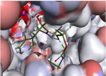
27
Figure 5: Shows the 3D structure of the native ligand Spiroepoxytriazole binds into the active site of the receptor (MetAP2).
Figure 6: 2D diagram showing various interactions such as Vander Waals, pi-sigma, Carbon
Hydrogen bond, etc, between the receptor Spiroepoxytriazole
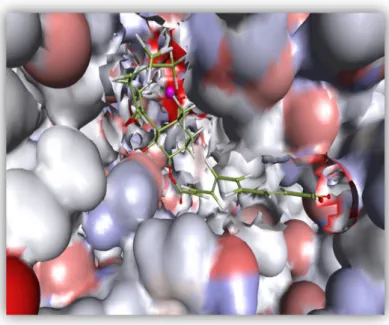
28 Figure 7: Shows the 3D structure of fumagillin binds into the active site of the enzyme (MetAP2).
Figure 8. 2D diagram that depicts various interactions such as Vander Waals, carbon Hydrogen bond, conventional Hydrogen bond, metal-Acceptor, pi-Alkyl ,etc, between the receptor and Fumagillin.
29
3.3 Screening the OTAVA’s Chemical Library using PyRx
The OTAVA’s Chemicals Library is one of the libraries of chemical compounds that contains more than three million, two hundred thousand (3,200,000) commercially “tangible compounds. “Tangible” is a term that signifies the synthesizability of these chemical compounds. All the compounds generated in this library are filtered in accordance with both Lipinski and Veber rules. By incorporating these rules implies that, these compounds are subject to the following properties: ClogP from 0 to 5, molecular weight from 160 to 500, number of Hydrogen -donors from 0 to 5, number of Hydrogen-acceptors from 0 to 9, number of NO2
groups from 0 to 2 and finally the number of rotatable bonds from 0 to 10. Several thousand of compounds initially in structural data file (SDF) format were converted to PDB file format and screened against the target protein using PyRx [42][43][44][45].
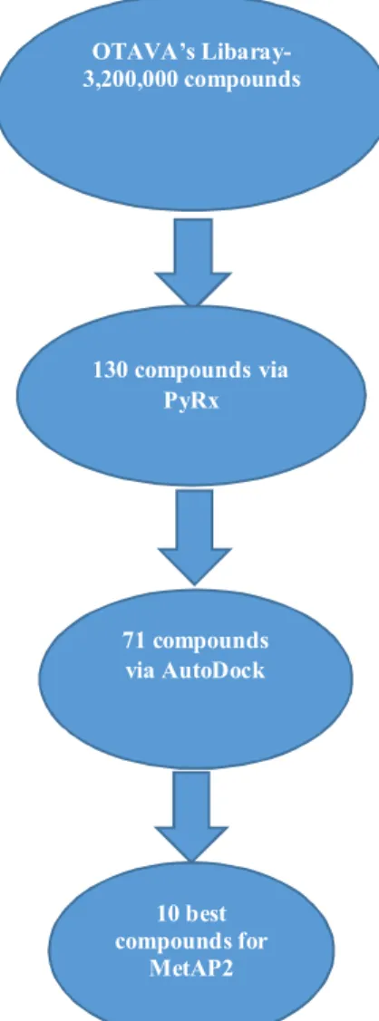
PyRx is an open source virtual screening software intended for computational Drug Discovery. PyRx empowers drug designers to screen libraries of compounds against potential drug targets typically a receptor or protein. While it is true that there is no “magic button” in drug discovery process, PyRx has a docking program which makes it worthwhile for Computer Aided Drug Design [46]. Here, PyRx was deployed to screen several thousand of compounds from the OTAVA’s Chemical Library. The AutoDock vina search space center was set to 23.2966, 25.1912, 13.8546 as the x, y, & z coordinates respectively while the dimensions in Angstrom (Å) was 250,000 for each (x, y, & z), and the exhaustiveness was 8. From PyRx’s result table, each candidate was selected based on the minimum threshold[46][47]. One hundred thirty candidates were selected out of the several thousand compounds that were screened. These selected candidates were redocked using AutoDock 4.2 for re-evaluation and further validation.
30
3.4 Redocking selected inhibitors using AutoDock.
In the redocking process, the grid parameter file (GPF) for each ligand was prepared with a grid box size of 60, 60, and 60 in each dimension. The x, y, & z coordinates were 26.53, 21.30, and 17.55 in the order given corresponding to the center of the receptors coordinates. The Lamarckian Genetic Algorithm was used in performing the docking simulation. All other parameters of the LGA were again set to their default values; for example, GA run was set to 10, a population size of 150, 27, 000 generations etc, except the docking simulation run that was set to twenty-five million (25, 000,000) energy evaluations.
31
Chapter 4
Result and Discussion
There have been numerous efforts to design a selective, and a potential inhibitor of MetAP2, but such efforts have not been fully materialized. The only known inhibitor of this enzyme is the fungal product (fumagillin) and its analogs – all of which has failed in numerous clinical trials due to their poor pharmacokinetics properties and toxicities. Apart from this natural product (fumagillin) and its derivatives such as beloranib, TN470, etc, no other inhibitor has shown selectivity and potency against MetAP2 enzymes both in-vitro and in-vivo. The effort to design or generate a new class of selective and potential inhibitors with fewer side effects remains a major challenge for drug designers or Medicinal Chemists. In this present research, due to these various liabilities of these natural products; the research intends to address this problem through an In Silico approach by screening a library of tangible-chemical compounds to select candidates based on inhibitory potential and energy values. It is postulated herein that these candidates would serve as a new class of inhibitors and/or lead compounds for the target receptor. To fulfill this plan, few native ligands were re-docked into the minimized receptor’s binding cavity to compare their docked inhibition constants with the experimental ones. Based on this analysis, the inhibitory potentials of the new class of inhibitors were evaluated. A deviation between native ligands experimental inhibition constants and docked ligands inhibition constants was observed with that of the docked ligands inhibition constants being lower than the experimental ones. Henceforth, the lowest experimental inhibition constant (Ki) value of these native ligands was considered based on which the new class of inhibitors for the target was selected.
32
In a drug discovery process, drug designers seek for a drug candidate that will work at a lower concentration to inhibit a target receptor without inhibiting other receptors simultaneously. If any drug molecule tends to target only a receptor of interest at low concentration with no effects on other biological targets or proteins, such drug molecule is indeed considered to be ideal inhibitor for that target. Here, by using In Silico screening and Molecular modelling the researcher aims to develop such ideal drugs having high potency and excellent pharmacokinetics for human MetAP-2. Also the ADMET values of these candidates should pass the required properties.
Presently, the well- known inhibitors of this enzyme are fumagillin and its derivatives but have all failed in clinical studies. To generate these new class of inhibitors for the target enzyme, several thousand of chemical compounds were screened from OTAVA’s Chemical Library via PyRx. After the screening process, one-hundred-thirty chemical compounds were selected based on a threshold of -9.0 kcal/mole inspired by the lowest experimental inhibition constant as a minimum criterion. These selected inhibitors were redocked into to the receptor’s binding cavity using AutoDock 4.2 for further validation and evaluation. The results of AutoDock 4.2 placed some candidates below and above the minimum value. All candidates whose inhibition constants were either above or the same as the minimum threshold were considered and those whose inhibition constants values were below the minimum were discarded and excluded. A total of seventy-one candidates were selected and a total of fifty-nine candidates were excluded, a situation that draws a special attention to the difference in scoring functions between the two (2) docking programs. AutoDock being one of the most favored among all docking tools, these Seventy-one 71 compounds were accepted and considered as potential candidates for the target receptor. To further limit the number of possible inhibitors, ten 10
33
best candidates were chosen based on their minimum energy values. Compound 1 and Compound 2 were selected in order to provide vivid analysis of the various binding interactions between the receptor and the inhibitors. A simulation and an experimentation of these compounds in the near future will be worthwhile to substantiate and validate their inhibitory potential. A summary of the selection process is given in the Figure 9 below.
Figure 9: Shows a summary of the selection process. OTAVA’s Libaray-3,200,000 compounds 130 compoundsvia PyRx 71 compounds via AutoDock 10 best compounds for MetAP2
34
All seventy-one selected candidates for Methionine Aminopeptidase 2 along with their various energy values, inhibition constants, run numbers and number of torsional angles are given in
Table 2.
Table 2. Depicts all 71 selected candidates along with their energy values, inhibition constants, run numbers and number of torsional angles.
Cod e
Structure of Inhibitor Run
Number Number of Torsional Angel ΔG Value Kcal/mole Inhibition Constant (Ki) nM 1 8 6 -9.23 171.80 2 9 4 -10.03 44.34 3 9 5 -10.47 21.13 4 2 4 -9.56 98.30 nM
35 5 4 4 -9.78 67.76 6 9 6 -9.53 103.90 7 8 4 -9.53 104.03 8 8 6 -9.29 153.71 9 6 4 -9.66 82.79
36
10 4 4 -9.32 146.88
11 10 5 -9.64 85.20
12 9 4 -9.22 174.23
37 14 7 7 -9.68 80.34 15 6 5 -9.53 103.14 16 8 6 -10.56 18.06 17 7 7 -9.06 229.90 18 7 5 -9.30 152.30
38
19 4 6 -9.21 177.12
20 2 6 -9.13 203.72
21 2 6 -9.11 211.79
39
23 7 4 -9.30 152.82
24 2 3 -10.25 30.61
25 4 3 -9.34 143.48
40 27 2 4 -9.00 254.04 28 3 4s -9.86 59.50 29 10 3 -9.27 161.30 30 10 5 -9.47 113.47 31 10 5 -9.51 106.27
41
32 6 3 -9.65 84.23
33 4 5 -10.39 24.12
34 -10.12 38.18
42
36 5 6 -9.49 110.34
37 6 6 -9.47 113.67
38 10 4 -9.48 113.09
43 40 7 5 -9.69 79.48 41 8 3 -9.26 162.37 42 4 5 -9.80 65.01 43 1 5 -10.42 23.16 44 7 5 -9.91 54.04
44
45 2 5 -11.22 6.00
46 3 4 -9.65 84.47
47 4 3 -9.98 48.29
45
49 1 6s -10.61 16.80
50 7 6 -9.98 48.13
51 3 3 -9.18 187.25
46
53 9 4 -9.71 76.28
54 7 4 -9.21 176.00
55 4 5 -9.27 159.48
47
57 5 5 -9.23 172.96
58 5 5 -10.60 17.08
59 3 5 -9.31 149.70
48
61 8 4 -9.49 110.93
62 5 4 -9.50 108.09
63 6 4 -9.31 150.90
49
65 10 4 -9.48 112.41
66 1 4 -9.68 80.91
67 7 3 -9.94 51.88
50
69 7 2 -9.20 179.44
70 8 2 -9.14 199.63
71 7 5 -9.31 149.53
From Table 2 above, ten best candidates were considered based on lowest Gibbs free energy values from -10.0 kcal/mole to – 11.22 kcal/mole.
51
Table 3: Shows the ten best candidates selected based on lowest Gibbs free energy values.
Code Structure of Compound Run Number Number of Torsional Angle (ΔG) Value In Kcal/mole Inhibition Constant (Ki) nM 1 2 5 -11.22 6.00 2 4 5 -10.39 24.12 3 1 6 -10.61 16.80 4 5 5 -10.60 17.08
52
5 8 6 -10.56 18.06
6 9 5 -10.47 21.13
7 1 5 -10.42 23.16
53
9 1 5 -10.12 38.18
10 9 4 -10.03 44.34
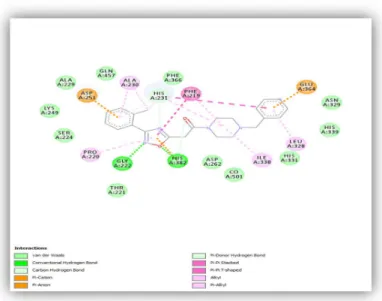
To provide vivid interactions of the docked poses of these ten (10) selected potential inhibitors of the target enzyme, Compound 1 and compound 2. As it is evident below in the diagram, an analysis of the best binding mode of compound 1, the compound with the lowest energy value (-11.22Kcal/mole) in the binding cavity of MetAP2 revealed that it is located in the vicinity of the cofactor (Co2+). It interacts with the amino acid residues in the lining of the binding cavity of the receptor as well as the di-metal cofactor (Co2+). The chemical structure analysis of compound 1 revealed the following chemical features: a molecular formula of C22H24N4O2 (52 atoms),
molecular weight of 376.461792g/mole, molecular volume of 313.988912g/L, with no valence electron and chiral center. It has a dipole moment of 4.615 Debye. In addition, it contains two benzene rings which are major functional groups, two nitrogen containing cyclohexane ring, a carbonyl group, and a 1, 3, 5 – oxadiazole ring –all of which is associated with different interactions with the amino acid residues in the active site of the receptor. The following important interactions between the receptor and the inhibitor were observed: two pi-anion
54
interactions, one between one of the benzene rings and the ASP251 and the other between the second benzene ring and the GLU364 as depicted in brown. Two conventional hydrogen bonds, one between the nitrogen of oxadiazole ring and the GLY222, and the other between the oxygen of the oxadiazole ring and the HIS382 as shown in green. A pi-cation interaction is observed between the oxadiazole ring and one of the most conserved residues -HIS382. The hydrophobic aliphatic amino acid Phenylalanine (PHE219) is involved with pi-pi stacked interactions with one of the benzene rings and the oxadiazole ring. Pi-Alkyl interactions are shown between ALA230 and one of the benzene rings, another between the other benzene ring and the LEU328, while PRO220 also has the same interaction with the oxadiazole ring. Alkyl interactions are observed between the methyl group on the benzene ring and ALA230 as well as the HIS231, and also between HIS231 and the nitrogen containing cyclohexane ring. Here, carbon-hydrogen bond interactions with the following residues are also depicted, between HIS231 and one of the benzene ring and the oxadiazole ring, and also between HIS382 and the carbonyl carbon. Vander der Waals interaction with the cofactor is observed along with various Vander Waals interactions among the rest of the molecules especially those hydrophobic amino acid residues
55 Figure 10: 2D diagram showing various interactions between the receptor and the inhibitor as depicted in
different colors.
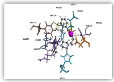
Figure 11: The 3D orientation of compound 1 in the binding cavity of MetAP-2 enzyme. Amino acid residues within the lining of the active site are shown as sticks labeled black, the inhibitor is depicted as a scaled ball and stick, and finally the divalent cofactor (Co2+) is shown as a CPK model (pink) in the
56 Figure 12: Compound 1 binding into MetAP2 active site.
Similar to compound 1, Compound 2 has similar chemical features and interactions with the receptor. There is a peculiar interaction that is overserved between the receptor and the inhibitor, that is, a metal-acceptor interaction. This interaction is between the cofactor and the nitrogen of the oxygen containing ring in which the nitrogen atom acts as a metal ion acceptor.
![Figure 2. Comparison of MetAPs primary structures. Conserved metal binding amino acids residues within the C-terminal catalytic domain are shown (DDHEE) [18]](https://thumb-eu.123doks.com/thumbv2/9libnet/4330363.71248/19.892.164.671.323.694/figure-comparison-metaps-structures-conserved-residues-terminal-catalytic.webp)
![Figure 4 In Silico Approach during Drug Discovery process[29]](https://thumb-eu.123doks.com/thumbv2/9libnet/4330363.71248/26.892.100.797.387.1019/figure-silico-approach-drug-discovery-process.webp)