http://journals.tubitak.gov.tr/medical/ © TÜBİTAK
doi:10.3906/sag-1403-24
Clinical outcomes and optical performance of four different
multifocal intraocular lenses
Başak BOSTANCI CERAN1,*, Tamer TAKMAZ2, İzzet CAN3, Gülizar DEMİROK4, Betül Seher UYSAL5 1Ulucanlar Eye Training and Research Hospital, Ankara, Turkey
2Department of Ophthalmology, Atatürk Training and Research Hospital, Ankara, Turkey 3Maya Eye Hospital, Ankara, Turkey
4Faculty of Medicine, Dr Rıdvan Ege Hospital, Ufuk University, Ankara, Turkey 5Department of Ophthalmology, Atatürk Training and Research Hospital, Ankara, Turkey
1. Introduction
Significant technical advances in ophthalmology have led cataract surgeries to evolve into refractive procedures. Increased life expectancy has brought more people with cataracts to consult their physicians regarding surgeries; this has enabled satisfactory vision without the need for spectacles. One of the most important reasons why monofocal intraocular lenses (IOLs) cannot mimic human crystalline lens entirely is their inability to accommodate. Postoperatively, while distance vision can be made clear, reading vision is compromised. To this end, multifocal IOLs offer an alternative for people seeking independence from spectacles (1–3).
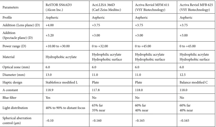
In this study, we evaluated clinical results and optical performances of four different multifocal IOLs. Eyes that received Acrysof ReSTOR SN6AD3 (Alcon Laboratories), Acri.LISA 366D (Carl Zeiss Meditec AG), Acriva Reviol
MFM 611 (VSY Biotechnology), and Acriva Reviol MFB 625 (VSY Biotechnology) IOLs were investigated in the present study. The main properties of the IOLs are shown in Table 1.
2. Materials and methods
The study was approved by the hospital’s ethics committee and was performed in accordance with the ethical principles as described in the Declaration of Helsinki.
In the scope of this retrospective study, charts of 90 eyes of 51 patients who received multifocal IOLs between October 2007 and July 2011 in Ataturk Training and Research Hospital were evaluated.
Patients who had previous eye surgery or eye diseases that could affect the final visual acuity like glaucoma, amblyopia, retinal or corneal pathologies, or corneal astigmatism higher than 1.00 D were excluded from
Background/aim: The purpose of the present study was to evaluate clinical outcomes and optical performance of 4 different multifocal
intraocular lenses (IOLs).
Materials and methods: Ninety eyes of 51 patients who received Reviol MFM 611, Reviol MFM 625, Acri.LISA, and ReSTOR SN6AD3
multifocal IOLs after cataract surgery were retrospectively evaluated. The patients were similar in terms of age, sex, cataract hardness and axial length. The mean outcome measures were uncorrected and corrected distance visual acuity (UDVA, CDVA), distance-corrected intermediate and near visual acuity (DCIVA, DCNVA), intra or postoperative complications, and contrast sensitivity (CS) results under mesopic conditions. The mean follow up period was 10.5 months (range: 6–12 months).
Results: All cases were within ± 0.75 D of emmetropia. Postoperative increase in UDVA and DCNVA was statistically significant in
all groups. The Acri.LISA group showed slightly lower DCIVA compared with the other IOLs. CS was clinically similar between the groups. None of the patients developed any early or late postoperative complication or neuroadaptation problem, which necessitated explantation of the lens.
Conclusion: All four multifocal lens designs provided satisfactory visual functions and CS results in patients who fulfilled the criteria
for multifocal lens implantation.
Key words: Cataract surgery, multifocal IOLs, phacoemulsification, premium IOLs, presbyopia
Received: 08.03.2014 Accepted/Published Online: 08.04.2015 Final Version: 19.04.2016 Research Article
the study. In addition, patients needing high contrast sensitivity (CS) for their occupation and people with meticulous personality were precluded since multifocal lens implantation is contraindicated in such patients. All patients enrolled in this study met the inclusion criteria and signed an informed consent agreement before any procedure was performed.
Thirty-nine patients underwent binocular surgery and 12 patients had unilateral cataract surgery. Sixteen patients had bilateral and 3 eyes had unilateral Reviol MFM 611 IOL, 14 eyes of 7 patients had Reviol MFB 625 IOL, 10 patients had bilateral and 8 had unilateral Acri.Lisa IOL, and 6 had bilateral and 1 had unilateral ReSTOR SN6AD3 IOL implantation.
Cataract hardness was assessed using the Lens Opacities Classification System III (LOCS III) scale (4). All eyes had group 2 to 4 nuclear or corticonuclear cataracts.
Keratometric evaluation, corrected distance and uncorrected distance visual acuity (CDVA, UDVA) measurement, detailed biomicroscopy, applanation tonometry, and funduscopic examination after dilation were performed with each patient preoperatively. Corneal toricity was measured using Keratron Scout corneal analyzer (Optikon 2000 SpA). The immersion method (Cinescan Ultrasound, Quantel Medical) was preferred for IOL power assessment.
A standard dilation regimen of cyclopentolate hydrochloride (1.0%), phenylephrine hydrochloride (2.5%), and ketorolac tromethamine (0.5%) were used preoperatively. All of the surgeries were performed by two experienced surgeons (İC, TT) using the same phacoemulsification system (Infiniti Vision Systems, Alcon Laboratories Inc.). Any perioperative complication was noted.
UDVA and CDVA were determined by the Early Treatment Diabetic Retinopathy Study (ETDRS) charts on day 1, week 1, month 1, month 3, and month 6 exams and transformed into logMAR units for statistical analysis. Distance-corrected near and intermediate visual acuities (DCNVA, DCIVA) were obtained using the Jaeger scale. Postoperative complications such as anterior chamber reaction, posterior capsular opacification (PCO), decentralization of the IOL, and tilt were recorded. CS measurement was performed on the 3rd month visit with and without a glare source (CSV 100E, Vector Vision) under mesopic conditions. Halo and glare incidence, as well as patient satisfaction, were investigated.
3. Statistical analyses
Data analyses were done by SPSS, for Windows, version 11.5. The Shapiro–Wilk test for normality was used to investigate the distribution of constant variables. Kruskal– Wallis, Conover’s nonparametric, Pearson’s chi-square,
Fisher’s exact t, and Wilcoxon’s signed rank tests were used. P values less than 0.05 were considered significant. In all multiple comparisons, in order to control type 1 error, Bonferroni correction was performed.
4. Results
The mean follow-up period of the patients was 10.4 ± 1.2 months (min: 6 max: 12). The average age of the patients was 52.3 ± 12.6 (min: 22 max: 75). Forty-nine percent of the patients (n = 25) were male, whereas 51% were female (n = 26). There was no statistical difference between the groups in terms of age and sex distribution (P = 0.492 and P = 0.810, respectively).
Mean preoperative UDVA of the patients was 0.38 ± 0.23 (0.54 ± 0.41 logMAR) and CDVA was 0.59 ± 0.29 (0.30 ± 0.33 logMAR). There was no statistical difference between the groups for UDVA and DCVA (P = 0.073 and P = 0.369, respectively).
The mean keratometric difference of the eyes was 0.6 ± 0.3 D preoperatively. The difference between the groups was statistically insignificant (P = 0.288). Groups were similar in terms of axial length, IOL power, and cataract hardness (P = 0.6 08, 0.345, and 0.051, respectively).
There was one posterior capsular rupture and 3 iris prolapse in the Reviol MFM 611 group. In the Reviol MFB 625 group, one case had an irregular capsulorrhexis. One case had a small posterior capsular rupture and 2 had iris prolapse in the Acri.LISA group. No complication occurred in the ReSTOR group. No vitreous loss was seen in these cases and all of the IOLs could be centralized in the bag.
Six months following surgery, the mean UDVA of the patients was 0.81 ± 0.19 (0.1 ± 0.11 logMAR), whereas the mean DCVA was 0.96 ± 0.12 (0.01 ± 0.03 logMAR). Postoperative increase in UDVA was statistically significant in all groups (Table 2). When in-group changes were evaluated, the difference between the pre- and postoperative UDVAs and CDVAs were statistically insignificant (P = 0.261 and P = 0.159, respectively).
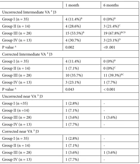
When intermediate visual acuities were evaluated, the Reviol MFM 611 group was found to have more cases reading J 1 and 2 compared with the Acri.LISA group. In the 6 month follow-up period, the difference between Acri.LISA and the other groups was significant (Table 3). No significant difference was found in terms of DCNVA and uncorrected near visual acuities (Table 3).
All of the cases were within ± 0.75 D emmetropia on month 1 and month 6 postoperative examinations. The change between the groups between month 1 and month 6 was insignificant.
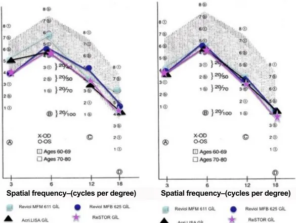
There was no significant difference between the groups in terms of CS with or without a glare source under mesopic conditions. CS results can be seen in Figure 1.
Table 1. Properties of IOLs.
Parameters ReSTOR SN6AD3(Alcon Inc.) Acri.LISA 366D(Carl Zeiss Meditec) Acriva Reviol MFM 611(VSY Biotechnology) Acriva Reviol MFB 625(VSY Biotechnology)
Profile Aspheric Aspheric Aspheric Aspheric
Addition (Lens plane) (D) +4.00 +3.75 +3.75 +3.75
Addition
(Spectacle plane) (D) +3.20 +3.00 +3.00 +3.00
Power range (D) +10.00 to +30.00 0 to +32.00 0 to +45.00 0 to +45.00 Material Hydrophobic acrylate Hydrophilic acrylateHydrophobic surface Hydrophilic acrylateHydrophobic surface Hydrophilic acrylateHydrophobic surface
Optical zone (mm) 6.0 6.0 6.0 6.0
Diameter (mm) 13.0 11.0 11.0 12.5
Haptic design Stableforce modified L Plate Plate Balance modified C
A constant 118.9 117.8 118.0 118.0
Blue filter Yes No No No
Light distribution 40% to 90% to distant focus 65% far35% near 60% far40% near 60% far40% near Spherical aberration
control (µm) –0.10 –0.160 –0.165 –0.165
Group 1: Reviol MFM 611, Group 2: Reviol MFB 625, Group 3: Acri.Lisa 366D, Group 4: ReSTOR SN6AD3.
Table 2. Preoperative and month 6 UDVAs and DCVAs.
Preoperative month 6 UDVA LogMAR Group I (n = 35) 0.4 (0.2–2.0)A 0.1 (0.0–0.5)A Group II (n = 14) 0.7 (0.0–2.0)A 0.05 (0.0–0.3)A Group III (n = 28) 0.3 (0.0–1.0)A 0.1 (0.0–0.4)A Group IV (n = 1 3) 0.5 (0.0–1.3)A 0.1 (0.0–0.4)A DCVA LogMAR Group I (n = 35) 0.1 (0.0–1.0)A 0.0 (0.0–0.5)A Group II (n = 14) 0.35 (0.0–2.0) 0.0 (0.0–0.0) Group III (n = 28) 0.2 (0.0–1.0)A 0.0 (0.0–0.2)A Group IV (n = 13) 0.3 (0.0–1.3) 0.0 (0.0–0.1)
Group 1: Reviol MFM 611, Group 2: Reviol MFB 625, Group 3: Acri.Lisa 366D, Group 4: ReSTOR SN6AD3.
A = difference between day 0 and month 6 is significant according to Bonferroni correction (P < 0.0042).
During the follow up period, PCO was observed in 20 cases. Only 3 cases had significant opacification that necessitated Nd-YAG laser capsulotomy. There was no significant difference in terms of PCO formation between the groups (Table 4).
At month 6, halo and glare incidence and quality of vision were investigated. Halo and glare rates are shown in Table 4; they were similar between the groups.
Two cases from the Acri.LISA group needed spectacles for computer use and 1 from ReSTOR needed eyeglasses for distance vision. All of the patients reported that they would recommend this surgery to a relative.
5. Discussion
Reading is an indispensable part of modern life. Many studies show that losing near vision diminishes quality of life (5–8). To address this issue a lot of effort has been focused on the development and improvement of cataract surgery techniques and IOL designs, as well as the introduction of various multifocal implants. In our clinic, patients who desire complete independence from spectacles and meet the criteria for multifocal IOL implantation have been receiving these IOLs since 2007. Various implants with different surface and design properties have been used.
Table 3. Month 1 and month 6 uncorrected and distance corrected intermediate and
near visual acuities.
1 month 6 months Uncorrected Intermediate VA ³ J3 Group I (n = 35) 4 (11.4%)B 0 (0%)B Group II (n = 14) 4 (28.6%) 3 (21.4%)C Group III (n = 28) 15 (53.5%)B 19 (67.8%)BCD Group IV (n = 13) 4 (30.7%) 3 (23.1%)D P value A 0.002 <0 .001 Corrected Intermediate VA ³ J3 Group I (n = 35) 4 (11.4%) 0 (0%)B Group II (n = 14) 1 (7.1%) 0 (0%)C Group III (n = 28) 10 (35.7%) 11 (39.3%)BC Group IV (n = 13) 3 (23.1%) 1 (7.7%) P value A 0.043 < 0.001 Uncorrected near VA ³ J3 Group I (n =35) 1 (2.8%) -Group II (n =14) 1 (7.1%) -Group III (n = 28) 1 (3.6%) 1 (3.6%) Group IV (n = 13) 1 (7.7%) -Corrected near VA ³ J3 Group I (n = 35) 1 (2.8%) -Group II (n = 14) 1 (7.1%) -Group III (n = 28) 1 (3.6%) 1 (3.6%) Group IV (n = 13) 1 (7.7%)
-Group 1: Reviol MFM 611, -Group 2: Reviol MFB 625, -Group 3: Acri.Lisa 366D, -Group 4: ReSTOR SN6AD3
A= the Bonferroni correction, P < 0.025, was significant.
B = the difference between Group I and III was significant (P < 0.001). C = the difference between Group II and III was significant (P < 0.01). D = the difference between Group III and IV was significant (P < 0.001).
The ReSTOR SN6AD3 IOL combines apodized diffractive and refractive technology for near and far vision. Rodriguez et al. (9,10) showed that it protects the retina from the short wavelength light without affecting CS and chromatic aberrations through its blue light-filtering surface.
In a study by Alfonso et al. (11), the BCVA of ReSTOR SN6AD3 patients 3 months postoperatively was 0.05 ± 0.09 logMAR and all of the cases had 20/25 VA and more; these results were similar to our findings.
Multifocal IOL implanted patients are reported to have difficulty in intermediate vision (84). Another study by Alfonso et al. (12) compared DCIVA of patients who received m-IOLs with low addition to high addition m-IOLs and reported a higher DCIVA. All patients with SN6AD3 IOL had 20/25 and higher DCIVA in that study.
Acri.LISA 366D IOL is an aspherical biconvex refractive diffractive lens whose diameter is the same as that of ReSTOR. Alfonso et al. (13) showed the BCVA and DCNVA of the cases to be 0.89 ± 0.77 and 0.96 ± 0.88,
Spatial frequency–(cycles per degree) Spatial frequency–(cycles per degree)
Figure 1. Contrast sensitivity results of the patients without and with the glare source under mesopic
conditions.
Table 4. PCO, halo, and glare rates.
PCO Halo Glare
Group I (n = 35) 7 (20.0%) 10 (28.6%) 8 (22.9%)
Group II (n = 14) 4 (28.6%) 4 (28.6%) 4 (28.6%)
Group III (n = 28) 7 (25.0%) 7 (25.0%) 6 (21.4%)
Group IV (n = 13) 2 (15.4%) 4 (30.8%) 1 (7.7%)
P value 0.845 0.980 0.522
Group 1: Reviol MFM 611, Group 2: Reviol MFB 625, Group 3: Acri.Lisa 366D, Group 4: ReSTOR SN6AD3.
respectively, and Kaymak and Mester (14) reported BCVA and DCNVA of 1.17 ± 0.81 and 0.91 ± 0.74, respectively. The BCVA of our patients was 0.98 ± 0.05. Furthermore, 96.2% of the patients had J 2 or 1 DCNVA.
Acriva Reviol MFM 611 IOL has similar properties to those of Acri.LISA IOL, but differs by its spherical aberration control, number of diffractive rings, and light distribution. In a previous study from our clinic we demonstrated that, through its soft transition steps, all patients had J 2 or 1 DCIVA and DCNVAs were also satisfactory (15).
Acriva Reviol MFB 625 is an IOL with balance modified haptics. Light distribution, aberration control, and number of rings are similar to the MFM 611 model. The target of balance modified haptics is preventing postoperative posterior capsular contraction.
The results of a multicenter trial regarding this m-IOL were presented by Akova et al. (16) at the European Society of Cataract and Refractive Surgeons’ meeting in 2012. Binocular UCVA, DCVA, UNVA, DCNVA, UIVA, and DCIVA were respectively determined as 0.92 ± 0.2; 1 ± 0.1; J 1.1 ± 0.3; J 1 ± 0.3; J 1.16 ± 0.5, and J 1.1 ± 0.4. Furthermore, they reported a PCO rate of 3.33% at the postoperative 6-month visit; only 0.4% of the patients had nd:YAG laser (16).
Preoperative astigmatism is an important measure for the m-IOL decision. There are a number of studies investigating the effect of cylindrical power in m-IOL implanted eyes. Ravalico et al. (17) reported lower visual quality in eyes having more than 1.0 D cylinder. Dick et al. (18) reported more halo in a similar group.
One of the reasons for the satisfactory postoperative visual acuity in our study may be due to excluding the cases over 1.0 D astigmatism for m-IOL implantation.
CS results of m-IOLs are reported to be in between normal ranges, but slightly lower than eyes having monofocal IOLs. The reason for the diminished CS is distribution of the light energy in two or more focal points. According to the multifocality principle, nonfocal images are reflected over the main focal image and decrease CS (19).
Alio et al. (20) evaluated quality of life of multifocal and monofocal IOL-implanted patients and reported that
the decrease in CS in the multifocal group was viewed by patients (and surgeons) as acceptable sacrifice in exchange for improved near vision.
Vingolo et al. (21) compared halo and glare incidence in patients with multifocal and monofocal IOLs. According to their study, 22% of the multifocal IOL group and 15% of the monofocal group had the halo symptom, while 28% of each group had glare.
Thirteen patients with unilateral cataracts received multifocal IOLs in our clinic. All of the patients were in the prepresbyopic age group and the lens in the other eye of each of these patients was clear. There are a number of studies reporting satisfactory results of unilateral multifocal IOL implantation; however, binocular summation loss is shown to be a disadvantage (22,23).
Moreover, unexpected satisfactory stereopsis results were obtained in a similar group of patients and no statistical differences were reported between such cases and monofocal IOL implanted eyes (24).
All of the patients with unilateral multifocal IOLs had 20/20 visual acuity in the 6-month postoperative period. None of the patients needed spectacles for near, intermediate, or distance vision. Only one patient reported mild glare. Cionni et al. (25) reported less glare in patients with unilateral multifocal IOLs compared with patients with bilateral implants, though this difference was insignificant.
None of the patients had any early or late postoperative complication that would necessitate IOL explantation. Careful patient selection for multifocal IOL implants plays an important role here. All of the patients were informed about the possible unwanted visual symptoms such as halo and glare that might occur after the surgery. Evaluation of the patient’s occupation, life style, and habits, as well as their personality is important before multifocal IOL decision.
When the results of multifocal IOL implantation in our clinic were evaluated, it was seen that by the help of advanced technology, IOL designs have evolved to increase patient satisfaction and visual quality. For patients who meet the criteria for multifocal IOL implantation and are interested to have independence from spectacle, these lenses provide satisfactory and safe results.
References
1. Mester U, Hunold W, Wesendahl T, Kaymak H. Functional outcomes after implantation of Tecnis ZM900 and Array SA40 multifocal intraocular lenses. J Cataract Refr Surg 2007; 33: 1033-1040.
2. Zelichowska B, Re Mickas M, Stankiewicz A, Cervino A, Montes-Mico R. Apodized diffractive versus refractive multifocal intraocular lenses: optical and visual evaluation. J Cataract Refr Surg 2008; 34: 2036-2042.
3. Alfonso JF, Fernandez-Vega L, Baamonde MB, Montes-Mico R. Prospective visual evaluation of apodized diffractive intraocular lenses. J Cataract Refr Surg 2007; 33: 1235-1243. 4. Chylack LT, Wolfe JK, Singer DM. The lens opacities
classification system III. Arch Ophthalmol 1993; 111: 831-836. 5. Richter-Mueksch S, Weghaupt H, Skorpik C, Velikay-Parel M,
Radner W. Reading performance with a refractive multifocal and a diffractive bifocal intraocular lens. J Cataract Refr Surg 2002; 28: 1957-1963.
6. Alio JL, Radner W, Plaza-Puche AB, Ortiz D, Neipp MC, QuilesnMJ, Rodriguez-Marin J. Design of short Spanish sentences for measuring reading performance: Radner-Vissum test. J Cataract Refr Surg 2008; 34: 638-642.
7. Radner W, Obermayer W, Richter-Mueksch S, Willinger U, Velikay-Parel M, Eisenwort B. The validity and reliability of short German sentences for measuring reading speed. Graefes Arch Clin Exp 2002; 240: 461-467.
8. Heutz WW, Eckhardt HB, Reohrig B, Grolmus R. Reading ability with 3 multifocal intraocular lens models. J Cataract Refr Surg 2006; 32: 2015-2021.
9. Rodriguez-Galietero A, Monte´s-Mico´ R, Munoz G, Albarran- Diego C. Comparison of contrast sensitivity and color discrimination after clear and yellow intraocular lens implantation. J Cataract Refr Surg 2005; 31: 1736-1740. 10. Rodriguez-Galietero A, Montes-Mico R, Munoz G, Albarran-
Diego C. Blue-light filtering intraocular lens patients with diabetes: contrast sensitivity and chromatic discrimination. J Cataract Refr Surg 2005; 31: 2088-2092.
11. Alfonso JF, Fernandez-Vega L, Valcarcel B, Montes-Mico R. Visual performances after Acrysof ReSTOR aspheric intraocular lens implantation. J Optom 2008; 1: 30-35.
12. Alfonso JF, Fernandez L, Puchades C, Montes,Mico R. Intermediate visual function with different multifocal intraocular lens models. J Cataract Refr Surg 2010; 36: 733-739.
13. Alfonso J, Fernandez-Vega L, Senaris A, Montes-Mico R. Prospective study of the Acri.LISA bifocal intraocular lens. J Cataract Refr Surg 2007; 33: 1930-1935.
14. Kaymak H, Mester U. First results with a new aberration correcting bifocal intraocular lens. Ophthalmologe 2007; 104: 1046-1051 (article in German with an abstract in English). 15. Can İ, Bostancı Ceran B, Soyugelen G, Takmaz T. Comparison
of clinical outcomes with 2 small-incision diffractive multifocal intraocular lenses. J Cataract Refr Surg 2012; 38: 60-67.
16. Akova Y, Arslan O, Baykara M, Cinh M, Takmaz AGT, Usta Y. Safety and effectiveness of Acriva Reviol MFB 625 multifocal intraocular lens: a prospective multicenter clinical study. In: The 30th Congress of the European Society of Cataract
and Refractive Surgeons; 8–12 September 2012; Milan, Italy: ESCRS; 2012.
17. Ravalico G, Parentin F, Baccara F. Effect of astigmatism on multifocal intraocular lenses. J Cataract Refr Surg 1999; 25: 804-807.
18. Dick HB, Krummenauer F, Schwenn O, Krist R, Pfeiffer N. Objective and subjective evaluation of photic phenomena after monofocal and multifocal intraocular lenses. Ophthalmology 1999; 106: 1878-1886.
19. Monte´s-Mico´R, Espana E, Bueno I, Charman WN, Menezo JL. Visual performance with multifocal intraocular lenses; mesopic contrast sensitivity under distance and near conditions. Ophthalmology 2004; 111: 85-96.
20. Alio JL, Plaza-Puche AB, Pinero DP, Amparo F, Rodriguez-Pratz J, Ayala MJ. Quality of life evaluation after implantation of 2 multifocal intraocular lens models and a monofocal model. J Cataract Refr Surg 2011; 37: 638-648.
21. Vingolo EM, Grenga PL, Iacobelli L, Grenga G. Visual acuity and contrast sensitivity: AcrySof ReSTOR apodized diffractive versus AcrySof SA60AT monofocal intraocular lenses. J Cataract Refr Surg 2007; 33: 1244-1247.
22. Gray PJ, Lyall MG. Diffractive multifocal intraocular lens implants for unilateral cataracts in prepresbyopic patients. Brit J Ophthalmol 1992; 76: 336-337.
23. Jacobi PC, Dietlein TS, Luke C, Jacobi FK. Multifocal intraocular lens implantation in prepresbyopic patients with unilateral cataract. Ophthalmology 2002; 109: 680–686. 24. Haring G, Gronemeyer A, Hedderich J, de Decker W.
Stereoacuity and aniseikonia after unilateral and bilateral implantation of the Array refractive multifocal intraocular lens. J Cataract Refr Surg 1999; 25: 1151-1156.
25. Cionni RJ, Osher R, Snyder ME, Nordlund ML. Visual outcome comparison of unilateral versus bilateral implantation of apodized diffractive multifocal intraocular lenses after cataract extraction Prospective 6-month study. J Cataract Refr Surg 2009; 35: 1033-1039.


