Accepted: 2013.02.21 Published: 2013.05.15
3651
4
—
34
Effects of carvedilol therapy on cardiac
autonomic control, QT dispersion, and
ventricular arrhythmias in children with
dilated cardiomyopathy
ABCDEFG 1
Mehmet Burhan Oflaz
ABCF 2
Sevket Balli
BEFG 3
Ayse Esin Kibar
BEFG 4
Ibrahim Ece
ADEG 5
Celal Akdeniz
ADEF 5
Volkan Tuzcu
Corresponding Author: Mehmet Burhan Oflaz, e-mail: [email protected] Source of support: Departmental sources
Background: The purpose of this study was to examine the effects of carvedilol therapy on autonomic control of the heart and QT-interval dispersion (QTd) among children with idiopathic dilated cardiomyopathy (DCM) whose symp-toms were not adequately controlled with standard congestive heart failure therapy.
Material/Methods: Patients with DCM who were treated with carvedilol were enrolled in the study. All patients had undergone carvedilol therapy in addition to standard therapy for at least 6 months. Clinical, echocardiographic, and elec-trocardiographic parameters, and 24-h Holter records of patients were retrospectively evaluated before and after carvedilol treatment.
Results: A total 34 patients (mean age: 7.4±4.3 years) with DCM were analyzed in the study. The median follow-up pe-riod was 9.5 months. After the 6 months of carvedilol therapy the clinical score significantly improved, left ven-tricular ejection fraction (LVEF) and fractional shortening (LVFS) significantly increased, and left ventricle end-diastolic dimensions and end-systolic dimensions significantly decreased. There were statistically significant increases in mean SDNN, SDANN, rMSSD, and pNN50 (p=0.002, p=0.001, p=0.008, and p=0.026, respectively). After the carvedilol therapy, SDNN was correlated with the clinical score, heart rate, LVEF, LVFS, and total pre-mature ventricular contractions (PVCs). In addition, rMSSD and pNN50 were correlated with heart rate, LVEF and LVFS. A significant reduction was observed in QTc-minimum, QTc-maximum, and QTd values (434.9±40.7
vs. 416.1±36.5, 497.8±43.6 vs. 456.3±41.7, 58.6±17.1 vs. 49.3±15.6; p<0.001, p=0.001, and p=0.008,
respec-tively). QTd was significantly related to PVCs (r=0.62, p=0.02).
Conclusions: We conclude that the addition of carvedilol to standard therapy can improve clinical symptoms and heart rate variability, and reduce in arrhythmia markers in children with DCM.
Key words: dilated cardiomyopathy • carvedilol • QT dispersion • heart rate variability • children
Full-text PDF: http://www.medscimonit.com/download/index/idArt/883911 Authors’ Contribution: Study Design A Data Collection B Statistical Analysis C Data Interpretation D Manuscript Preparation E Literature Search F Funds Collection G
1 Department of Pediatric Cardiology, Cumhuriyet University Faculty of Medicine, Sivas, Turkey
2 Department of Pediatric Cardiology, Ataturk Hospital, Balikesir, Turkey 3 Department of Pediatric Cardiology, Mersin Children’s Hospital, Mersin, Turkey 4 Department of Pediatric Cardiology, Yuzuncu Yil University Faculty of Medicine,
Van, Turkey
5 Department of Pediatric Cardiology-Electrophysiology, Istanbul Medipol University Faculty of Medicine, Istanbul, Turkey
Background
Dilated cardiomyopathy (DCM) is a rare disease in children, with an annual incidence of 0.34/100 000 persons [1]. Recent studies have suggested that multiple neuroendocrine factors, includ-ing the reduction in parasympathetic activity and activation of sympathetic nervous and renin-angiotensin-aldosterone (RAA) system, play an important role in the genesis and progression of congestive heart failure (CHF) [2–4]. In addition, adrenergic stimulation may predispose to ventricular tachyarrhythmia and sudden cardiac death [5,6]. Such observations have led to the use of neurohormonal antagonists such as aldosterone antag-onists, angiotensin-converting enzyme inhibitors (ACEI), and b-adrenergic blockers for the treatment of CHF [1,7–9]. Reduced measures of heart rate variability (HRV), considered a marker of tonic sympathetic and vagal outflow, have been shown to be related with prognosis in heart failure. Several investigators have demonstrated that measurements of HRV can be used to evaluate the effect of ACEI and b-adrenergic receptor antago-nists on cardiac autonomic activity [7,10,11]. Dysregulation of autonomic nervous system function and impaired homogenei-ty of myocardial repolarization are 2 major mechanisms for the genesis of ventricular arrhythmias [12]. Increased QT-interval
dispersion (QTd) and decreased HRV have been reported in adult patients with CHF, and are considered as potential markers for arrhythmogenicity and for use in predicting mortality [12–14]. Carvedilol is a nonselective b-blocker that also has b-blocker and antioxidant effects. It decreases the chronic adrenergic overstimulation of the myocardium and improves myocardial function, and has been shown to improve survival, decrease morbidity, and improve quality of life in adults with CHF [7,15]. Although some studies have suggested that carvedilol therapy decreased QTd and had a beneficial effect on HRV parameters in adult patients, the effects of carvedilol therapy on inhomo-geneity of ventricular repolarization and autonomic nervous regulation are not clear [7,12,16,17]. To the best of our knowl-edge, there have been few published studies about the effect of carvedilol on arrhythmias in dilated cardiomyopathy in pe-diatric patients, and there are no published data available about the effects of carvedilol therapy on heart rate dynamics and arrhythmia markers in children. Therefore, the aim of this study was to examine the effects of carvedilol therapy on au-tonomic control of the heart and QT-interval dispersion as an arrhythmia marker among children with DCM whose symptoms were not adequately controlled with standard CHF therapy.
Score (points)
0 1 2
History
Diaphoresis Head only Head and body during exercise Head and body at rest
Tachypnea Rare Several times Frequent
Physical examination
Breathing Normal Retractions Dyspnea
Respiratory rate (respirations/minute)
0–1 year <50 50–60 >60
1–6 year <35 35–45 >45
7–10 year <25 25–35 >35
11–14 year <18 18–28 >28
Heart rate (beats/minute)
0–1 year <160 160–170 >170
1–6 year <105 105–115 >115
7–10 year <90 90–100 >100
11–14 year <80 80–90 >90
Hepatomegaly (liver edge from right
costal magrin, centimeter) <2 2–3 >3
Material and Methods
Patients
After obtaining approval from the institutional review board of our institution, we retrospectively reviewed the hospital re-cords of the 34 patients who were followed-up in our pediat-ric cardiology clinic with DCM and treated with carvedilol in addition to standard therapy of digoxin, diuretics, and ACEI. Data collected included: age at diagnosis, therapy prior to carvedilol, time between diagnosis and initiation of carvedilol, age, weight, symptoms at carvedilol initiation and last follow-up, dosage, and adverse effects of the drug. All patients who received carvedilol in addition to standard therapy were fol-lowed-up for at least 6 months. The diagnosis of dilated car-diomyopathy was defined as a child having both left ventric-ular (LV) contractility 2 SD below the normal mean and LV end-diastolic dimension 2 standard deviations above the nor-mal mean, which was not caused by dysrhythmia or any oth-er structural heart disease [18].
Clinical assessment
We used a modified scoring system of CHF signs and symptoms described by Ross and Reithmann et al. [19,20] (Table 1). Each sign or symptom was graded on a scale of 0, 1, or 2 points ac-cording to the severity. The sum of points formed the clinical score (range 0–12 points), with a higher score corresponding to more severe heart failure.
The inclusion criteria were: patients with DCM whose symp-toms were not adequately controlled with standard CHF ther-apy (clinical score of 5 or more), and whose systemic ventricle is morphologic left ventricle and left ventricular ejection frac-tion (LVEF) ≤0.40. The exclusion criteria were: congenital heart defect, atrial fibrillation, sustained or symptomatic ventricular dysrhythmias, sinus or AV node dysfunction, bradycardia, acute myocarditis, bronchial asthma, obstructive or severe regurgi-tative valvular disease, significant renal, hepatic, gastrointesti-nal disease, endocrine disorders, taking any drugs influencing QT dispersion, and use of antiarrhythmic drugs. Patient char-acteristics including age, sex, weight, height and concomitant medications were collected for all patients. Clinical, echocar-diographic, electrocardiographic parameters, and 24-h Holter records of patients were retrospectively evaluated before and after carvedilol treatment.
Echocardiographic assessment
Echocardiographic data obtained before and after carvedilol therapy were retrospectively reviewed from patient medical records. For estimates of left ventricular dimension, function
and shortening fraction were reported with the standard meth-od of 2-dimensionally directed M-mmeth-ode measurements [18].
Electrocardiography and QTd analysis
Standard 12-lead electrocardiography was obtained simulta-neously using a recorder set at 50 mm/s paper speed and cal-ibration of 1milivolt/centimeter, in a comfortable supine posi-tion. QT intervals (QTc=QT/√RR, maximum and minimum QTc intervals) were also measured. QT dispersions were manually measured in all electrocardiograms (ECG) by the same inves-tigator. All measurements were repeated by a second investi-gator who was blinded to the demographic information and therapy. QT intervals were measured from the beginning of the QRS complex to the end of the T wave, which was defined as return to baseline in each ECG lead. When U waves were pres-ent, the QT interval was measured to the nadir of the curve between the T and U waves [12,21]. For each lead, 2 or more consecutive cycles were measured and the arithmetic mean of the QT interval for that lead was used in all calculations for QTd. QTd was calculated as the difference between the longest and shortest QT interval measured in each individual ECG lead [13,14]. Premature ventricular contractions (PVCs) were char-acterized by the following: ectopic, premature, and bizarrely shaped QRS complexes, usually wider than 120 msec; absence of P waves preceding a QRS complex; the T wave is usually large, and its direction is opposite the major deflection of the QRS. Couplets were characterized by 2 consecutive premature ventricular contractions. Nonsustained ventricular tachycardia (VT) was characterized by 3 or more consecutive beats lasting less than 30 seconds, at a rate >100/min.
HRV analysis
All records of the 24-Holter monitoring before and after carvedilol therapy were obtained from the computer-based electronic Holter archive of our institution. Holter studies were considered adequate for interpretation if there was greater than 16 hours of analyzable data for the 24-hour recording. The tapes were manually reviewed by an independent observ-er who was blinded to the patient’s identity and study treat-ment. Total numbers of PVC and number of episodes of VT were calculated. We also performed HRV analysis by using 24-hour Holter ECG monitoring (DMS 300 Holter recorder; DMS Inc., New York, NY, USA). Abnormal beats and areas of artifact were automatically and manually identified and excluded from the analysis. We analyzed HRV in the time domain by the following 5 standard 24-hour time-domain measures: SDNN (standard deviation of all normal sinus R-R intervals during 24 hours), SDNNi (mean of the standard deviation of all normal sinus R-R intervals for all 5-minute segments), SDANN (standard devi-ation of the average normal sinus R-R intervals for all 5-min-ute segments), rMSSD (root mean square of the successive
normal sinus R-R interval difference), and pNN50 (percentage of successive normal sinus R-R intervals longer than 50 ms).
Statistical analysis
Descriptive analysis was performed for demographic and clin-ical characteristics of the patients. Continuous variables were reported as the mean ± standard deviations. The Kolmogorov-Smirnov test was used to assess evidence of deviation from normality. Changes from pre- to post-carvedilol therapy were assessed using paired t-tests for continuous variables when the differences were approximately normally distributed, and by using Wilcoxon signed rank tests for differences with skewed distributions. The relationship among variables was evaluat-ed by Pearson’s correlation coefficient. A p-value <0.05 was considered to be significant. Analyses were performed with the software package SPSS 11.0 (SPSS, Inc, Chicago, IL, USA).
Results
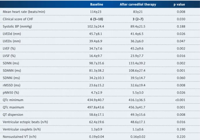
A total 34 patients (18 male, 16 female, mean age: 7.4±4.3 years, range 32 months to 14 years) with DCM were analyzed in the study. All patients had undergone carvedilol therapy in addition to standard therapy for at least 6 months. The medi-an follow-up period was 9.5 months (rmedi-ange 6.4–13.7 months) after the initiation of carvedilol therapy. Baseline patient char-acteristics are shown in Table 2.
Clinical characteristics
The initial mean carvedilol dose was 0.14±0.07 mg/kg/day, and 0.46±0.28 mg/kg/day at 6 months. All patients tolerated the highest dose of carvedilol. As standard treatment, 34/34 (100%) were on digoxin, 32/34 (94.1%) were on furosemide, and 32/34 (94.1%) were on ACEI. The average heart rate was significantly reduced after carvedilol treatment (114±23 vs. 89±21 beat/min, p=0.008). Systolic blood pressure tended to decrease after carvedilol therapy, but did not reach statistical
significance. After 6 months of carvedilol treatment, the Ross clinical score significantly improved from 6 to 3 (p=0.03) (Table 3). The most common adverse events were dizziness (22%), vomiting (13%), hypotension (8%), and headache (5%). No se-rious adverse effects were observed that necessitated discon-tinuation of carvedilol therapy.
Echocardiography
LVEF significantly increased from 34.7±7.6% (range 22–40%) to 45.2±9.6% (range 29–61%) following carvedilol treatment (p=0.002). After carvedilol treatment, the left ventricular frac-tional shortening (LVFS) significantly increased from 16.4±9.7% (range 10–33%) to 23.9±7.7% (range 16–37%) (p=0.016), left ventricle end-diastolic dimensions (LVEDd) significantly de-creased from 45.7±8.1 mm (range 31–60 mm) to 41.4±6.5 mm (range 26–58 mm) (p=0.026) and the left ventricle end-systol-ic dimensions (LVEDs) signifend-systol-icantly decreased from 39.4±6.9 mm (range 24–47 mm) to 34.2±6.0 mm (range 20–43 mm) (p=0.047) (Table 3).
Heart rate variability parameters
There were significant increases in mean SDNN, SDANN, rMS-SD, and pNN50 after carvedilol therapy (p=0.002, p=0.001,
p=0.008, and p=0.026, respectively). A trend toward an
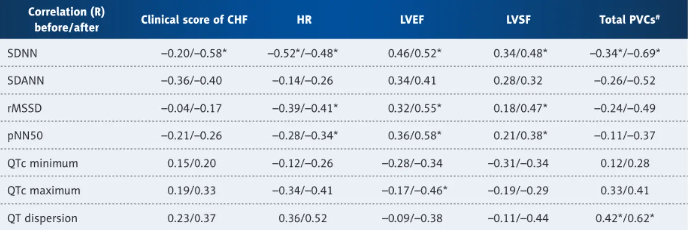
in-crease in SDNNi did not achieve statistical significance after 6 months. Baseline SDNN was significantly correlated with base-line heart rate and total PVCs. After carvedilol therapy, SDNN was correlated with the clinical score of CHF, heart rate, LVEF, LVSF, and total PVCs. In addition, rMSSD and pNN50 were cor-related with heart rate, LVEF, and LVSF after carvedilol therapy. The comparison of clinical, hemodynamic, heart rate variabil-ity, and ventricular arrhythmia parameters of patients receiv-ing carvedilol therapy at baseline and after the treatment are shown in Table 3, and the correlation between changes in HRV in the time domains and changes in hemodynamic parame-ters are presented in Table 4.
QT interval and ventricular arrhythmias
Intra- and interobserver variability were assessed in 18 ran-domly chosen patients; all intra- and interobserver variability for ECG parameters ranged from 3.1 to 4.8%. A significant re-duction was observed in maximum and minimum QTc inter-val, QTc, and QTd values after carvedilol treatment. QTd was slightly higher in patients with a lower clinical score than in those with a higher clinical score, but the difference was not statistically significant. QTd was significantly related to total PVCs, but QTd was not related to age, sex, clinical score of CHF, heart rate, LVEF, LVSF, or LVEDd (Table 4). No patient had a history of a previous abnormal heart rhythm. Although 8 pa-tients (23.5%) had PVCs before treatment, they disappeared
Age (mean, [range]) (years) 7.4±4.3 [32 months–14 years] Gender (male/female) 18 male/16 female Body weight (kg) 39.6±10.4 ACEI 32/34 (94.1%) Diuretics 32/34 (94.1%) Digoxin 34/34 (100%) Carvedilol (mean dose, mg/kg) 0.46±0.28
Table 2. Initial characteristics of patients (n=34).
Data are expressed as mean ± standard deviation or number (%) of patients. ACEI, angiotensin-converting enzyme inhibitors.
in 4 patients and PVC decreased in 2 patients after treatment. Sustained ventricular tachycardia was not observed in any pa-tients. Before carvedilol therapy, 5 patients (14.7%) had tricular couplets and 2 patients (5.8%) had nonsustained ven-tricular tachycardia. A trend toward a decrease in venven-tricular couplets and nonsustained ventricular tachycardia did not reach statistical significance after 6 months. Patients with DCM who had arrhythmic events during follow-up had significantly greater QTd than those without arrhythmic events (59.7±16.8
vs. 51.4±15.6, p=0.026).
Discussion
The neurohumoral mechanisms of CHF involve activation of the sympathetic nervous system and the RAA system, leading to intrinsic myocardial dysfunction, apoptosis, and remodeling
[22,23]. Stimulation of a-receptors increases oxygen consump-tion of the myocardium by increasing the afterload, which causes peripheral and coronary vasoconstriction that results in accu-mulation of calcium in the myocyte, leading to cell death, and contributes to remodeling of the heart with fibrosis and hyper-trophy [4,24]. Carvedilol is a third-generation b-blocking agent that at therapeutic target doses blocks all 3 adrenergic recep-tors that decrease the chronic adrenergic overstimulation of the myocardium and improve myocardial function, and it has been shown to inhibit free radical induced cardiac contractile dysfunction [25,26]. Therefore, it is important to examine the clinical effect of a b-adrenergic blocker therapy on DCM (e.g., carvedilol) to verify its efficacy in children. There are limited data concerning the use of carvedilol in children with ventricular dys-function [22,27]. We have shown that oral carvedilol added to standard drug therapy improved ventricular function and clin-ical symptom scores in children with DCM, and we also found
Baseline After carvedilol therapy p value
Mean heart rate (beats/min) 114±23 83±21 0.008 Clinical score of CHF 6 (5–10) 3 (2–7) 0.030 Systolic BP (mmHg) 102.3±24.4 89.4±21.5 0.188 LVEDd (mm) 45.7±8.1 41.4±6.5 0.026 LVEDs (mm) 39.4±6.9 36.2±6.0 0.047 LVEF (%) 34.7±7.6 45.2±9.6 0.002 LVSF (%) 16.4±9.7 23.9±7.7 0.016 SDNN (ms) 98.7±35.6 133.4±39.2 0.002 SDANN (ms) 81.3±38.2 108.6±27.4 0.001 SDNNi (ms) 34.2±10.3 39.5±14.7 0.060 rMSSD (ms) 23.6±15.2 32.6±19.4 0.008 pNN50 (%) 4.7±2.9 5.5±3.0 0.026 QTc minimum 434.9±40.7 416.1±36.5 <0.001 QTc maximum 497.8±43.6 456.3±41.7 0.001 QT dispersion 58.6±17.1 49.3±15.6 0.008 Ventricular ectopic beats (n/h) 62.4±19.6 48.6±17.1 0.016 Ventricular couplets (n/h) 1.3±0.9 1.1±0.6 0.190 Nonsustained VT (n/h) 0.19±0.04 0.16±0.02 0.220
Table 3. Comparison of clinical, hemodynamic, heart rate variability and ventricular arrhythmia parameters of patients receiving carvedilol at baseline and after the treatment.
Data are expressed as mean ± standard deviation and median (range); p value is given for comparison between baseline and after carvedilol therapy. CHF – congestive heart failure; BP – blood pressure; LVEDd – left ventricular end diastolic diameter; LVEDs – left ventricular end systolic diameter; LVEF – left ventricular ejection fraction; LVSF – left ventricular fractional shortening; SDNN – standard deviation of all normal RR intervals during 24 h; SDANN – standard deviation of 5-minute mean RR intervals; SDNNi – mean of all 5-minute standard deviation of RR intervals; rMSSD – root-mean-square of difference of successive RR intervals; pNN50 – percentage of adjacent RR intervals >50 ms different; QTc – corrected QT interval; VT – ventricular tachycardia.
a significant correlation between changes in LVEF and time do-main parameters of HRV, including SDNN, rMSSD, and pNN50. In accordance with the results obtained from adults, the im-provement of the autonomic function seen after the initiation of carvedilol therapy is likely to play an important role in chil-dren with DCM. Recently, some authors have observed a sta-tistically significant association between sudden cardiac death and depressed SDNN in patients with DCM. However, Bilchick et al. [28] demonstrated that SDNN has a strong and indepen-dent association with mortality in patients with moderate-to-severe CHF [29]. In this context, the UK-Heart prospective study [10] has recently demonstrated that reduced SDNN was the best noninvasive independent predictor of cardiac death in patients with CHF. In our study, SDNN and SDANN were found to be in-creased after the addition of carvedilol to standard medical therapy; these results may have important clinical implications. The pNN50 and rMSSD predominantly reflect parasympathet-ic activity and are independent of long-term trends [30]. In the present study, improvements in rMSSD and pNN50 were not-ed, and pNN50 and rMSSD correlated to improvement in LVEF. Our findings confirm that carvedilol treatment has a beneficial effect on the mechanisms that sustain the harmful hyperadren-ergic state and may improve prognosis in children with DCM. QTd has been found to be a significant, noninvasive prognos-tic marker of inhomogeneity of myocardial repolarization in several disease settings, and increased QTd may predispose to arrhythmic events [12,31]. In a retrospective study of adult patients with CHF, Fu et al. [32] found a larger QTd in patients who died suddenly or had spontaneous ventricular tachycardia than in survivors. However, only limited data is available re-garding the effects of carvedilol on QTd in children with DCM. One retrospective study reported significant increases in QTd
values in patients with LV systolic dysfunction [13]. Our study showed that carvedilol therapy decreased QT dispersion and QTc parameters, and improved ventricular repolarization char-acteristics in children with DCM after 6 months of follow-up. A randomized trial in adult patients with DCM showed a signif-icant effect of carvedilol in reducing ventricular arrhythmias [9]. SDNN and SDANN could predict ventricular tachycardia on Holter monitoring of patients with CHF [30]. Our study results suggest that the increase in SDNN and reduction in QTd are related with decrease in total PVCs. The increase in SDNN and reduction in QTd under carvedilol treatment may be partly due to an adren-ergic blocking effect. Moreover, antiapoptotic effects and inhi-bition of chronic remodeling of the myocardium may indirectly contribute to the observed homogenization of the ventricular repolarization process and prevention of induction of arrhyth-mia in patients with CHF [12]. We observed that ventricular ec-topic beats disappeared in 4 patients and decreased in 2 pa-tients with carvedilol treatment. Reduced heart rate at rest may lead to better oxygen supply, lower the risk for life-threaten-ing arrhythmia, and slow the myocardial remodellife-threaten-ing process; thus, carvedilol may have also been effective in the control of ventricular ectopic beats [33,34]. Our data support the evidence that the increase in heart rate variability reflects improved au-tonomic regulation of heart rate, and show a significant corre-lation between heart rate variability changes and hemodynam-ic improvement with carvedilol therapy in children with DCM.
Conclusions
We conclude that the addition of carvedilol to standard med-ical regimens can improve clinmed-ical symptoms and heart rate Correlation (R)
before/after Clinical score of CHF HR LVEF LVSF Total PVCs#
SDNN –0.20/–0.58* –0.52*/–0.48* 0.46/0.52* 0.34/0.48* –0.34*/–0.69* SDANN –0.36/–0.40 –0.14/–0.26 0.34/0.41 0.28/0.32 –0.26/–0.52 rMSSD –0.04/–0.17 –0.39/–0.41* 0.32/0.55* 0.18/0.47* –0.24/–0.49 pNN50 –0.21/–0.26 –0.28/–0.34* 0.36/0.58* 0.21/0.38* –0.11/–0.37 QTc minimum 0.15/0.20 –0.12/–0.26 –0.28/–0.34 –0.31/–0.34 0.12/0.28 QTc maximum 0.19/0.33 –0.34/–0.41 –0.17/–0.46* –0.19/–0.29 0.33/0.41 QT dispersion 0.23/0.37 0.36/0.52 –0.09/–0.38 –0.11/–0.44 0.42*/0.62*
Table 4. Correlation between HRV-QT parameters and clinical-hemodynamic-PVC parameters at before and after carvedilol treatment.
# In quantifying total PVCs, each of the complexes occurring in clusters as couplets or runs of ventricular tachycardia was counted
individually and contributed to the total PVC count. * p<0.05. SDNN – standard deviation of all normal RR intervals during 24 h; SDANN – standard deviation of 5-minute mean RR intervals; rMSSD – root-mean-square of difference of successive RR intervals; pNN50 – percentage of adjacent RR intervals >50 ms different; QTc – corrected QT interval; HR – heart rate; LVEF – left ventricular ejection fraction; LVSF – left ventricular fractional shortening; PVCs – premature ventricular contractions.
variability in association with improved left ventricular func-tion, and reduce arrhythmia markers in children with DCM. A randomized, controlled, prospective trial is required to deter-mine the true efficacy of carvedilol on the progression of con-gestive heart failure and to more clearly define its role in the cardiac autonomic dysfunction and rhythm disorders of chil-dren with DCM.
Conflict of interest
None.
References:
1. Arola A, Jokinen E, Ruuskanen O et al: Epidemiology of idiopathic cardiomy-opathies in children and adolescents. Am J Epidemiol, 1997; 146: 385–93 2. Doughty RN, Whalley GA, Gamble G et al: Left ventricular remodeling with
carvedilol in patients with congestive heart failure due to ischemic heart disease. J Am Coll Cardiol, 1997; 29: 1060–66
3. Eckberg DL, Drabinsky M, Braunwald E: Defective cardiac parasympathet-ic control in patients with heart disease. N Engl J Med, 1971; 285: 877–83 4. Rusconi P, Gómez-Marín O, Rossique-González M et al: Carvedilol in chil-dren with cardiomyopathy: 3-year experience at a single institution. J Heart Lung Transplant, 2004; 23: 832–38
5. Podrid PJ, Fuchs T, Candinas R: Role of the sympathetic nervous system in the genesis of ventricular arrhythmia. Circulation, 1990; 82(2 Suppl.): I103–13
6. Thayer JF, Yamamoto SS, Brosschot JF: The relationship of autonomic im-balance, heart rate variability and cardiovascular disease risk factors. Int J Cardiol, 2010; 141: 122–31
7. Bullinga JR, Alharethi R, Schram MS et al: Changes in heart rate variability are correlated to hemodynamic improvement with chronic carvedilol ther-apy in heart failure. J Card Fail, 2005; 11: 693–99
8. Chiu KM, Chan HL, Chu SH, Lin TY: Carvedilol can restore the multifractal properties of heart beat dynamics in patients with advanced congestive heart failure. Auton Neurosci, 2007; 132: 76–80
9. Olsen SL, Gilbert EM, Renlund DG et al: Carvedilol improves left ventricular function and symptoms in chronic heart failure: a doubleblind randomized study. J Am Coll Cardiol, 1995; 25: 1225–31
10. Nolan J, Batin PD, Andrews R et al: Prospective study of heart rate variabil-ity and mortalvariabil-ity in chronic heart failure: Results of the United Kingdom Heart Failure Evaluation and Assessment of Risk Trial (UK-Heart). Circulation, 1998; 98: 1510–16
11. Yi G, Goldman JH, Keeling PJ et al: Heart rate variability in idiopathic di-lated cardiomyopathy: relation to disease severity and prognosis. Heart, 1997; 77: 108–14
12. Akdeniz B, Guneri S, Savas IZ et al: Effects of carvedilol therapy on arrhyth-mia markers in patients with congestive heart failure. Int Heart J, 2006; 47: 565–73
13. Bonnar CE, Davie AP, Caruana L et al: QT dispersion in patients with chron-ic heart failure: beta blockers are associated with a reduction in QT disper-sion. Heart, 1999; 81: 297–302
mia risk in patients with long QT intervals. Br Heart J, 1990; 63: 342–44 15. Shaddy RE, Boucek MM, Hsu DT et al: Carvedilol for children and
adoles-cents with heart failure: a randomized controlled trial. JAMA, 2007; 298: 1171–79
16. Mortara A, La Rovere MT, Pinna GD et al: Nonselective beta adrenergic blocking agent carvedilol, improves arterial baroflex gain and heart rate variability in patients with stable chronic heart failure J Am Coll Cardiol, 2000; 36: 1612–18
17. Yildirir A, Sade E, Tokgozoglu L, Oto A: The effects of chronic carvedilol ther-apy on QT dispersion in patients with congestive heart failure. Eur J Heart Fail, 2001; 3: 717–21
18. McMahon CJ, Nagueh SF, Eapen RS et al: Echocardiographic predictors of adverse clinical events in children with dilated cardiomyopathy: a prospec-tive clinical study. Heart, 2004; 90: 908–15
19. Reithmann C, Reber D, Kozlik-Feldmann R et al: Post-receptor defect of ad-enylyl cyclase in severely failing myocardium from children with congeni-tal heart disease. Eur J Pharmacol, 1997; 330: 79–86
20. Ross RD: Grading the severity of congestive heart failure in infants. Pediatr Cardiol, 1992; 13: 72–75
21. Postema PG, De Jong JS, Van dB I, Wilde AA: Accurate electrocardiograph-ic assessment of the QT interval: teach the tangent. Heart Rhythm, 2008; 5: 1015–18
22. Askari H, Semizel E, Bostan OM, Cil E: Carvedilol therapy in pediatric pa-tients with dilated cardiomyopathy. Turk J Pediatr, 2009; 51: 22–27 23. Eichhorn EJ, Bristow MR: Medical therapy can improve the biologic
prop-erties of the chronically failing heart. Circulation, 1996; 94: 2285–96 24. Packer M: Pathophysiology of chronic heart failure. Lancet, 1992; 340: 88–92 25. Blume ED, Canter CE, Spicer R et al: Prospective single-arm protocol of
carvedilol in children with ventricular dysfunction. Pediatr Cardiol, 2006; 27: 336–42
26. Flesch M, Maack C, Cremers B et al: Effect of beta-blockers on free radi-calinduced cardiac contractile dysfunction. Circulation, 1999; 100: 346–53 27. Azeka E, Ramires JA, Valler C, Bocchi EA: Delisting of infants and children
from the heart transplantation waiting list after carvedilol treatment. J Am Coll Cardiol, 2002; 40: 2034–38
28. Bilchick KC, Fetics B, Djoukeng R et al: Prognostic value of heart rate vari-ability in chronic congestive heart failure (Veterans Affair’s Survival trial of Antiarrhythmic Therapy in Congestive Heart Failure). Am J Cardiol, 2002; 90: 24–28
29. Grutter G, Giordano U, Alfieri S et al: Heart rate variability abnormalities in young patients with dilated cardiomyopathy. Pediatr Cardiol, 2012; 33: 1171–74
30. Ponikowski P, Anker SD, Chua TP et al: Depressed heart rate variability as an independent predictor of death in chronic congestive heart failure sec-ondary to ischemic or idiopathic dilated cardiomyopathy. Am J Cardiol, 1997; 79: 1645–50
31. Kuo CS, Munakata K, Reddy CP, Surawicz B: Characteristics and possible mechanism of ventricular arrhythmia dependent on the dispersion of ac-tion potential duraac-tion. Circulaac-tion, 1983; 67: 1356–67
32. Fu GS, Meissner A, Simon R: Repolarization dispersion and sudden cardiac death in patients with impaired left ventricular function. Eur Heart J, 1997; 18: 281–89
33. Fujita B, Franz M, Goebel B et al: Prognostic relevance of heart rate at rest for survival and the quality of life in patients with dilated cardiomyopathy. Clin Res Cardiol, 2012; 101: 701–7
34. Erdoğan I, Ozer S, Karagöz T et al: Treatment of dilated cardiomyopathy with carvedilol in children. Turk J Pediatr, 2009; 51: 354–60
![Table 1. Clinical score modified from Ross and Reithmann et al. [19,20].](https://thumb-eu.123doks.com/thumbv2/9libnet/5443285.104500/2.850.82.774.614.1096/table-clinical-score-modified-ross-reithmann-et-al.webp)