129
C677T
and
A1298C
polymorphisms
and
Turkish
women
with
recurrent
pregnancy
loss
Irem Yengel, Tulay Yorulmaz, Murat Api
DepartmentofObstetrics& Gynaecology,SchoolofMedicine,IstanbulMedipolUniversity,İstanbul, Turkey
Corresponding author:
Irem Yengel
Medipol Sefaköy Hospital Tevfikbey M. Maslakçeşme C. No:30 Küçükçekmece, İstanbul Phone: +90 530 4675260; Fax: +90 2129122626; E-mail: [email protected] ORCID ID: https://orcid.org/ 0000-0002-1971-4092 Original submission: 29 July 2019; Revised submission: 28 September 2019; Accepted: 03 December 2019 doi: 10.17392/1062-20
Med Glas (Zenica) 2020; 17(1):129-135
ABSTRACT
Aim Recurrent pregnancy loss (RPL) poses a challenge in
repro-ductive medicine because the etiology is often unknown. Here we investigated the frequency of mutations in the Factor V Leiden (FVL), prothrombin (FII), and methylene tetrahydrofolate reducta-se (MTHFR) genes in women with RPL and healthy women.
Methods Blood samples were obtained from patients with ≥2
consecutive pregnancy losses and no identifiable etiology before 12 weeks of pregnancy (n=145). The control group comprised 105 age-matched women with ≥2 live births.
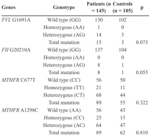
Results The frequency of homozygotes for FVL 1691AA was 15
(10.3%) in patients and three (2.86%) in controls (p=0.073), whi-le for FII 20210AA it was eight (5.5%) and one (0.9%), respec-tively (p=0.055). For two polymorphisms in MTHFR, genotype frequencies of 89 (61.4%) were found in patients and 55 (52.4%) in controls for 677TT (p=0.322), and 89 (61.4%) and 62 (59%) for 1298CC, respectively (p=0.810).
Conclusion Despite a trend towards significance for FII G20210A,
no significant differences in genotype frequencies of these polymorphisms between patients and controls was found. No evi-dence of the role of FVL G1691A, MTHFR C677T, and MTHFR A1298C in RPL in our Turkish cohort was found; however, further investigation of FII as a culprit gene in RPL is warranted.
Key words: gene polymorphisms, habitual abortions, MTHFR A1298C, MTHFR C677T polymorphisms, methylenetetrahydro-folate reductase, thrombophilia
130
INTRODUCTION
Recurrent pregnancy loss (RPL) is defined as a loss of two or more pregnancies, and represents a signi-ficant clinical problem affecting 2%-5% of couples (1). The RPL is one of the most frustrating challen-ges in reproductive medicine because its etiology is often unknown and there are few evidence-based diagnostic and treatment strategies. Although stu-dies on the etiology, evaluation, and management of RPL are often flawed (2), a number of genetic, infective, anatomical, and endocrine factors as well as immune thrombophilia defects have been postulated as causes for RPL (3). However, despi-te detailed investigations, as many as 80% of all cases remain unexplained (4-6). Thrombophilia has been identified as the main cause of RPL in up to 40% of cases, and in particular, early RPL (7). The contribution of specific thrombophilic genes to the pathophysiology of RPL has remained contro-versial. Hereditary thrombophilias are a group of genetic disorders of blood coagulation resulting in a hypercoagulable state, which in turn can result in abnormal placentation. In early pregnancy, this may manifest as spontaneous loss (8,9). Various studies in recent years have examined the inci-dence of specific thrombophilic gene mutations in women with unexplained pregnancy loss. Some of these studies demonstrated an association between thrombophilic gene mutations and RPL (10,11), while others have not found evidence of such an association (12).
Presence of the FII 20210A or FVL 1691A muta-tion increases the risk of early RPL, with an odds ratio (OR) of 2.49 for FII G20210A, and 2.71 and 1.68 for homozygous and heterozygous carriers of FVL, respectively. In contrast, there is no signifi-cant increase related to homozygosity for MTHFR 677T (OR = 1.40, 95% confidence interval (CI) 0.77–2.55) (13). The A1298C polymorphism is another common mutation in MTHFR, and in con-trast to C677T in which homozygosity (TT) is asso-ciated with a significant increase in total plasma homocysteine level, the CC genotype of A1298C is not associated with changes to homocysteine plasma level (14,15). Further, although the pre-sence of MTHFR mutations is significantly more common in women with a history of miscarriage (16), current evidence fails to robustly support an association between these polymorphisms and increased risk for recurrent miscarriage (17).
The EPCOT study showed that the risk for still-birth (but not miscarriage) is highest in women with combined thrombophilic defects (18). However, investigation of even minor throm-bophilic mutations for evidence of an association with recurrent miscarriage has resulted in hete-rogeneous and inconsistent results (19). Further, most mutation carriers will not develop any cli-nical signs and remain undiagnosed because the-se conditions contribute to a small absolute risk towards clinically significant thrombosis. Howe-ver, when carriers are exposed to additional risk factors, such as pregnancy or possibly oral con-traceptives, the risk of life-threatening thrombotic events is significantly increased and may become clinically evident (20,21). Since most carriers are otherwise asymptomatic, the diagnosis would usually be missed. Importantly, preliminary stu-dies of thromboprophylaxis during pregnancy in carriers suggest that treatment may significantly improve pregnancy outcome (8,22,23).
The aim of this study was to determine the frequ-ency of FVL, FII, and MTHFR mutations in Tur-kish women with recurrent pregnancy loss.
PATIENTS AND METHODS Patients and study design
An observational case-control study was carri-ed out at the Department of Gynaecology and Obstetrics in the Numune Education and Re-search Hospital Adana, Turkey. The study was approved by the Ethics Committee for Medical Research at the Numune Education and Resear-ch Hospital in Adana, Turkey.
During the period from March 2008 to May 2012, 145 women with at least two recurrent pregnancy losses were recruited as the case group, and 105 healthy women with at least two successful delive-ries and no miscarriages were recruited as the con-trol group. Informed consent was obtained from all individual participants included in the study. Pregnancy losses occurred before the 12th week of gestation, based on the last menstrual period. For women with RPL, anatomic, hormonal and chromosomal abnormalities, as well as antiphos-pholipid syndrome were excluded (patients with a history of arterial and venous thromboemboli-sm and/or patients with anti-cardiolipin and lu-pus anticoagulants were excluded from the
stu-131
dy. In addition, 3D ultrasound was performed to identify any genital tract abnormalities, and pati-ents and their parpati-ents were karyotyped to exclude known chromosomal abnormalities. Further, me-dical disorders, endocrine disorders (i.e. diabetes, thyroid dysfunction, hyperprolactinemia, and lu-teal insufficiency), infection, intermarriage, and venous thromboembolism were investigated and also excluded. Women with no known medical disorders or previous thrombosis were included in the control group.
Informed consent was obtained from all partici-pants included in the study.
Methods
Peripheral blood samples (5 mL) were collected from participants in tubes containing ethylenedia-minetetraacetic acid (EDTA) for DNA extraction. Genomic DNA was extracted using a magnetic bead extraction technique, DNA was obtained extracted from EDTA blood samples collected in
EDTA tubes, with Roche Manga Pure Compact 1.0 Automatic DNA Isolation Device (CH-6343 Rotkreuz Switzerland) and Manga Pure Compact Nucleic Acid Isolation Kit.
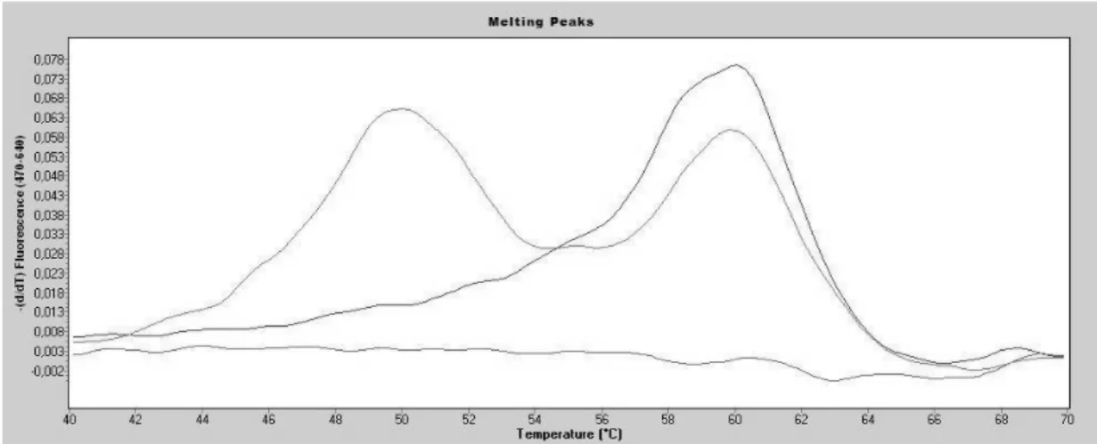
Obtained DNA samples were analysed by the real-time PCR method, applying melting curve analysis with hybridization probes. Melting tem-perature (Tm) is the specific temtem-perature at which half of the DNA molecule becomes a single helix. The base temperature is largely determined by the melting temperature, and the Tm values of DNA molecules having different sequences are different from each other. In the melting curve analysis, the temperature of the double helix DNA sample is gradually increased to form a graph showing the temperature dependent change of the signal: tm tempatures in factor II G20210A (Figure 1), factor V (Leiden) G1691A (Roche, Indianapolis, IN, USA) (Figure 2 ), MTHFR C677T (TIB Mo-lbiol, Berlin, Germany) (Figure 3), and MTHFR A1298C (TIB Molbiol) (Figure 4).
Figure 1. Factor II Melting curve analysis
19
Figure 1. Factor II Melting curve analysis
Figure 2. Factor V Melting curve analysis
20
132
Melting curves of mutant and healthy alleles were analysed separately for each patient; only those with a wild-type melting curve were considered to be ‘normal’ and those with both a wild-type and mutant melting curve were ‘heterozygous mutant’ and patients with only a mutant-type melting cur-ve were considered ‘homozygous mutant.
Statistical analysis
As the mean age of patients (30.5±6.5 years) did not differ from that of controls (30.5±6.7 years) (p=0.989) indicating a normal distribution, re-sults were expressed as mean and standard de-viation. Comparison of FVL, FII, and MTHFR genotype frequencies between cases and controls was performed using the χ2 test. A p< 0.05 was considered to indicate statistical significance.
RESULTS
The frequencies of individuals heterozygotes (AG) for the FVL polymorphism in case and control groups were 14 (9.65%) and 3 (2.86%),
respectively, whereas the frequencies of indivi-duals homozygotes (AA) for the FVL risk allele in case and control groups were one (0.7%) and 0, respectively (Table 1) (p=0.073).
Genes Genotype Patients (n = 145) (n = 105)Controls p
FVL G1691A Wild type (GG) 130 102
Homozygous (AA) 1 0
Heterozygous (AG) 14 3
Total mutation 15 3 0.073
FII G20210A Wild type (GG) 137 104
Homozygous (AA) 0 0 Heterozygous (AG) 8 1 Total mutation 8 1 0.055 MTHFR C677T Wild type (CC) 56 50 Homozygous (TT) 21 11 Heterozygous (CT) 68 44 Total mutation 89 55 0.322
MTHFR A1298C Wild type (AA) 56 43
Homozygous (CC) 25 15
Heterozygous (AC) 64 47
Total mutation 89 62 0.810
Table 1. Genotype distribution of FVL G1691A, FII G20210A,
MTHFR C677T and MTHFR A1298C polymorphisms in women
with recurrent miscarriage and normal controls Figure 3. MTHFR 677 Melting curve analysis
21
Figure 3. MTHFR 677 Melting curve analysis
Figure 4. MTHFR 1298 Melting curve analysis
22
133
The frequencies of individual heterozygotes (AG) for the FII G20210A polymorphism in case and control groups were eight (5.5%) and one (0.9%), respectively (Table 1), while no indivi-dual from either cohort was homozygous (AA) for the FII G20210A risk allele (p=0.055). The proportions of individuals heterozygotes (CT) for MTHFR C677T were 68 (46.9%) and 44 (41.9 %) in the case and control groups, respectively, and the proportion of individuals homozygotes (TT) for the risk allele were 21 (14.5%) and 11 (10.5%), respectively (Table 1). Although the frequency of homozygous TT cases was increased compared to that found in controls, the difference was not statistically significant (p=0.322). In addi-tion, 64 (44.14%) cases and 47 (44.76%) controls were heterozygous (AC) for the MTHFR A1298C polymorphism, while 25 (17.24%) cases and 15 (14.28%) controls were homozygous (CC) for the A1298C risk allele (Table 1). There were no signi-ficant differences in A1298C genotype frequenci-es between our cohorts (p=0.810).
DISCUSSION
We carried out multiple analyses to determine whether certain polymorphisms in three candi-date genes are associated with increased risk for RPL. We evaluated genotype frequencies of the FVL G1691A, FII G20210A, MTHFR C677T, and MTHFR A1298C polymorphisms in women with first-trimester RPL. Although we found some differences in genotype frequencies of the-se polymorphisms, the results were not statisti-cally different between cohorts.
Based on these findings, we conclude that these mutations have no major role in the etiology of first-trimester recurrent abortion in our Turkish cohort. The lack of an association between the FII G20210A polymorphism and RPL in our stu-dy is similar to that in recent studies that demon-strated that FII G20210A as a solitary defect is not associated with increased risk of RPL. Howe-ver, considering that we found a trend towards significance, the possibility cannot be ruled out that the mutation is a risk factor for certain types of RPL (24,25).
Case-control studies and meta-analysis have shown that there is a high prevalence of FVL in women with RPL (26-28). According to a syste-matic review of the literature, there is an OR of
2 between RPL and FVL (29,30). Reznikoff-Etiévant et al. reported that 10.3% among a total of 260 Caucasian women with a history of ≥2 unexplained pregnancy losses at less than 10 weeks gestation were positive for FVL compa-red to 4.6% among controls (27) concluding that FVL was significantly associated with RPL befo-re 10 weeks of gestation. A study by Grandone et al. also supports this conclusion (28). Furthermo-re, Rey et al. found in a meta-analysis that FVL was associated with early and late RPL, as well as late non-recurrent fetal loss (26).
On the other hand, Ivanov et al. performed a study including 94 women with embryonic loss prior to 10 weeks of gestation and 59 women with pre-gnancy loss between 10 and 14 weeks of gesta-tion compared to a control group of 100 healthy women with a history of at least one uncomplica-ted full-term pregnancy and found that FVL valence was not significantly associated with pre-gnancy loss prior to 10 weeks of gestation (9.6%) compared to the controls (7%); however, there was evidence of an association in women with postembryonic loss (10–14 weeks of gestation) exhibiting an FVL prevalence of 18.6%. Further, prevalence of FII G20210A was significantly higher in both groups with embryonic (17%) and early fetal loss (16.9%) compared to that in con-trols (3%). In addition, FII G20210A was signifi-cantly associated with an increased risk of early recurrent pregnancy loss throughout the entire first trimester, while FVL was only significantly higher in the early phase of pregnancy correspon-ding to the beginning of placentation, but was not associated with embryonic-stage RPL. These results suggest that the first trimester should be viewed rather as a heterogeneous interval, with different relations to FVL in the embryonic and postembryonic fetal period (29).
Yousefian et al. found no significant difference in MTHFR 677TT and A1298C polymorphisms ge-notype frequency between patient and control gro-ups in 204 women with ≥3 consecutive pregnancy losses before 22 weeks of pregnancy; similarly, there was no statistically significant difference in the proportion of homozygous MTHFR 1298CC individuals between patient and control cohorts (12.3% vs. 8% respectively) indicating that MTHFR mutations are not associated with RPL in the recruited cohort (30). Hashimoto et al.
evalu-134
REFERENCES
1. Vettriselvi V, Vijayalakshmi K, Paul SF, Venka-tachalam P. ACE and MTHFR gene polymorphisms in unexplained recurrent pregnancy loss. J Obstet Gynaecol Res 2008; 34:301-6.
2. Christiansen OB, Nybo Andersen AM, Bosch E, Daya S, Delves PJ, Hviid TV, Kutteh WH, Laird SM, Li TC, van der Ven K. Evidence-based investi-gations and treatments of recurrent pregnancy loss. Fertil Steril 2005; 83:821.
3. Altintas A, Pasa S, Akdeniz N, Cil T, Yurt M, Ayyil-diz O, Batun S, Isi H. Factor V Leiden and G20210A prothrombin mutations in patients with recurrent pregnancy loss: data from the southeast of Turkey. Ann Hematol 2007; 10:727-31.
4. Regan L, Rai R. Thrombophilia and pregnancy loss. J Reprod Immunol 2002; 55:163-80.
ated the FVL mutation in a group of 52 Japanese women with a history of three or more idiopathic first-trimester miscarriages and 41 of their partners and found no differences compared to that in pa-rous women without obstetric complications (31). Chatzidimitriou et al. reported the impact of 12 thrombophilic polymorphisms as risk factors for RPL, among 48 Greek women with a history of RPL, vs 27 healthy parous women using multi-plex PCR and in situ hybridization on nitrocellu-lose films. Heterozygous FV Leiden, homozygo-us PAI-1 4G/4G, heterozygohomozygo-us MTHFR C677T, homozygous MTHFR A1298C, as much as the combined thrombophilic genotypes MTHFR 677T + ACE Ι/D, MTHFR 677T/1298C + ACE D/D, ACE I/D + b-fibrinogen -455 G/A, FV HR2 + b-fibrinogen -455 G/A showed a correlation as risk factors for RPL, whereas the rest of the inve-stigated polymorphisms and their combinations did not render statistically significant differences between the two groups in study (32).
Bigdeli R et al. found no significant associati-on between FII (A20210G) and FV (A4070G) polymorphism and RPL investigating the frequ-ency and association between ten polymorphisms of seven thrombophilia genes and RPL in an Ira-nian population (on 200 women with recurrent pre-gnancy loss and also on 200 women with at least one successful pregnancy as the control group) (33). As a limitation to our study, other congenital thrombophilic defects could not be analysed be-cause of a lack of genetic laboratory facilities required. Nevertheless, based on our findings, we did not find evidence of an association between FVL, FII, and MTHFR polymorphisms and
first-trimester recurrent fetal loss, and thus we conclu-de that genetic testing of these variants is not an absolute necessity for pregnant women. Howe-ver, because we found a trend towards signifi-cance for association between FII G20210A and RPL, which has been reported in other studies, further investigation of the putative role of this gene in RPL is warranted.
In conclusion, despite the fact that there have been many relevant studies performed, yet there is no consensus on the role of culprit genes and polymorphisms in RPL. However, many institu-tions offer genetic testing in cases of RPL and clinicians take the results into consideration. Further, a number of clinicians adopt a prudent approach administering LMWH to their patients for several weeks antenatally under the presump-tion there may be other thrombotic mutapresump-tions that are either not part of routine genetic testing or not known. Hopefully, as studies continue to be performed in this field and there is a better un-derstanding, we may be able to change current practices accordingly, providing better, more in-formed treatment and care for women with RPL.
ACKNOWLEDGMENTS
The authors wish to thank Doctor Ozge Ozalp Yuregir for help with the mutation analysis and thank Doctor Emre Yengel for proofreading.
FUNDING
No specific funding was received for this study.
TRANSPARENCY DECLARATION
Conflicts of interest: None to declare.
5. Sarig G, Younis JS, Hoffman R, Lanir R, Blumen-feld Z, Brenner B. Thrombophilia is common in wo-men with idiopathic pregnancy loss and is associa-ted with late pregnancy wastage. Fertil Steril 2002; 77:342-7.
6. Wramsby ML, Sten-Linder M, Bremme K. Primary habitual abortions are associated with high frequ-ency of Factor V Leiden mutation. Fertil Steril 2000; 74:987-91.
7. Brenner B, Sarig G, Weiner Z, Younis J, Blumen-feld Z, Lanir N. Thrombophilic polymorphisms are common in women with fetal loss without apparent cause. Thromb Haemost 1999; 82:69.
8. Younis JS, Ohel G, Brenner B, Ben-Ami M. Fa-milial thrombophilia - the scientific rationale for thrombophylaxis in recurrent pregnancy loss? Hum Reprod 1997; 12:1389-90.
135
9. Brown HL. Antiphospholipid antibodies and recu-rrent pregnancy loss. Clin Obstet Gynecol 1991; 34:17-26.
10. Sarig G, Younis JS, Hoffman R, Lanir R, Blumen-feld Z, Brenner B. Thrombophilia is common in wo-men with idiopathic pregnancy loss and is associa-ted with late pregnancy wastage. Fertil Steril 2002; 77:342-7.
11. Rey E, Kahn SR, David M, Shrier I. Thrombophi-lic disorders and fetal loss: a meta-analysis. Lancet 2003; 361:901-8.
12. Carp H, Salomon O, Seidman D, Dardik R, Rose-nberg N, Inbal A. Prevalence of genetic markers for thrombophilia in recurrent pregnancy loss. Hum Re-prod 2002; 17:1633-7.
13. Robertson L, Wu O, Langhorne P, Twaddle S, Clark P, Lowe GD, Walker ID, Greaves M, Brenkel I, Re-gan L, Greer IA. Thrombophilia in pregnancy: a systematic review. Br J Haematol 2006; 132:171-96. 14. Haviv YS, Shpichinetsky V, Goldschmidt N, Atta
IA, Ben-Yehuda A, Friedman G. The common muta-tions C677T and A1298C in the human methylene-tetrahydrofolate reductase gene are associated with hyperhomocysteinemia and cardiovascular disease in hemodialysis patients. Nephron 2002; 92:120-6. 15. Zetterberg H, Regland B, Palmer M, Ricksten A,
Palmqvist L, Rymo L, Arvanitis DA, Spandidos DA, Blennow K. Increased frequency of combined methylenetetrahydrofolate reductase C677T and A1298C mutated alleles in spontaneously aborted embryos. Eur J Hum Genet 2002; 10:113-8. 16. Coulam CB, Jeyendran RS, Fishel LA, Roussev R.
Multiple thrombophilic gene mutations rather than specific gene mutations are risk factors for recurrent miscarriage. Am J Reprod Immunol 2006; 55:360-8. 17. Inbal A, Muszbek L. Coagulation factor deficiencies
and pregnancy loss. Semin Thromb Hemost 2003; 29:171-4.
18. Girolami A, Scandellari R, Lombardi AM, Girolami B, Bortoletto E, Zanon E. Pregnancy and oral contra-ceptives in factor V deficiency: a study of 22 patients (five homozygotes and 17 heterozygotes) and review of the literature. Haemophilia 2005; 11:26-30. 19. Hohlagschwandtner M, Unfried G, Heinze G, Huber
JC, Nagele F, Tempfer C. Combined thrombophilic polymorphisms in women with idiopathic recurrent miscarriage. Fertil Steril 2003; 79:1141-8.
20. Martinelli I, Sacchi E, Landi G, Taioli E, Duc F, Mannucci PM. High risk of cerebral-vein thrombo-sis in carriers of a prothrombin-gene mutation and in users of oral contraceptives. N Engl J Med 1998; 338:1793-7.
21. Rosendaal FR, Koster T, Vandenbroucke JP, Reitsma PH. High risk of thrombosis in patients homozygous for factor V Leiden (activated protein C resistance). Blood 1995; 85:1504-8.
22. Brenner B, Hoffman R, Blumenfeld Z, Weiner Z, Younis JS. Gestational outcome in thrombophilic women with recurrent pregnancy loss treated by enoxaparin. Thromb Haemost 2000; 83:693-7. 23. Younis JS, Ohel G, Brenner B, Haddad S, Lanir
N, Ben-Ami M. The effect of thrombophylaxis on pregnancy outcome in patients with recurrent pre-gnancy loss associated with factor V Leiden muta-tion. BJOG 2000; 107:415-9.
24. Resch B, Gallistl S, Kutschera J, Mannhalter C, Muntean W, Mueller WD. Thrombophilic polymor-phisms--factor V Leiden, prothrombin G20210A, and methylenetetrahydrofolate reductase C677T mutations--and preterm birth. Wien Klin Wochen-schr 2004; 116:622-6.
25. Altintas A, Pasa S, Akdeniz N, Cil T, Yurt M, Ayyil-diz O. Factor V Leiden and G20210A prothrombin mutations in patients with recurrent pregnancy loss: data from the southeast of Turkey. Ann Hematol 2007; 86:727-31.
26. Rey E, Kahn SR, David M, Shrier I. Thrombophi-lic disorders and fetal loss: a meta-analysis. Lancet 2003; 361:901-8.
27. Reznikoff-Etiévan MF, Cayol V, Carbonne B, Ro-bert A, Coulet F, Milliez J. Factor V Leiden and G20210A prothrombin mutations are risk factors for very early recurrent miscarriage. BJOG 2001; 108:1251-4.
28. Grandone E, Margaglione M, Colaizzo D, d’Addedda M, Cappucci G, Vecchione G, Sciannamé N, Pavo-ne G, Di Minno G. Factor V Leiden is associated with repeated and recurrent unexplained fetal losses. Thromb Haemost 1997; 77:822-4.
29. Ivanov PD, Komsa-Penkova RS, Konova EI, Ko-vacheva KS, Simeonova MN, Popov JD. Associati-on of inherited thrombophilia with embryAssociati-onic and postembryonic recurrent pregnancy loss. Blood Co-agul Fibrinolysis 2009; 2:134-40.
30. Yousefian E, Kardi MT, Allahveisi A. Methyle-netetrahydrofolate reductase C677T and A1298C polymorphism in Iranian women with idiopathic re-current pregnancy losses. Iran Red Crescent Med J 2014; 16:e16763.
31. Hashimoto K, Shizusawa Y, Shimoya K, Ohashi K, Shimizu T, Azuma C, Murata Y. The factor V Leiden mutation in Japanese couples with recurrent sponta-neous abortion. Hum Reprod 1999; 14:1872-4. 32. Chatzidimitriou M, Chatzidimitriou D, Mavridou M,
Anetakis C, Chatzopoulou F, Lialiaris T, Mitka S. Thrombophilic gene polymorphisms and recurrent pregnancy loss in Greek women. Int J Lab Hematol 2017; 39:590-5.
33. Bigdeli R, Younesi MR, Panahnejad E, Asgary V, Heidarzadeh S, Mazaheri H, Aligoudarzi SL. Asso-cition between thrombophilia gene polymorphisms and recurrentpregnancy loss risk in the Iranian popu-lation. Syst Biol Reprod Med 2018 64:274-82.