Radiation Dose Reduction in Thorax CT
Toraks Bilgisayarlı Tomografide Radyasyon Doz Azaltımı
Gizem Gül Koç1, Ali Kokangül2, Tahir Hanalioğlu1,
1
TOBB University of Economics and Technology, Department of Industrial Engineering-ANKARA
2Çukurova University, Department of Industrial Engineering. ADANA
Cukurova Medical Journal 2013; 38 (3): 422-427.
ABSTRACT
Computerized Tomography is a method of working with X-rays and is increasing in application day by day. Scientists continue to conduct studies by reducing the dose of X-ray that patients are exposed to in order to reduce the cancer risk. The aim is to get the most effective image by the most minimal dose. In this review, the importance of CT radiation dose reduction and the methods in adults are presented.
Key Words: CT,dose,x-ray,adult.
ÖZET
Bilgisayarlı Tomografi X-ışınlarıyla çalışan ve günümüzde kullanımı giderek artan bir yöntemdir. Kanser riskini azaltmak amacıyla hastaların maruz kaldığı X-ışın dozunu azaltmaya yönelik çalışmalar devam etmektedir. Amaç mümkün olan en düşük dozla en etkin görüntüyü elde etmektir. Bu makalede erişkinlerde BT radyasyon dozunu azaltmanın önemi ve yöntemleri sunulmaktadır.
Anahtar Kelimeler: BT,doz,x-ray,erişkin
Computerized Tomography (CT) is an X-ray method used for the diagnosis and follow up of diseases. In recent years, with developments in CT technology, the numbers of patients having CT examination are increasing rapidly around the world1. Paralleling this, cancer risk is on the agenda in terms of public health because of the total x-ray dose of patients exposed. For this reason, reducing radiation doses by optimising CT protocols has become an important research subject1,2.
There are many factors that affect the dose the patient is exposed to and what the CT scanner produces (Table 1). First, is the structure of the CT and the programs used for image development, and second, are the characteristics of the human
body. Generally, while the structure of the equipment (gantry and tube feature, the detector structure and the sensitivity of the x-ray filters) and the programs used for image development and dose reduction cannot be changed other than by the manufacturer, CT examination parameters can be appropriately selected for each patient. There are several methods used for dose reducing; they are: 1) Reducing the tube flow or reducing the dose by automatic tube flow modulation (automatic exposure control). 2) Reducing the tube voltage or reducing the dose by automatic tube voltage selection (adaptive kolovoltage)2,3. 3) Dose reduction by using iterative reconstruction algoritm instead of the filtered back projection (FBP) which is a conventional image producing protocol4-7 (for
instance, ASIR adaptive statistical image reconstruction, IRIS iterative reconstruction in image space, SAFIRE Sinogram-Affirmed Iterative
Reconstruction, AIDR 3D three dimensional
adaptive iterative dose reduction)8,17. 4) Special protective shielding for organs sensitive to radiation such as the breasts and eyes. 5) Dose reduction appropriate for body size. 6) Dose reduction technique for special clinical
practices9,10. 7) Model-based iterative reconstruction (MBIR) is the latest development and provides advanced dosage reduction13.
CT image quality and dose reduction are issues that should be mutually considered. When the dose is reduced, image quality is compromised. In other words, dose reduction is possible if there is a sufficient image quality .
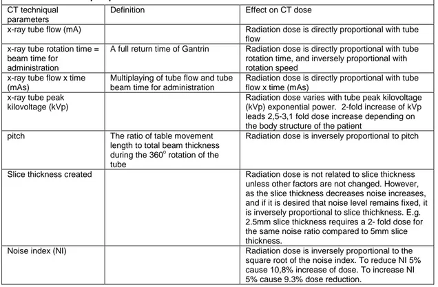
Table 1. CT techniqual parameters that affect Radiation dose
CT techniqual parameters
Definition Effect on CT dose
x-ray tube flow (mA) Radiation dose is directly proportional with tube flow
x-ray tube rotation time = beam time for
administration
A full return time of Gantrin Radiation dose is directly proportional with tube rotation time, and inversely proportional with rotation speed
x-ray tube flow x time (mAs)
Multiplaying of tube flow and tube beam time for administration
Radiation dose is directly proportional with tube flow x time (mAs)
x-ray tube peak kilovoltage (kVp)
Radiation dose varies with tube peak kilovoltage (kVp) exponential power. 2-fold increase of kVp leads 2,5-3,1 fold dose increase depending on the body structure of the patient
pitch The ratio of table movement length to total beam thickness during the 360o rotation of the tube
Radiation dose is inversely proportional to pitch
Slice thickness created Radiation dose is not related to slice thickness unless other factors are not changed. However, as the slice thickness decreases noise increases, and if it is desired that noise level remains fixed, it is inversely proportional to slice thichkness. E.g. 2.5mm slice thickness requires a 2- fold dose for the same noise ratio compared to 5mm slice thickness.
Noise index (NI) Radiation dose is inversely proportional to the square root of the noise index. To reduce NI 5% cause 10,8% increase of dose. To increase NI 5% cause 9.3% dose reduction.
While choosing the CT examination parameters for dose reduction, it is important to know patient characteristics such as age, weight, height, body mass index [weight/height2 (kg/m2) ] or the thickness or density of the body area examined. In accordance with patient characteristics and the type of examination, CT examination parameters such as x-ray tube flow (miliamper; mA), tube voltage (peak kilovoltaj; kVp), pitch value that can be described as screening density (The ratio of table movement length to total beam thickness
during the 360o rotation of the tube), and number of screening (how many times/phases the involved body region scanned), automatic tube voltage selection, automatic tube flow modulation (automatic exposure control) x-ray filters, iterative reconstruction algoritms can be applied, different detector size can be chosen, dose reduction can be obtained by single phase imaging and imaging based on indication.
Schindera ST et al (2013)3 investigated the effect of automatic tube voltage selection in
Abdominal CT angiography on image quality and radiation dose with a phantom study. They have simulated small, medium-sized and large patients by placing the abdominal aort phantom filled with contrast agent into various sized cylindrical water containers. Phantoms were screened by standard 120 kilovolt abdominal CT angiography and also optimized tube voltage protocol (automatic tube voltage modulation technique). Attenuation in the abdominal aorta (X-ray attenuation), ground and image noise were measured, contrast-noise ratio was calculated and three independent readers evaluated the total image quality. In various simulated body sizes, a significant radiation dose reduction was obtained with automatic tube voltage selection image quality without detoriation in image quality when compared to standard 120 kilovolt abdominal CT angiography.
Chang W. et al (2013)4 evaluated dose reduction in liver CT examination by the model based iterative reconstruction algorhytm (MBIR) compared to image quality. In 103 patients, CT imaging was requested due to liver metastasis suspicion; half dose iterative reconstruction algorhytm (MBIR), reference dose (filtered back projection-FBP), half dose FBP and adaptive statistical iterative reconstruction (ASIR) were used. In 73 of the patients, images were created by reference dose CT, FBP and half dose portal phase CT, both FBP and MBIR, and in the rest of the 30 patients, images were created by half dose CT, FBP, ASIR and MBIR protocols. CT attenuation coefficient (X-ray attenuation) and mean image noise were measured from different sites (liver, aorta, the main portal vein and subcouteneous adipose tissue), and additionally contrast-noise ratios were calculated. Half dose MBIR protocol showed the lowest image noise, a higher noise ratio, and better noise quality than half dose ASIR and half dose FBP. All these protocols have shown less image noise, high contrast-noise ratio and similar image quality when compared to classic FDP.
Kataria B et al (2013)5 screened 45 cases both with standard low dose and 30% reduced abdominal CT technique twice, and the images were created both with iterative reconstruction and filtered back projection (FBP). Four radiologists evaluated the image quality independent of each other by using five visual criteria. They found that the iterative reconstruction algorhytm increased CT image quality but dose reduction was relatively low. Therefore, they reported that the exact effectiveness of this algorithm could not be determined.
Hu XH et al (2011)6 evaluated that radiation dose is reduced 40% with iterative reconstruction algorythm in non-contrast thorax CT. CT phantoms created by water were taken as reference at 120 kVp /150 miliamper x seconds (mAs) and 100 kVp/270 mas, and images were obtained as the tube flow was decreased in increments of 10% up to 40%. Image noise was evaluated for the images created with FBP and iterative reconstruction in image space (IRIS). In addition to the phantoms, 90 patients were evaluated with contrast CT, and 30 of them were evaluated with standard protocol (120 kVp/120 mAs), 30 of them with low dose protocol (100 kVp/110 mAs), and the other 30 patients were evaluated with low dose (120 kv/67mAs). All the images were created seperately and B30 and I30 were used as kernel. Signal noise ratio and contrast noise ratio were measured from two different sites and subjective and objective image qualities were evaluated. Consequently, when compared to FBP, IRIS have protected or reduced the image quality and provided 30% dose reduction in non-contrast thorax CT.
Willemink MJ. Et al (2013)7 conducted a
metaanalysis through medline, EMBASE with the original research including iterative reconstruction between the years 2006-2012. They concluded that iterative reconstruction reduces radiation dose subjectively and objectively as they increased the image quality. They reported that iterative
reconstruction should be researched in regard to dose reduction in future studies.
Singh S et al (2013)8 investigated dose reduction with non-linear adaptive filters (NLAF). Additionally, they observed four different image serials with 150, 110, 75 and 40 mAs. They screened an area of 10 cm in twenty-four with 64 sections of CT. NLAF’s were used to create images in three low doses (100, 75 and 40 mAs). Two separate radiologists blind to the doses evaluated filtered and non-filtered image qualities. Objective noise, CT atennuation values, weight of the patients and transfer diameters, CT dose index (CTDIvol), DLP (doz lengh product) values were
measured and recorded. It was demonstrated that tube flow was reducted to 40 mas in Thorax CT by protecting lesion detectability and image quality wth NLAF application.
Al-Hinnawi AR et al (2013)9 examined dose reduction by using half dose, bilateral, non-linear filters in thorax-pelvic CT. They applied two different protocols from thorax to pelvis. They compared full dose CT and half dose CT with BF. It was determined that BF lead to 50% dose reduction and increased image quality.
Shuman WP et al (2013)11 compared image quality in liver CT by using MBIR, ASIR and FBP protocols. They obtained routine 3 phase CT images of 36 patients. To compare the obtained images they measured lesion diameter, density, surrounding tissue density, and image noise. In the evaluations performed by two radiologists, MBIR, ASIR and FBP showed a ratio of comparable lesion characteristics and subjective lesion visualization with low ground noise and high contrast noise ratio. Since it can create a more qualified image with the same dose, it was shown that this protocol allows more dose reduction than other protocols.
Vardhanabhuti V et al (2013)13 investigated image quality in standard and low dose thorax CT with protocols using FBP, ASIR and MBIR algorythm. Thirty cases were screened once with normal and 2 times with low dose and all three
images were obtained by using FBP, ASIR and MBIR protocols. DLP and effective doses were recorded by comparing objective and subjective image qualities. MBIR provided a high level of noise reduction and improvement in image quality when compared to other protocols. Thereby, it was reported that it made reducing the dosage possible.
Pickhardt PJ et al (2012)15 applied very low dose CT to 45 adult patients just after the standard dose application. Very low dose images created by FBP, ASIR and MBIR were compared with the images obtained by standard dose FBP. The images obtained by MBIR protocol were found superior to the images obtained by the other two methods.
Lee KH et al (2012)16 used attenuation based
automatic tube voltage selection and tube flow modulation together for contrated CT examinations in order to investigate whether it led to dose reduction or not. They stated that combined use of automatic tube flow selection and automatic tube flow modulation allowed dose reduction by protecting image quality compared to automatic tube flow modulation.
Ohno Y et al (2012)17 compared image quality with standard dose and three dimensional adaptive iterative dose reduction (AIDR 3D) in reduced and low dose thorax CT. They used different protocols in 37 patients that they divided into two groups. While the images were obtained by standard dose (150 mAs) without AIDR 3 in the first protocol, in the second group CT images were obtained by both using and not using AIDR 3 with low dose (25 mAs) and reduced dose (50 mAs). They have demonstrated that AIDR 3D is effective in reducing image noise and improving image quality.
Consequently, although there are several studies in literature about dose reduction
3-9,11,13,15-17
, we think this issue is currently under development. Recently, it has been demonstrated that low and medium dose reduction is possible by CT protocols and developed softwares1-6,8-17 , and the recent expectation is to provide advanced dose
reduction7. It is important to determine the necessary parameters and to know its contribution to dose reduction in order to be able to develop techniques and software programs for CT dose reduction. We suggest that it is possible to provide an advanced level of radiation dose reduction by optimizing CT protocols. Determing the contributing parameters that affect dose will allow the development of new protocols and reveal the importance of previously unused parameters. This information will enable the development of new software. Thus, it will be possible to reduce the damage to humans by radiation.
REFERENCES
1. Callahan MJ. CT dose reduction in practice. Pediatr Radiol. 2011;41 Suppl 2:488-92.
2. Sodickson A. Strategies for reducing radiation exposure in multi-detector row CT. Radiol Clin North Am. 2012;50:1-14.
3. Schindera ST, Winklehner A, Alkadhi H, Goetti R, Fischer M, Gnannt R, Szucs-Farkas Z. Effect of automatic tube voltage selection on image quality and radiation dose in abdominal CT angiography of various body sizes: a phantom study. Clin Radiol. 2013;68:e79-86.
4. Chang W, Lee JM, Lee K, Yoon JH, Yu MH, Han JK, Choi BI. Assessment of a Model-Based, Iterative Reconstruction Algorithm (MBIR) Regarding Image Quality and Dose Reduction in Liver Computed Tomography. Invest Radiol. 2013;48:598-606. 5. Kataria B, Smedby O. Patient dose and image quality
in low-dose abdominal CT: a comparison between iterative reconstruction and filtered back projection. Acta Radiol. 2013:March 10. (Epub ehead of Pirnt) 6. Hu XH, Ding XF, Wu RZ, Zhang MM. Radiation dose
of non-enhanced chest CT can be reduced 40% by using iterative reconstruction in image space. Clin Radiol. 2011;66:1023-9.
7. Willemink MJ, Leiner T, de Jong PA, de Heer LM, Nievelstein RA, Schilham AM, Budde RP. Iterative reconstruction techniques for computed tomography part 2: initial results in dose reduction and image quality. Eur Radiol. 2013;23:1632-42.
8. Singh S, Digumarthy SR, Back A, Shepard JA, Kalra MK. Radiation dose reduction for chest CT with non-linear adaptive filters. Acta Radiol. 2013;54:169-74 9. Al-Hinnawi AR, Daear M, Huwaijah S. Assessment of
bilateral filter on 1/2-dose chest-pelvis CT views. Radiol Phys Technol. 2013;6:385-98.
10. Chang KJ, Yee J. Dose reduction methods for CT colonography. Abdom Imaging. 2013;38:224-32. 11. Shuman WP, Green DE, Busey JM, et al.
Model-Based Iterative Reconstruction Versus Adaptive Statistical Iterative Reconstruction and Filtered Back Projection in Liver 64-MDCT: Focal Lesion Detection, Lesion Conspicuity, and Image Noise. AJR Am J Roentgenol. 2013;200:1071-6
12. Maldjian PD, Goldman AR. Reducing radiation dose in body CT: a primer on dose metrics and key CT technical parameters. AJR Am J Roentgenol. 2013;200:741-7
13. Vardhanabhuti V, Loader RJ, Mitchell GR, et al. Image quality assessment of standard- and low-dose chest CT using filtered back projection, adaptive statistical iterative reconstruction, and novel model-based iterative reconstruction algorithms. AJR Am J Roentgenol. 2013;200:545-52
14. Mayo J, Thakur Y. Pulmonary CT angiography as first-line imaging for PE: image quality and radiation dose considerations. AJR Am J Roentgenol. 2013;200:522-8.
15. Pickhardt PJ, Lubner MG, Kim DH, et al. Abdominal CT with model-based iterative reconstruction (MBIR): initial results of a prospective trial comparing ultralow-dose with standard-ultralow-dose imaging. AJR Am J Roentgenol. 2012;199:1266-74
16. Lee KH, Lee JM, Moon SK, et al. Attenuation-based automatic tube voltage selection and tube current modulation for dose reduction at contrast-enhanced liver CT. Radiology. 2012; 265:437-47
17. Ohno Y, Takenaka D, Kanda T, et al. Adaptive iterative dose reduction using 3D processing for reduced- and low-dose pulmonary CT: comparison with standard-dose CT for image noise reduction and radiological findings. AJR Am J Roentgenol. 2012;199:W477-85,
Yazışma Adresi / Address for Correspondence:
Dr. Gizem Gül Koç
TOBB University of Economics and Technology Department of Industrial Engineering
ANKARA
Email: [email protected] Geliş tarihi/Received on: 23.09.2013 Kabul tarihi/ Accepted on:24.10.2013
![A [5]Rotaxane-Based photosensitizer for photodynamic therapy](data:image/gif;base64,R0lGODlhAQABAIAAAP///wAAACH5BAEAAAAALAAAAAABAAEAAAICRAEAOw==)