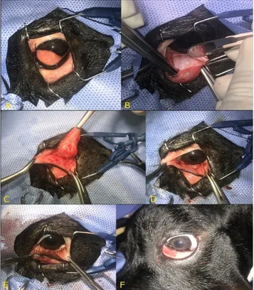
Surgical Treatment of Prolapse of the Third Eyelid Gland in Dogs using Modified Morgan Pocket Technique
Tam metin
Şekil
Benzer Belgeler
• Medical treatment can be used after surgery to prevent recurrence of the endometriosis..
The ultrasound-guided transvaginal ovarian interstitial laser treatment may be an effective new method to manage anovulation in PCOS patients. Ovarian
The upcoming part of this paper discusses about the works related for authentication and key management scheme for smart meters, proposed protocol architecture and results
We present a case of vagus nerve stimulator infection treated successfully with a single surgical debridement of the infected wound and six weeks of antibiotic treatment, but
Diabete bagh periferik fasiyal paraliziye nedeni ile iist goz kapagma altm plak yerle§tirilmesi yanmda, alt goz kapagmda ektropium nedeni ile kama rezeksiyon
[22] compared T-tube drainage and primary closure techniques following LCBDE and concluded that the cost, operation time, postoperative complication and biliary complication
Methods: The Leibinger ® titanium multiperforated titanium plate and screw system described for craniofacial fixation was used together with steel wires for re-correction and
Commonly seen in young, giant breed dogs. Eversion is corrected
