Corresponding Author: Baki Erdem E-mail: [email protected] Received: 18.10.2017 Accepted: 19.01.2018 DOI:10.5152/eamr.2018.20082
INTRODUCTION
The presence of abnormal adnexal mass is a common clinical problem that can affect women of all ages. Although most of the adnexal masses are benign, the primary objective in the diagnostic evaluation is the exclusion of malignancy. Currently, there is no effective screening method for ovarian cancer (1). Ovarian cancer is the second most common gynecological cancer in the world and is the main cause of death due to gynecologic malignancy in women. The lifetime risk of epi-thelial ovarian cancer, which constitutes the majority of ovarian cancers, is 1.3%, and 70% of the patients are diagnosed in an advanced stage (2).
Ovarian masses may be functional or neoplastic, and neoplastic tumors may be benign or malig-nant. Most (80%-85%) of the ovarian tumors are benign, and two-thirds are seen in women aged between 20 and 44 years (3). Adnexal masses are evaluated through history, physical examination, laboratory tests, and imaging methods preoperatively (4).
In this study, we aimed to analyze the adnexal masses that were referred to the gynecologic oncology clinic of our hospital because of malignancy suspicion.
METHODS
This retrospective study was conducted in accordance with the 2008 Declaration of Helsinki princi-ples. We routinely request consent from patients we are going to operate on at our clinic, therefore Cite this article as:
Erdem B, Peker N, Seyhan NA, Aşıcıoğlu O, Temel Yüksel İ, Ülker V, et al. Analysis of Adnexal Masses which are Operated in Gynecologic Oncology Clinic. Eur Arch Med Res 2018; 34 (4): 231-4. ORCID IDs of the authors: B.E. 0000-0002-6407-8718; N.P. 0000-0002-4854-3851; N.A.S. 0000-0003-0543-6180; O.A. 0000-0002-0363-424X; İ.T.Y. 0000-0002-7337-9977; V.E. 0000-0002-8912-8160; Ö.A. 0000-0002-2699-4969
Original Article
Original Article
231
1
Eur Arch Med Res 2018; 34 (4): 231-4
Analysis of Adnexal Masses Which
are Operated in Gynecologic
Oncology Clinic
Baki Erdem
1, Nuri Peker
2, Niyazi Alper Seyhan
1, Osman Aşıcıoğlu
1,
İlkbal Temel Yüksel
1, Volkan Ülker
1, Özgür Akbayır
11Department of Gynecologic Oncology, Kanuni Sultan Suleyman Training and Research Hospital, İstanbul, Turkey
2Department of Obstetrics and Gynecology, İstinye University Liv Hospital, İstanbul, Turkey
Abstract
Objective: Abnormal adnexal masses are a common gynecologic problem that affects women of all ages. Although many of these masses are benign, the primary aim of diagnostic evaluation is to exclude malignancy. In this trial, we aimed to analyze adnexal masses that are referred and operat-ed in our gynecologic oncology clinic.
Methods: A total of 1,152 patients operated in our clinic due to suspicious malignancy between 2004 and 2017 were included in this trial. Patient data from the database of the gynecologic oncol-ogy clinic were analyzed retrospectively.
Results: Among all included patients, benign pathological results were determined in 529 (45.92%) patients, whereas malignant and borderline tumors were determined in 505 (43.83%) patients and 118 (10.24%) patients, respectively. The median cancer antigen 125 level in the malignancy group was 150 (7-15,554) IU/mL, whereas that in the benign and borderline groups, it was 19 and 29 IU/ mL, respectively.
Conclusion: Many benign masses imitate ovarian cancer. Although some of them can be separated, pre-operative careful examination and histological examination are the established diagnostic procedures. Keywords: Carcinoma, ovarian neoplasms, pathology
Content of this journal is licensed under a Creative Commons Attribution-NonCommercial 4.0 International License.
written informed consent was obtained from each participant. Ethical approval was not obtained because of the retrospective nature of the study between 2004 and 2017, a total of 1,152 patients with adnexal masses who were operated for the suspi-cion of malignancy in our gynecological oncology clinic were included in the study. The patients who were operated in an external center with the consideration that they had benign adnexal masses, whose pathology result was malignant, and who were referred to us for complementary surgery were excluded from the study. The patients were analyzed retrospectively from the database of the gynecologic oncology clinic. Demographic characteristics of the patients and pathological features of the masses were recorded.
Statistical Analysis
Statistical Package for Social Sciences, version 22.0 (IBM SPSS Corp.; Armonk, NY, USA) program was used for the statistical analysis.
RESULTS
A total of 254 patients underwent laparoscopy, and 898 patients underwent laparoscopy. Out of 529 adnexial masses, benign pathology was observed in 529 (45.92%) patients, malign pathology in 505 (43.83%) patients, and borderline pathology in 118 (10.24%) patients. While the mean age of the patients with malignant pathology was 50.27±12.17, the mean age of the patients with borderline pathology was 42.38±13.9. While the median cancer antigen (CA) 125 level was observed as 150 U/ mL (7-15,554) in the patients with malignant adnexal mass, it was found as 19 IU/mL in the benign group and as 29 IU/mL in the borderline group. Of the patients with malignant adnexal mass, 59% were in the postmenopausal period. It was found that 27% of borderline patients and 32% of benign patients were in the postmenopausal period (Table 1). While the accura-cy rate between frozen and final pathology in malignant adnex-al masses was 85%, it was 80% in the borderline group. In the benign group, the accuracy rate between frozen and final pathology was higher than the other two groups (95%).
The results of the final pathology in the benign group showed that 123 patients (23.25%) had mucinous cystadenoma, 90 patients (17.01%) had serous cystadenoma, 32 patients (6.04%) had fibroma, 70 patients (13.23%) had fibrothecoma, 98 patients (18.52%) had dermoid cyst, and 97 patients (18.33%) had endo-metrioma (Table 2).
In the borderline group, 50 patients (42.37%) had serous bor-derline tumors, and 68 patients (57.62%) had mucinous border-line tumors (Table 3).
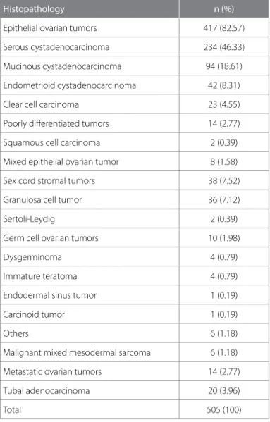
The results of the pathological examination in the malignancy group showed that 417 patients (82.57%) had epithelial ovarian tumor, 38 patients (7.52%) had sex cord stromal tumor, 10 patients (1.98%) had germ cell tumors, 14 patients (2.77%) had metastatic ovarian tumor, and 20 patients (3.96%) had tubal adenocarcinoma.
DISCUSSION
Ovarian and tubal cancers are the gynecologic cancers with highest mortality rates in developed countries (5). In their study conducted on 4,848 patients with adnexal mass, Dirk et al. reported that the rate of malignancy in oncology centers was 43% and that the rate of malignancy in other centers was 17% (48). In our study, we found the rate of malignancy as 43.83% and the rate of borderline tumor as 10.24%. Of ovarian cancers, 80%-85% have epithelial origin. High-grade serous tumors are the most common histological type (5). In our study, we most commonly (46.33%) found the histology of serous epithelial Table 1. General characteristics of patients with adnexal mass
n (%) 1152 Age (mean) mean±SD 46.23 Parity (mean) mean±SD 2.76 Ca 125 (median) U/mL 77.45 Largest tumor diameter cm (median) 9.0 Postmenopausal status Benign 529 (45.92) 43.25±14.8 2.74±2.48 19 (2-2697) 8 (2-42) %32 Borderline 118 (10.24) 42.38±13.9 2.40±2.33 29 (2-15346) 10 (4-48) %27 Malignant 505 (43.83) 50.27±12.17 2.89±2.28 150 (7-15554) 10 (2-50) %59
Table 2. The distribution of benign adnexal masses
Histopathology n (%) Mucinous cystadenoma 123 (23.25) Serous cystadenoma 90 (17.01) Fibroma 32 (6.04) Fibrothecoma 70 (13.23) Dermoid cyst 98 (18.52) Struma ovarii 2 (0.37) Endometrioma 97 (18.33) Pelvic abscess 11 (2.07) Benign brenner 6 (1.13) Total 529 (100)
Table 3. Distribution of borderline tumors
Histopathology n (%)
Serous borderline tumors 50 (42.37)
Mucous borderline tumors 68 (57.62)
Total 118 (100)
Eur Arch Med Res 2018; 34 (4): 231-4
232
1
ovarian adenocarcinoma in the malignant group, which is con-sistent with the literature.
Sex cord stromal ovarian tumors account for approximately 7% of malignant ovarian tumors, and the most common malignant tumors in this group are granulosa cell tumors (7). We found that the rate of granulosa cell tumors in the malignant adnexial group was 7.12%. Malignant germ cell tumors account for up to 5% of all malignant ovarian neoplasms, and the majority are seen in the first two decades (8). Because of the small number of patients less than 18 years of age admitted to our clinic, we detected malignant germ cell tumors only in 10 (1.98%) patients. Most commonly, we detected dysgerminoma and immature teratoma. Of the ovarian tumors, 5%–6% are fre-quently caused by metastases from other organs such as other genital organs, gastrointestinal tract, or breast (9). In this study, we found the rate of metastatic ovarian tumors as 2.77%. Nowadays, it is accepted that the most common high-grade serous carcinoma is caused mainly by fimbria and tuba uterine, and it is accepted that there are tuba uterine and peritoneal cancers at the etiology (10). In our study, the rate of primary tubal adenocarcinoma was found to be 3.96%. Borderline
tumors constitute up to 10%-15% of epithelial ovarian tumors (11). In our study, we found the rate of borderline tumor as 10.24%.
The shape, boundaries, and diameter of the ovary mass, wheth-er it is cystic or solid, single or bilatwheth-eral, and the features such as the presence of acid should be determined through ultrasonog-raphy (USG). Tumor markers, especially human chorionic gonad-otropin, alpha fetoprotein, and CA-125, are important in the dif-ferential diagnosis. However, the definitive diagnosis in the ovarian masses is made by histopathological examination of the operation material (12). In the results of USG, physical examina-tion, and tumor markers, cystadenoma, fibroma, fibrotekom, dermoid cyst, and endometrioma were found to be the major benign masses mimicking malignancy in our study. We found that squamous cell carcinoma developed on the basis of der-moid cyst only in two patients, and carcinoid tumor developed in one patient. Although there may be malignant changes in endometriosis, it can sometimes be found together with ovarian tumors without malignant changes; ovarian cancers, which are most commonly accompanied by endometriosis, are clear cell and endometrioid type cancers (13). In our study, we found three endometrioid adenocarcinomas and three clear cell carci-nomas.
CONCLUSION
Many benign masses can mimic ovarian cancer, and some of these masses can be differentiated from malignancy with careful preoperative examination, but definitive diagnosis is made by histopathological examination.
Ethics Committee Approval: Authors declared that the research was conducted according to the principles of the World Medical Association Declaration of Helsinki “Ethical Principles for Medical Research Involving Human Subjects”, (amended in October 2013).
Informed Consent: Written informed consent was obtained from the patients who participated in this study.
Peer-review: Externally peer-reviewed.
Author Contributions: Concept - B.E.; Design - B.E., O.A.; Supervision - N.P.; Data Collection and/or Processing - İ.T.Y., N.A.S.; Analysis and/or Interpretation - B.E, Ö.A.; Literature Search - B.E.; Writing Manuscript - B.E.; Critical Review - V.Ü., Ö.A.
Conflict of Interest: The authors have no conflicts of interest to declare.
Financial Disclosure: The authors declared that this study has received no financial support.
REFERENCES
1. Siegel RL, Miller KD, Jemal A. Cancer statistics, 2016. CA Cancer J Clin 2016: 66: 7-30. [CrossRef]
2. Kuznia AL, Roett MA. Genital Cancers in Women: Ovarian Cancer. FP Essent 2015; 438: 24-30.
3. Koonings PP, Campell K, Mishell DR, Grimes DA. Relative frequen-cy of primary ovarian neoplasms: a 10-year review. Obstet Gynecol 1989: 74: 921-6.
4. ACOG Committee on Practice Bulletins. Management of Adnexal Masses. Washington, DC: American College of Obstetricians and Gynecologists, 2007. Number 83. Reaffirmed 2015.
Table 4. Distribution of malignant adnexal tumors
Histopathology n (%)
Epithelial ovarian tumors 417 (82.57)
Serous cystadenocarcinoma 234 (46.33)
Mucinous cystadenocarcinoma 94 (18.61) Endometrioid cystadenocarcinoma 42 (8.31)
Clear cell carcinoma 23 (4.55)
Poorly differentiated tumors 14 (2.77)
Squamous cell carcinoma 2 (0.39)
Mixed epithelial ovarian tumor 8 (1.58)
Sex cord stromal tumors 38 (7.52)
Granulosa cell tumor 36 (7.12)
Sertoli-Leydig 2 (0.39)
Germ cell ovarian tumors 10 (1.98)
Dysgerminoma 4 (0.79)
Immature teratoma 4 (0.79)
Endodermal sinus tumor 1 (0.19)
Carcinoid tumor 1 (0.19)
Others 6 (1.18)
Malignant mixed mesodermal sarcoma 6 (1.18)
Metastatic ovarian tumors 14 (2.77)
Tubal adenocarcinoma 20 (3.96)
Total 505 (100)
Erdem et al. Adnexal Masses Eur Arch Med Res 2018; 34 (4): 231-4
5. Seidman JD, Horkayne-Szakaly I, Haliba M, Boice CR, Kurman RJ, Ronnett BM. The histologic type and stage distribution of ovarian carcinomas of surface epithelial origin. Int J Gynecol Pathol 2004; 23: 41-4. [CrossRef]
6. Timmerman D, Van Calster B, Testa A, Savelli L, Fischerova D, Frowman W, et al. Predicting the risk of malignancy in adnexal mass-es based on the Simple Rulmass-es from the International Ovarian Tumor Analysis Group. Am J Obstet Gynecol 2016; 214: 424-37. [CrossRef]
7. Malmstrom H, Hogberg T, Risberg B, Simonsen E. Granulosa cell tumor of the ovary: Prognostic factors and outcome. Gynecol Oncol 1994; 52: 50-5. [CrossRef]
8. Scully RE, Young RH, Clement PB. Tumours of the ovary, maldevel-oped gonads, fallopian tube and broad ligament. In: Atlas of Tumor Pathology: 3rd series, Fascicle 23. Washington, DC: Armed Forces İnstitute of Pathology 1998: 169-498.
9. Petru E, Pickel H, Heydarfadai M, Lahousen M, Haas J, Schaider H, et al. Nongenital cancers metastatic to the ovary. Gynecol Oncol 1992; 44: 83-6. [CrossRef]
10. Crum CP, Drapkin R, Miron A, Ince TA, Muto M, Kindelberger DW, et al. The distal fallopian tube: A new model for pelvic serous car-cinogenesis. Curr Opin Obstet Gynecol 2007; 19: 3-9. [CrossRef]
11. Barnhill DR, Kurman RJ, Brady MF, Omura GA, Yordan E, Given FT, et al. Preliminary analysis of the behavior of stage 1 ovarian serous tumors of low malignant potential: A Gynecologic Oncology Group study. J Clin Oncol 1995; 13: 2752-6. [CrossRef]
12. Akercan F, Cirpan T, Yildiz PS, Ozsener S, Karadadas N, Ulukis M. Approaches To The Dıagnosıs and Treatment Of Bening Adnexial Masses. Ege Tıp Dergisi 2005; 44:151-4.
13. Orezzoli JP, Russell AH, Oliva E, Del Carmen MG, Eichhorn J, Fuller AF. Prognostic implication of endometriosis in clear cell carcinoma of ovary. Gynecol Oncol 2008; 110: 336-44. [CrossRef]
Eur Arch Med Res 2018; 34 (4): 231-4