Istanbul Aydın University Journal of Faculty of Dentistry
Yıl 2 Sayı 3 - 2016 ISSN 2149-5572 ORIGINAL ARTICLES
Pull-Out Strength of Fiber Posts Luted with Different Self-Adhesives Cem ŞAHİN, Simel AYYILDIZ
Mutagenic Potential of a Self-Adhesive Flowable Composite Tuğba TOZ, Zeliha AYDOĞAN, Duygu TUNCER, Emel KARAMAN
CASE REPORTS
Aesthetic Management of Anterior Teeth with Direct Composite Veneers and Bleaching After Fixed Orthodontic Treatment Engin Fırat ÇAKAN, Fatma YILDIRIM, Ahu TOPKARA
Endodontic Treatment of Mandibular Premolar with Three Canals Using CBCT Seda AYDEMİR, Alper SİNANOĞLU, Işıl Kaya BÜYÜKBAYRAM, Göze ARUKASLAN
Single No-Prep Porcelain Laminate Veneer Restorations: 2 Case Reports Volkan TURP, Simel AYYILDIZ, Deniz ŞEN, Gülcan BAHARDIRLI
Clinical and Radiological Evaluation of the Problems Occured Due to Inappropriate Treatment of a Patient with Anterior Open Bite: A Malpractice Case
Orhan AKSOY, Sercan KÜÇÜKKURT
Proliferative Verrucous Leukoplakia: Five Years Follow-Up Behçet EROL, Sercan KÜÇÜKKURT, Tuğçe BİÇER AYTUGAR, Nihan AKSAKALLI
REVIEWS
Chairside Cerec System and CAD/CAM Materials Işıl Kaya BÜYÜKBAYRAM, Engin Fırat ÇAKAN, Mağrur KAZAK
Nanoceramics and Hybrid Materials used in CAD/CAM Systems
Nesrin CEREN, Volkan TURP, Faruk EMİR, Gökhan AKGÜNGÖR, Simel AYYILDIZ, Deniz ŞEN
AYDIN DENTAL
Year 2 Number 3 - 2016
ISTANBUL AYDIN UNIVERSITY
JOURNAL OF FACULTY OF DENTISTRY
ISTANBUL AYDIN UNIVERSITY JOURNAL OF FACULTY OF DENTISTRY
AYDIN DENTAL
Ahu URAZ Gazi University, Turkey Ali ZAIMOGLU Istanbul Aydın University, Turkey Arzu ATAY Gulhane Military Medical Academy, Turkey Aylin BAYSAN The London School of Medicine and Dentistry, London, U.K.
Ayşen NEKORA AZAK Istanbul Aydın University, Turkey Behçet EROL Istanbul Aydın University, Turkey
Berna TARIM, Istanbul University, Turkey Bilgin GİRAY Istanbul Aydın University, Turkey Bora ÖZDEN Ondokuz Mayıs University, Turkey
Can DÖRTER Istanbul University, Turkey Cansu ALPASLAN Gazi University, Turkey
Cem TANYEL Istanbul University, Turkey Cemal ERONAT Ege University, Izmir, Turkey Didem ÖNER ÖZDAŞ Istanbul Aydın University, Turkey Elif KALYONCUOĞLU Ondokuz Mayıs University, Turkey
Engin Fırat CAKAN, Istanbul Aydın University, Turkey Erman BULENT TUNCER Istanbul Aydın University, Turkey Ersin YILDIRIM Gulhane Military Medical Academy, Turkey
Esra SOMTÜRK Istanbul Aydın University, Turkey Feyza OTAN ÖZDEN Ondokuz Mayıs University, Turkey Fulya TOKSOY TOPÇU Gulhane Military Medical Academy, Turkey
Gülce ALP Istanbul Aydın University, Turkey
Günseli GÜVEN POLAT Gülhane Military Medical Academy, Turkey Hakan ÖZBAŞ Istanbul University, Turkey
Handan ERSEV Istanbul University, Turkey Kadriye DEMİRKAYA Gulhane Military Medical Academy, Turkey
Kemal SÜBAY Istanbul Aydın University, Turkey Korkud DEMİREL Istanbul University, Turkey Leyla KURU Marmara University, Istanbul, Turkey Mehmet CUDİ BALKAYA Istanbul Aydın University, Turkey Işıl Kaya BÜYÜKBAYRAM, Istanbul Aydın University, Turkey
Raif ERİŞEN Istanbul University, Turkey Rezzan ÖZER Dicle University, Turkey Rüdiger JUNKER Danube Private University, Austria
Sedat ÇETİNER Gazi University, Turkey Sema BELLİ Selçuk University, Turkey Sema ÇELENK Dicle University, Turkey Semih BERKSUN Ankara University, Turkey Serap KARAKIŞ Istanbul Aydın University, Turkey
Serdar CİNTAN Istanbul University, Turkey Simel AYYILDIZ Gulhane Military Medical Academy, Turkey
Şeniz KARAÇAY Gulhane Military Medical Academy, Turkey Ümit KARAÇAYLI Gulhane Military Medical Academy, Turkey
Vesela STEFANOVA Medical University of Plovdiv, Bulgaria Proprietor - Sahibi
Mustafa Aydın
Editor-in-Chief - Yazı İşleri Müdürü Nigar Çelik
Editor - Editör
Jülide Özen Editorial Board - Yayın Kurulu Jülide Özen
Sercan Küçükkurt
Academic Studies Coordination Office (ASCO) Akademik Çalışmalar Koordinasyon Ofisi İdari Koordinatör - Administrative Coordinator Nazan Özgür
Technical Editor - Teknik Editör Hakan Terzi
Language - Dili English - Türkçe
Publication Period - Yayın Periyodu Published twice a year - Yılda iki kere yayınlanır April and October - Nisan ve Ekim
Correspondence Address - Yazışma Adresi Beşyol Mahallesi, İnönü Caddesi, No: 38 Sefaköy, 34295 Küçükçekmece/İstanbul Tel: 0212 4441428 - Fax: 0212 425 57 97 web: www.aydin.edu.tr - e-mail: [email protected] Printed by - Baskı
ISSN: 2149-5572
Scientific Board
İstanbul Aydın Üniversitesi, Diş Hekimliği Fakültesi, Aydın Dental Dergisi özgün bilimsel araştırmalar ile uygulama çalışmalarına yer veren ve bu niteliği ile hem araştırmacılara hem de uygulamadaki akademisyenlere seslenmeyi amaçlayan hakem sistemini kullanan bir dergidir.
Istanbul Aydın University, Journal of the Faculty of Dentistry, Aydın Dental is a double-blind peer-reviewed journal which provides a platform for publication of original scientific research and applied practice studies. Positioned as a vehicle for academics and practitioners to share field research, the journal aims to appeal to both researchers and academicians.
İÇİNDEKİLER - CONTENTS
ORIGINAL ARTICLES
Pull-Out Strength of Fiber Posts Luted with Different Self-Adhesives
Cem ŞAHİN, Simel AYYILDIZ... 1
Mutagenic Potential of a Self-Adhesive Flowable Composite
Tuğba TOZ, Zeliha AYDOĞAN, Duygu TUNCER, Emel KARAMAN... 9 CASE REPORTS
Aesthetic Management of Anterior Teeth with Direct Composite Veneers and Bleaching After Fixed Orthodontic Treatment
Engin Fırat ÇAKAN, Fatma YILDIRIM, Ahu TOPKARA... 17
Endodontic Treatment of Mandibular Premolar with Three Canals Using CBCT
Seda AYDEMİR, Alper SİNANOĞLU, Işıl Kaya BÜYÜKBAYRAM, Göze ARUKASLAN... 25
Single No-Prep Porcelain Laminate Veneer Restorations: 2 Case Reports
Volkan TURP, Simel AYYILDIZ, Deniz ŞEN, Gülcan BAHARDIRLI... 29
Clinical and Radiological Evaluation of the Problems Occured Due to Inappropriate Treatment of a Patient with Anterior Open Bite: A Malpractice Case
Orhan AKSOY, Sercan KÜÇÜKKURT... 35
Proliferative Verrucous Leukoplakia: Five Years Follow-Up
Behçet EROL, Sercan KÜÇÜKKURT, Tuğçe BİÇER AYTUGAR, Nihan AKSAKALLI... 41
REVIEWS
Chairside Cerec System and CAD/CAM Materials
Işıl Kaya BÜYÜKBAYRAM, Engin Fırat ÇAKAN, Mağrur KAZAK... 47
Nanoceramics and Hybrid Materials used in CAD/CAM Systems
1
© 2016 Published by Istanbul Aydın University, Faculty of Dentistry. All rights reserved. Aydın Dental - Year 2 Number 3 - 2016 (1-7)
PULL-OUT STRENGTH OF FIBER POSTS
LUTED WITH DIFFERENT SELF-ADHESIVES
Cem ŞAHİN1*, Simel AYYILDIZ21* School of Health Services, Dental Prosthetics Technology, Hacettepe University, Ankara, Turkey
2 Department of Prosthodontics, Center for Dental Sciences, Gulhane Military Medical Academy, Ankara, Turkey.
ABSTRACT
Introduction: Dislodging from the root canals is the most common failure of bonded fiber restorations. Self-adhesive system has the advantage of reduced cementation procedure. The aim of this study was to evaluate the bond strengths of fiber posts with three different self-adhesive/resin systems to coronal and apical thirds of the post space dentine
Materials and methods: Thirty freshly extracted single-root human teeth were selected for this study. The specimens were then randomly divided into 3 subgroups each containing 10 samples. Fiber posts (RelyX, 3M, ESPE, UK) were inserted into canals using RelyX Unicem 3M ESPE, St. Paul, MN, USA), G-Cem (GC Corporation, Tokyo, Japan) and Maxcem Elite (Kerr Hawe Neos Orange,CA, USA) adhesives. Two disc shaped sections perpendicular to the long axis of each root sample were obtained for push-out tests. Failure load value was recorded. Statistical analysis was performed using One-way ANOVA and Kruskall-Wallis tests.
Results: RelyX Unicem exhibited the highest mean push-out bond strength values at the coronal section of all samples. G-Cem exhibited higher push-out bond strength values at apical section The overall bonding data of RelyX Unicem was also observed to be the highest among groups.
Conclusion: Self-adhesive systems are of practical ways of intra-canal cementation. It does not require multiple steps for success. All the systems offered acceptable retention ability. Keywords: Self-adhesive systems, root-canal cementation, post-core
Keywords: Self-adhesive systems, root-canal cementation, post-core
ÖZET
Giriş: Bağlantı sonrası fiber restorasyonların kök kanallarından ayrılması en sık karşılaşılan başarısızlıktır. Self adeziv sistemler azaltılmış simantasyon prosedür avantajına sahiptir. Üç farklı self-adeziv rezin sistemin kullanıldığı bu çalışmanın amacı fiber postların koronal ve apikal üçlülerdeki bağlanma dayanımını değerlendirmektir.
Materyal ve metod: Bu çalışma için 30 adet yeni çekilmiş tek köklü insan dişi seçildi. Örnekler 10!arlı rasgele gruplandı. Fiber postlar (RelyX, 3M, ESPE, UK) RelyX Unicem (3M ESPE, St. Paul, MN, USA), G-Cem (GC Corporation, Tokyo, Japan) ve Maxcem Elite (Kerr Hawe Neos Orange,CA, USA) adezivler kullanılarak kanallara yerleştirildi. İtme testleri için örneklerden diş uzun eksenine dik ve birbirine paralel 2 disk şeklinde kesit alındı. İstatistiksel analizler için tek yönlü varyans analizi ve Kruskall-Wallis testleri kullanıldı.
Sonuçlar: RelyX Unicem tüm örneklerde koronal bölgede en yüksek itme değerleri ortaya çıkardı. G-Cem ise apikal bölgede en yüksek değerlere ulaştı. Tüm veriler değerlendirildiğinde RelyX-Unicem yine en yüksek değerleri verdi.
Karar: Kanal içi simantasyonunda self-adeziv sistemler pratik çözüm yoludur. Çoklu aşama gerektirmezler. Çalışmada kullanılan bütün sistemler kabul edilebilir bağlanma değerleri sunmaktadır.
Anahtar Kelimeler: Self-adeziv sistemler, Kök-Kanal Simantasyonu, Post-Kor
Pull-Out Strength of Fiber Posts Luted with Different Self-Adhesives
2
INTRODUCTION
Root filled teeth with excessive loss of coronal tooth structure are frequently reconstructed with posts and cores1. Most clinical failures
with endodontic post systems are related with the decementation of the post and/or root fractures2, 3. Conventional metal post
restorations may result an unaesthetic gray discoloration when used with all-ceramic restorations. Besides, fiber posts have better vision and generally offer advantages about application over conventional cast posts or prefabricated metallic posts4-6. The most
common failure of restorations bonded with fiber posts is dislodging of the posts from the root canals7, 8. Various luting agents
and adhesive systems have been proposed for luting fiber posts to root canal dentine9.
Contemporary resin cements, frequently used to lute fiber posts, may be divided into three subgroups according to the adhesive approach; etch-and-rinse adhesive systems, self-etching primers, self-adhesive cements10.
The self-adhesive systems eliminate the need for separate etching, priming, and bonding steps in an attempt to simplify the cementation procedure11. However, information on fiber
post cementation technique is still confusing and inadequate in the literature10, 12. Besides, as
the number of dentine tubules decreases from coronal to the apical direction, the success of bonding to dentine may vary at these different regions of even the same root canal9.
Therefore, the aim of this study was to evaluate the bond strengths of fiber posts with three different adhesive/resin systems to coronal and apical thirds of the post space dentine.
MATERIALS AND METHODS
Thirty freshly extracted single-root human teeth were selected for this study. Soft tissue and remnants were cleaned gently from the root surfaces. The crown of each tooth was removed with a diamond disc 1 mm apical from the cemento-enamel junction. After gently removing the pulpal residuals working length was determined as 1 mm shorter than the canal length. The canals were instrumented using a crown-down technique with rotary ProTaper instruments (Dentsply, Maillefer, Ballaigues, Switzerland) to size of finishing F3. The canals were irrigated with 2% sodium hypochlorite (NaOCl) among each file size and then the root canals were dried with #30 sterile paper points (Spident, Incheon, Korea). All root canals were obturated with lateral compaction technique. Single cone ProTaper Gutta-percha (#30) (F3) (Dentsply, Maillefer, Ballaigues, Switzerland) with AH-26 sealer was used as a master cone. The samples were then stored at 100 % humidity for 7 days at 37 ºC to allow complete setting of the sealers. To prepare 8-mm-deep fiber post spaces ISO size-90 preparation instrument (Dentsply, Maillefer, Ballaigues, Switzerland) was used. Spaces were irrigated with 2% sodium hypochlorite solution and were dried with paper points. The specimens were then randomly divided into 3 subgroups each containing 10 samples. Fiber posts (RelyX, 3M, ESPE, UK) were inserted into canals using RelyX Unicem 3M ESPE, St. Paul, MN, USA), G-Cem (GC Corporation, Tokyo, Japan) and Maxcem Elite (Kerr Hawe Neos Orange,CA, USA) adhesives. All materials were used according to the manufacturers’ recommendations. All the posts were then seated to full depth in the prepared spaces using finger pressure. The excess luting agent
Cem SAHİN, Simel AYYILDIZ
3
Aydın Dental - Year 2 Number 3 - 2016 (1-7)
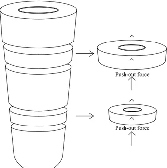
material was immediately removed with a small cotton brush. After initial setting, the resin luting cements were polymerized with a light cure for 40 s. Thirty minutes after the cementation procedures, all samples were stored in distilled water for 24 h. Two disc shaped sections perpendicular to the long axis of each root sample were obtained for push-out tests as shown in the figure 1. Using a low-speed saw (Micromet M; Remet S.p.A., Casalecchio di Reno, Italy).
Figure 1: Schematic view of obtaining coronal and apical sections
The thickness of each specimen was measured and recorded by a digital caliper (Insize, Insize Co., LTD, USA) having the accuracy of 0.001 mm. Apical side of each slice (1.0 ± 0.1 mm) was marked with an indelible marker.
Push-out tests were performed by applying a compressive load at a crosshead speed of 0.5 mm/min to the apical aspect of each slice via a cylindrical plunger mounted on a Universal Testing Machine (Lloyd LR 30K; Lloyd Instruments Ltd, UK). Load was performed
until the breakdown occur. Maximum failure load value was recorded.
Statistical analysis
The statistical difference between the push-out bond strength values of the groups were analyzed using One-way ANOVA. Then Kruskall-Wallis tests were performed to assess the significance among groups
RESULTS
Push-out test results are shown in Table 1. RelyX Unicem exhibited the highest mean push-out bond strength values at the coronal section of the samples. On the other hand Maxcem Elite exhibited the least mean push-out bond strength values at the apical section of the samples.
G-Cem exhibited higher push-out bond strength values at apical section than both RelyX unicem and Maxcem Elite adhesives, however this was not statistically significant at 0,05 confidence level, conversely the push-out values at the coronal third obtained with RelyX Unicem adhesive was significantly higher than other groups. (p<0.05).
The overall bonding data of RelyX Unicem was also observed to be the highest among groups. However the differences among all groups were not statistically significant (p>0.05).
DISCUSSION
Dual-cure adhesive systems need multi-clinical steps and do require pretreatment at dentin/enamel surface however, self-adhesive systems do not require any pretreatment. Self-adhesive systems have methacrylate monomer formulation with phosphoric acid
Push-out force
Pull-Out Strength of Fiber Posts Luted with Different Self-Adhesives
4
esters, which produces polymerization in an acidic environment13. Methacrylate monomers
infiltrate into dentin while the acidic esters demineralize the surface. To balance the acidic activity of the esters during and after the process glass-ionomer formulations are added into many of the self-adhesive systems. The interaction between the hydroxyapatite crystal of dentin and the methacrylate monomers are altered by such compositional changes. In addition, it should be reminded that the retentive data may vary depending on the
composition of the adhesive system itself. As the particle size of the filler ingredient increase (for cohesive strength) the flowing ability will degrease which is essential for self-adhesive systems to move through the retentive areas. Therefore, the activity of the material should be evaluated carefully for a detailed discovery of functional ability.
Several studies reported controversial results regarding bond strengths of different luting agents in the root canals7, 14-16. Kerstin and
Table 1: Mean push-out bond strengths for experimental groups
RelyX Unicem G-Cem Maxcem Elite
Coronal third 17.53 ± 3.01 N 15.43 ± 4.17 N 14.91 ± 3.46 N
Apical third 14.77 ± 2.72 N 15.07 ± 3.23 N 14.22 ± 3.82 N
Overall 16.15 ± 2.94 N 15.25 ± 3.76 N 14.56 ± 3.22 N
Sebastian17 found that RelyX self-adhesive
cement pointed out significantly higher push-out bond strength values than Panavia and Variolink II dual cure adhesive systems. They speculated that the moisture tolerance of these systems may explain the favorable adhesion of RelyX1. This result is also compatible with
ours, that RelyX Unicem showed significantly higher bonding values in the coronal section (p<0.05). The degree of moisture is known to be difficult to control inside the root canal. However, etch-and-rinse based adhesives need to be applied on the restrained moist dentin. Moreover, this is essential for the use of dual cure materials inside the root canal. The success of RelyX Unicem in our study may also be attributed to the optimum film thickness of this material18. Because of the
ingredients and composition of adhesive materials, it may be very difficult to produce thinner film thickness which is essential to avoid cohesive failures. Cohesive failures in adhesive materials induce failure of the
restoration even the penetration into dentin or restoration is quite acceptable. On the other hand, degree of conversion rates of this adhesive system is claimed to be very high19.
It is known that for the completion of the polymerization (adequate bond strengths and mechanical properties) 24 hours is requisite at the areas without energy of light20. In our
study all the samples were stored in water for sufficient polymerization for 24 hours.
On the contrary, Maxcem Elite represented significantly lower bond strength values than G-Cem and RelyX Unicem in both apical and coronal sections in our study (p<0.05). The 37% phosphoric acid activity of this system may not be enough to decalcify and remove dentin from the surface. The excess smear-like substrate is un-favored situation for adhesive activity.
The acidic activity of the material is a very effective factor which is the first step of
Cem SAHİN, Simel AYYILDIZ
5
Aydın Dental - Year 2 Number 3 - 2016 (1-7)
penetration of the resin into dentinal canals or irregular surfaces. It is known that irregular surfaces and/or dentinal canals are necessary for retention of the resin material. Higher pH concentration may completely deform the dentinal canal formation which is never desired. On the other hand, lower pH values may not be enough for required decalcification. Therefore, optimum acidic concentration is essential. Self-adhesive systems have a self-etch process that provide rough dentin surface which is essential for adhesive penetration as mentioned. The self-etch process of these systems cannot be inspected visually and may sometimes be inadequate. According to our results Maxcem Elite represented the lowest test values at both sections with respect to RelyX Unicem and G-Cem.
Investigations about the composition and structure of apical and coronal parts of root canal dentine also depicted conflict results. While some of the investigators reported higher bond strengths to root canal dentin in the apical one-third21, 22, others claimed that
higher values were obtained at the coronal one-third23, 24. In our study we obtained higher
bond strength values at coronal sections of each sample than apical sections. The difference was statistically significant for Relyx Unicem samples (p<0.05) but not for Maxcem Elite and G-cem samples.
Dual cure adhesive systems polymerize both with light and chemical activation. It is clear that during a root canal cementation because of non-light areas, chemical curing specialty should be assumed as more important than the light curing. However, the process starts with the light activity. Within the limitations of this study the samples for push-out tests were obtained from apical and coronal sections which are impossible to be light activated.
The lower bonding values at each sample may be attributed to this issue as mentioned. Polymerization shrinkage is one of the most important issue for light activated or dual cured resin systems. Researchers claimed that25, 26 chemical cured systems produces
higher bonding strength than dual cured systems. Therefore, push-out data of samples that are chemically cured may be different from dual cured.
CONCLUSIONS
Self-adhesive systems are of practical ways of intra-canal cementation. It does not require multiple steps for success. All the systems offered acceptable retention ability.
Conflict of Interest
We the authors declare that we do not have any conflict of interest
REFERENCES
[1] Bitter K, Meyer-Lueckel H, Priehn K, Kanjuparambil JP, Neumann K, Kielbassa AM. Effects of luting agent and thermocycling on bond
strengths to root canal dentine. Int Endod J. 2006;39:809-18.
[2] Bergman B, Lundquist P, Sjogren U, Sundquist G. Restorative and endodontic results after treatment with cast posts and cores. J Prosthet Dent. 1989;61:10-5. [3] Bitter K, Priehn K, Martus P, Kielbassa
AM. In vitro evaluation of push-out bond strengths of various luting agents to tooth-colored posts. J Prosthet Dent. 2006;95:302-10.
[4] Fokkinga WA, Kreulen CM, Vallittu PK, Creugers NH. A structured analysis of in vitro failure loads and failure modes of fiber, metal, and ceramic post-and-core systems.
Pull-Out Strength of Fiber Posts Luted with Different Self-Adhesives
6
Int J Prosthodont. 2004;17:476-82.
[5] Cormier CJ, Burns DR, Moon P. In vitro comparison of the fracture resistance and failure mode of fiber, ceramic, and conventional post systems at various stages of restoration. J Prosthodont. 2001;10:26-36.
[6] Newman MP, Yaman P, Dennison J, Rafter M, Billy E. Fracture resistance of endodontically treated teeth restored with composite posts. J Prosthet Dent. 2003;89:360-7.
[7] Ohlmann B, Fickenscher F, Dreyhaupt J, Rammelsberg P, Gabbert O, Schmitter M. The effect of two luting agents, pretreatment of the post, and pretreatment of the canal dentin on the retention of fiber-reinforced composite posts. J Dent. 2008;36:87-92. [8] Victorino KR, Kuga MC, Duarte MA,
Cavenago BC, So MV, Pereira JR. The effects of chlorhexidine and ethanol on push-out bond strength of fiber posts. J Conserv Dent. 2016;19:96-100.
[9] D’Arcangelo C, Zazzeroni S, D’Amario M, Vadini M, De Angelis F, Trubiani O, et al. Bond strengths of three types of fibre-reinforced post systems in various regions of root canals. Int Endod J. 2008;41:322-8. [10] Radovic I, Mazzitelli C, Chieffi N, Ferrari
M. Evaluation of the adhesion of fiber posts cemented using different adhesive approaches. Eur J Oral Sci. 2008;116:557-63.
[11] Saskalauskaite E, Tam LE, McComb D. Flexural strength, elastic modulus, and pH profile of self-etch resin luting cements. J Prosthodont. 2008;17:262-8.
[12] Rezende EC, Gomes GM, Szesz AL, Bueno CE, Reis A, Loguercio AD. Effects of Dentin Moisture on Cementation of Fiber Posts to Root Canals. J Adhes Dent. 2016;18:29-34.
[13] Gerth HU, Dammaschke T, Zuchner H, Schafer E. Chemical analysis and bonding reaction of RelyX Unicem and Bifix composites--a comparative study. Dent Mater. 2006;22:934-41.
[14] Cury AH, Goracci C, de Lima Navarro MF, Carvalho RM, Sadek FT, Tay FR, et al. Effect of hygroscopic expansion on the push-out resistance of glass ionomer-based cements used for the luting of glass fiber posts. J Endod. 2006;32:537-40.
[15] Sadek FT, Goracci C, Monticelli F, Grandini S, Cury AH, Tay F, et al. Immediate and 24-hour evaluation of the interfacial strengths of fiber posts. J Endod. 2006;32:1174-7. [16] Kremeier K, Fasen L, Klaiber B, Hofmann
N. Influence of endodontic post type (glass fiber, quartz fiber or gold) and luting material on push-out bond strength to dentin in vitro. Dent Mater. 2008;24:660-6.
[17] Bitter K, Paris S, Pfuertner C, Neumann K, Kielbassa AM. Morphological and bond strength evaluation of different resin cements to root dentin. Eur J Oral Sci. 2009;117:326-33.
[18] Kious AR, Roberts HW, Brackett WW. Film thicknesses of recently introduced luting cements. J Prosthet Dent. 2009;101:189-92. [19] Arrais CA, Giannini M, Rueggeberg FA.
Kinetic analysis of monomer conversion in auto- and dual-polymerizing modes of commercial resin luting cements. J Prosthet Dent. 2009;101:128-36.
[20] Al-Assaf K, Chakmakchi M, Palaghias G, Karanika-Kouma A, Eliades G. Interfacial characteristics of adhesive luting resins and composites with dentine. Dent Mater. 2007;23:829-39.
[21] Gaston BA, West LA, Liewehr FR, Fernandes C, Pashley DH. Evaluation of regional bond strength of resin cement to endodontic surfaces. J Endod. 2001;27:321-4.
Cem SAHİN, Simel AYYILDIZ
7
Aydın Dental - Year 2 Number 3 - 2016 (1-7)
[22] Muniz L, Mathias P. The influence of sodium hypochlorite and root canal sealers on post retention in different dentin regions. Oper Dent. 2005;30:533-9.
[23] Yoshiyama M, Matsuo T, Ebisu S, Pashley D. Regional bond strengths of self-etching/ self-priming adhesive systems. J Dent. 1998;26:609-16.
[24] Bouillaguet S, Troesch S, Wataha JC, Krejci I, Meyer JM, Pashley DH. Microtensile bond strength between adhesive cements and root canal dentin. Dent Mater. 2003;19:199-205. [25] Vaz RR, Hipolito VD, D’Alpino PH,
Goes MF. Bond strength and interfacial micromorphology of etch-and-rinse and self-adhesive resin cements to dentin. J Prosthodont. 2012;21:101-11.
[26] D’Arcangelo C, De Angelis F, D’Amario M, Zazzeroni S, Ciampoli C, Caputi S. The influence of luting systems on the microtensile bond strength of dentin to indirect resin-based composite and ceramic restorations. Oper Dent. 2009;34:328-36.
9
Aydın Dental - Year 2 Number 3 - 2016 (9-15)
1 Department of Restorative Dentistry, Faculty of Dentistry, TC Istanbul Medipol University , Istanbul, Turkey 2 Department of Molecular Biology , Faculty of Science. Hacettepe University, Ankara, Turkey
3 Department of Restorative Dentistry, Faculty of Dentistry, Başkent University, Ankara, Turkey 4* Department of Restorative Dentistry, Faculty of Dentistry, 19 Mayıs University, Samsun, Turkey
MUTAGENIC POTENTIAL OF A SELF-ADHESIVE
FLOWABLE COMPOSITE
Tuğba TOZ1, Zeliha AYDOĞAN2, Duygu TUNCER3, Emel KARAMAN4*
ABSTRACT
Objective: The aim of this study was to assess the potential mutagenic effects associated to extracts from current self-adhesive flowable resin composite (Vertise Flow,Kerr Corp, Orange, CA, USA) that allows skipping the time-consuming adhesive processes. Study design: The materials were eluted in dimethyl sulphoxide and the extracts were tested either after 1 day or 7 day incubation period at 370C. Mutagenic effects of the materials were tested on Salmonella typhipmurium strain TA 100 using the standard plate incorporation assay in the absence of S9 fraction from rat liver. The data were statistically analyzed using two-way variance analysis (p<0.05).
Results: The dose of the material and incubation as well as the interactions between these factors exhibited varying degrees of influences on the Salmonella typhipmurium colony number. However no mutagenic effect was detected for the self-adhesive restorative material.
Conclusion: It can be concluded that the adhesive restorative material tested in this study has no mutagenic potential.
Keywords: mutagenicity, AMES test, Salmonella tphymiruim, self-adhesive composite, bulk fill flowables, adhesive resin
ÖZET
Amaç: Bu çalışmanın amacı, zaman alıcı adeziv prosedürleri kısaltan self-adeziv akışkan rezin kompozit (Vertise Flow,Kerr Corp, Orange, CA, ABD) ekstrelerinin potansiyel mutajenik etkisinin değerlendirilmesidir.
Çalışma dizaynı: Materyal örnekleri dimetil sülfoksit içerisinde bekletilmişler ve 370C’ deki inkübasyon sürelerinin 1. ve 7. günlerinde test edilmişlerdir. Bu materyallerin mutajenik etkileri, Salmonella typhipmurium TA 100 suşunda, standart plak korporasyon yöntemi ile S9 fraksiyonu eksikliğinde değerlendirilmiştir. Veriler iki yönlü varyans analizi yardımı ile istatistiksel olarak analiz edilmiştir (p<0.05). Bulgular: Materyal dozu ve inkübasyon süresi ve birbirleri ile ilişkileri Salmonel-la typhipmurium koloni sayıları üzerinde farklı etkiler göstermiştir. Bununla birlikte self-adeziv restoratif materyal için mutajenik etki gözlenmemiştir.
Sonuç: Bu çalışmada test edilen adeziv restoratif materyalin mutajenik etkisi bulunmamıştır.
Anahtar Kelimeler: mutajenisite, AMES testi, Salmonella tphymiruim, self-adeziv kompozit, bulk fill akışkanlar, adeziv rezin
© 2016 Published by Istanbul Aydın University, Faculty of Dentistry. All rights reserved.
Mutagenic Potential of a Self-Adhesive Flowable Composite
10
INTRODUCTION
The introduction of adhesion to dentistry around the mid of the last century can be accepted as a big revolution.This revolution and patients attitude due to their esthetic demands has been contributed to the development of new adhesive restoratives. With adhesive technology, the dental clinicians have met with total etching concept and adhesive systems. After the Buonocore attitude about adhesion,various researchers developed new procedures to enhance handling and quality of adhesive restorative materials.The earlier adhesives include three steps in order to achieve bonding to tooth tissue named as the 3-step etch and rinse systems. The aim was later turned into 2-step etch and rinse and the adhesive systems were classified into two general categories such as two step etch and rinse systems that combine primer and bond together applied after etching; and two step self-etch systems, that etch and prime tooth tissue with primer before the application of bond. These materials were accepted as user-friendly and less technique sensitive. With further laboratory efforts 1step self-etch systems were introduced with dental technology that has the possibility to achieve all the steps. These materials are more effective in minimizing technique sensitivity, showed simultaneous demineralization and resin infiltration and reduce postoperative sensitivity however they still requires the polymerization step1.
Current dental technology goal is combining the benefits of adhesive and composite materials into a self-adhesive restorative. However, it is the biggest question to overcome the hydrophobic–hydrophilic mismatch between restorative material and especially dentin tissue 2.
The latest developments in self-adhesive restorative materials are promoted as materials require neither etching nor a bonding agent3.
Special phosphate dimethacrylate monomers, like glycerol phosphate dimethacrylate (GPDM), allows chemical interaction of the phosphonate groups with calcium ions of tooth tissue can be accepted as a way to reproduce self-adhesive restorative material 4. Firstly
introduced self-adhesive flowable composites were introduced mainly for cavity sealing and restoration. They include monomers mediating adhesion with tooth tissue thus they do not require any adhesive pretreatment5. These
materials did not improve the bond strength to enamel when compared to etch-and-rinse adhesives6 however they have similar shear
bond strength with self-etch and etch and rinse adhesive systems on superficial and deep dentin4. Vertise Flow (VF; Kerr, Orange, CA,
USA)was a self-adhesive flowable composite that includes the monomer glycerol phosphate dimethacrylate (GPDM) designed to bond to tooth tissue without a separate adhesive and etching step according to these contributions
2.
The continuous advancements in dental technology have raised questions about the biological safety of new materials and techniques. There are several investigations on the biocompatibility of dental materials generally focused on the characterization of cytotoxic effects in vitro 7.However, until
recently the efforts to obtain information on the key events leading to cell damage have been scarce. One of the well-known major consequences of dental monomers on living tissue is their induction of DNA mutations, which, if not repaired, could lead to birth defects or malignant transformation of the
Tuğba TOZ,Zeliha AYDOĞAN, Duygu TUNCER, Emel KARAMAN
11
Aydın Dental - Year 2 Number 3 - 2016 (9-15)
tissue as indicated by the induction of genotoxic effects. The Salmonella typhimurium/ microsome assay (Salmonella test; Ames test) is a widely accepted short-term bacterial assay for identifying substances that can produce genetic damage that leads to gene mutations. Also its low cost, simplicity, and speed make the Ames test an important and widespread part of biological examinations of dental materials and of standardization protocols8.
The objective of this study was to determine the mutagenic potential of this self-adhesive flowable composite Vertise Flow related to its monomer ingredients.
MATERIALS AND METHODS
Chemicals, positive mutagens and tester strains
D-glucose, d-biotin, crystal violet, and sodium chloride were purchased from Sigma Chemicals (Sigma Aldrich, Deisenhofen, Germany), ampicillin trihydrate and dimethyl sulphoxide (DMSO) was from Fluca (Sigma Aldrich, Deisenhofen, Germany), Oxoid agar, Oxoid nutrient broth no. 2 from Oxoid Ltd. ( Oxoid Ltd. , Hampshire, England), and citric acid monohydrate,sodium ammonium phosphate, sodium hydrogen phosphate were
obtained from Merck (Merck, Darmstadt, Germany). The positive mutagens sodium azide (NaN3) was purchased from Sigma Chemicals (Sigma Chemicals, Deisenhofen, Germany), and Daunomicina was purchased from Deva Holding (Deva Holding, Istanbul, Turkey). Sodium azide was used on S. typhimurium TA 100 and Daunomicina was used on S. typhimurium strains in the absence of a metabolically active microsomal fraction from rat liver (S9). S. typhimurium TA 100 was kindly provided by Dr. Bruce N Ames (University of California, Berkeley, CA, USA).
Preparation of test substances
Five disc shaped specimens (Table 1) were prepared by placing Vertise Flow into teflon molds according to the manufacturers’ instructions in laminar flow (Bioair, Siziano, Italy) to obtain sterile conditions . The dimensions of the discs were 5 mm in diameter and 2 mm and applied as bulk fill; than the surfaces were covered with transparent strip to prevent the formation of air-inhibited surface layer and light cured with LED (Elipar Free Light, 3 M ESPE, AG, Germany, 1007 mW/ cm2) 40 s.
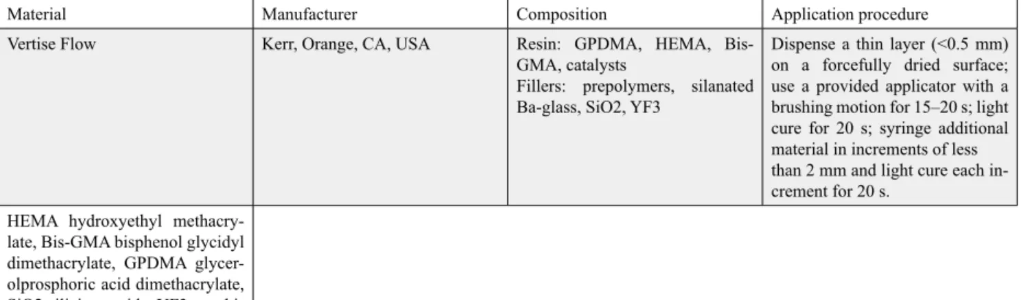
Table 1: Chemical composition and application procedure of Vertise Flow
Material Manufacturer Composition Application procedure Vertise Flow Kerr, Orange, CA, USA Resin: GPDMA, HEMA,
Bis-GMA, catalysts
Fillers: prepolymers, silanated Ba-glass, SiO2, YF3
Dispense a thin layer (<0.5 mm) on a forcefully dried surface; use a provided applicator with a brushing motion for 15–20 s; light cure for 20 s; syringe additional material in increments of less than 2 mm and light cure each in-crement for 20 s.
HEMA hydroxyethyl methacry-late, Bis-GMA bisphenol glycidyl dimethacrylate, GPDMA glycer-olprosphoric acid dimethacrylate, SiO2 silicium oxide, YF3 ytterbi-um tri-fluoride
Mutagenic Potential of a Self-Adhesive Flowable Composite
12
Extract Preparation
The specimens were eluted in 10 mL dimethyl sulphoxide (DMSO) and the extracts were tested after an incubation period of 24 h at 370C and 168 h at 370C in a fully humidified
air atmosphere incubator containing 5% CO2 (n=15). The ratio of sample surface area to the volume of the culture medium was adjusted to
approximately 3cm2/mL as recommended by
ISO.
Cytotoxicity testing
Prior to mutagenicity testing, cytotoxic amounts of the adhesive material extracts were evaluated. The rationale behind this test was to determine whether the test concentrations of the materials would have any cytotoxic effect 0.1 ml of a diluted of an overnight bacterial culture (TA 100 strain) was added to 2.5 ml top agar along with different concentrations of the tested chemicals. The top agar was poured onto nutrient agar plates and assessment of cytotoxicity was performed after 24 h incubation at 370C9.
Mutagenicity tests
Mutagenicity tests were conducted by the standard plate incorporation test as previously described by Maron and Ames10. Two test
strains of Salmonella typhimurium and TA 100 were used to detect frame-shift and base-pair mutation, respectively. Dimethyl sulfoxide samples of 25, 50, 75, 100 µL were plated into minimal agar plates with 2.5 ml of top agar previously supplemented with 0.05 mM histidine-biotin solution had been previously added. Overnight culture of TA 100 (0.1 ml) and the contents were mixed and poured on agar plates. Oxoid nutrient broth no. 2 was used for overnight culture. For plate incorporation assays, 0.1 ml of bacterial tester strain, and different concentrations of test extracts from different adhesive materials
were added separately to 2.5 ml of molten top agar. For this assay, Daunomicina and Sodium azide (NaN3) (known mutagens) without S9 were used as positive control of the TA 100 strains, respectively in the absence of S9 fraction. The negative control was DMSO that we used as solvent. Three plates were conducted for each dose group and each experiment repeated independently two or three times. The strains were checked routinely for ampicillin resistance, ultraviolet-light sensitivity, crystal-violet sensitivity, histidine requirement and spontaneous reversion rate. After 72 h of incubation, revertant colonies were counted. The mutagenicity was expressed as the number of revertants per plate.
Statistical analysis
The data were analyzed by one way ANOVA. Doses higher than the mean of the control group and consequent mutagenic condition were defined as ‘‘mutagenic’’, whereas an increase in dose approaching to, but not reaching a two-fold increase was defined as ‘‘weak mutagenic’’.
RESULTS
For a substance to be considered mutagenic in the Ames Test, the number of revertant colonies per plate containing the test material must be at least two fold higher than the revertant colony number of the vehicle control or there must be an increase in the revertant colony number in a dose dependent manner in plates containing test substance in compare to the vehicle control plates. As a sequel of cytotoxicity tests, it was found that none of the test doses of the materials exhibited cytotoxic effects, even for increased doses. The number of revertants in the positive control group was significantly increased in comparison with the negative control (solvent) which
Tuğba TOZ,Zeliha AYDOĞAN, Duygu TUNCER, Emel KARAMAN
13
Aydın Dental - Year 2 Number 3 - 2016 (9-15)
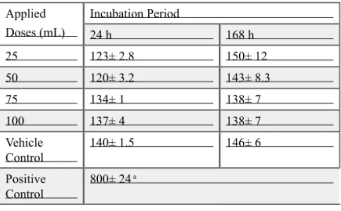
verified the conducted assay.The mutagenic effects of the materials had correlations with applied dose, and the incubation procedure as well as the combination of these parameters for some materials. Considering the bacterial colonization on the test plates of the materials with different doses and incubation periods it was seen that neither the incubation period nor the applied doses have significant effect on Vertise Flow’s mutagenicity (p > 0.05) (Table 2).
Table 2 : Mutagenicity of Vertise Flow
Applied Doses (mL) Incubation Period 24 h 168 h 25 123± 2.8 150± 12 50 120± 3.2 143± 8.3 75 134± 1 138± 7 100 137± 4 138± 7 Vehicle Control 140± 1.5 146± 6 Positive Control 800± 24 a
a The correlation between dose, and incubation
period is significant in comparison with the control group (p<0.05)
*DMSO was used as the vehicle control. *NaN3 was used as positive control.
DISCUSSION
Since the question whether biomaterials have adverse effects on the body is of major concern. Incomplete polymerization of dental resin composites and resin-based bonding agents under clinical conditions result in unreacted resin monomers that may be released from the resin matrix into the aqueous environment of oral cavity. An immediate question is whether the released monomers can reach sufficient concentration to induce a significant cellular effect.There have been several evidences
showing genotoxicity of resin monomers. Direct interaction between nucleotides and resin monomers, production of DNA damaging intermediates, or inhibition of DNA repair systems might be responsible for the mutagenicity of resin monomers 11. Released
monomers can reach sufficient concentration to induce a significant cellular effect. It has been estimated that the concentrations of some monomers released from the dentinal adhesives can be in the millimolar range after diffusion through the dentin layer. For instance, HEMA leaching from dentin adhesives may reach concentrations as high as 1.5–8 mmol/l 12. Therefore, the concentration
of dental monomers in the pulp may be in the millimolar range, high enough to be considered as potentially harmful for pulp cells. Ames test used in this study has been recommended as the mutagenesis screening test for chemicals and environmental samples because of its extensive database and good correlation with carcinogenicity. This assay which was specifically developed to detect chemically induced mutagenesis developed by Bruce Ames13 is generally preferred as
an initial screen to determine the mutagenic potential of new chemical technology as the most rapid, simple, sensitive and economical screening method. Ames test has also a good correlation with carcinogenicity14 thus
this method generally used to detect the possible mutagenic and genotoxic effects of dental materials. An Ames test with
Salmonella typhimurium revealed that a
monomer ingredients of resin composites glycidyl methacrylate (GMA) could cause mutagenicity through base-pair substitution and frame shift mutation in the genetic code15. None of the article was assessed the
mutagenic effects of Vertise Flow, only in one literature it was mentioned that Vertise Flow may cause cellular damage in gingival
Mutagenic Potential of a Self-Adhesive Flowable Composite
14
and pulp fibroblasts in vitro16. This material
has been generally used for cavity lining and restorative treatments applied directly to tooth tissues without any adhesive procedure 5. It is
already seriously mentioned in the literature that chemical activity of adhesive restorative materials is important for the restored tooth prognosis 17. Like all adhesive materials,
Vertise Flow may release components with possible harmful effects to dental structures especially pulp tissue and cause a wide spectrum of pulpa dentinal reactions. Thus biological safety and monomer release of this self-adhesive material remains in close contact with living dental tissue over a long period of time are very important. Vertise Flow includes GPDMA, HEMA and BisGMA as monomer component. In the literature no assay was evaluated the biocompatibility of GPDMA, however it was observed that HEMA and Bis-GMA could induce the enhancement of DNA migration in human lymphocytes18 and they
have genotoxic effects at chromosome level in V79 cells19. These monomers’ genotoxic
effect was also showed in the in vitro Mammalian Cell Gene Mutation Test (HPRT Test) in CHO (Chinese hamster ovary) cells20.
Nevertheless, hydroxylated metabolites of Bis-GMA monomer were not found to be mutagenic in L929 cells or Salmonella
typhimurium 21. However we could not
observed any mutagenic effect of Vertise Flow including HEMA and BisGMA in our study. The Ames Salmonella/microsome test generally detects 83% of the carcinogens as mutagenic with completed protocol. This ratio pointed out that Ames’ test is not able to state all carcinogens10. Mutagenicity tests
may exhibit false-positive results, andit is not possible to draw a conclusive statement based solely on a single study22. In addition to this
the mutagenic potential of Vertis Flow was tested in the absence of S9 fraction.Within
this limitation this material did not lead to mutagenicity, but also a possible mutagenic effect could be detected in the presence of S9. Thus this test must be repeated in the presence of S9.
CONCLUSIONS
In the light of the results of this study, Vertise Flow can be considered safe in terms of mutagenicity within these parameters. However all clinicians must consider the possible mutagenic potential of all dental restorative materials in their clinical practice.
REFERENCES
[1] Peumans M, De Munck J, Van Landuyt K, Lambrechts P, Van Meerbeek B. Five-year clinical effectiveness of a two-step self-etching adhesive. The journal of adhesive dentistry. 2007;9(1):7-10.
[2] Poitevin A, De Munck J, Van Ende A, Suyama Y, Mine A, Peumans M, et al. Bonding effectiveness of self-adhesive composites to dentin and enamel. Dental materials : official publication of the Academy of Dental Materials. 2013;29(2):221-30.
[3] Fu J, Kakuda S, Pan F, Hoshika S, Ting S, Fukuoka A, et al. Bonding performance of a newly developed step-less all-in-one system on dentin. Dental materials journal. 2013;32(2):203-11.
[4] Czasch P, Ilie N. In vitro comparison of mechanical properties and degree of cure of a self-adhesive and four novel flowable composites. The journal of adhesive dentistry. 2013;15(3):229-36.
[5] Eliades A, Birpou E, Eliades T, Eliades G. Self-adhesive restoratives as pit and fissure sealants: a comparative laboratory study. Dental materials : official publication of the Academy of Dental Materials. 2013;29(7):752-62.
Tuğba TOZ,Zeliha AYDOĞAN, Duygu TUNCER, Emel KARAMAN
15
Aydın Dental - Year 2 Number 3 - 2016 (9-15)
[6] Frankenberger R, Lohbauer U, Roggendorf MJ, Naumann M, Taschner M. Selective enamel etching reconsidered: better than etch-and-rinse and self-etch? The journal of adhesive dentistry. 2008;10(5):339-44.
[7] Malkoc S, Corekci B, Ulker HE, Yalcin M, Sengun A. Cytotoxic effects of orthodontic composites. The Angle orthodontist. 2010;80(4):571-6.
[8] Mortelmans K, Zeiger E. The Ames Salmonella/ microsome mutagenicity assay. Mutation research. 2000;455(1-2):29-60.
[9] Dean BJ, Brooks TM, Hodson-Walker G, Hutson DH. Genetic toxicology testing of 41 industrial chemicals. Mutation research. 1985;153(1-2):57-77.
[10] Maron DM, Ames BN. Revised methods for the Salmonella mutagenicity test. Mutation research. 1983;113(3-4):173-215.
[11] Schweikl H, Spagnuolo G, Schmalz G. Genetic and cellular toxicology of dental resin monomers. Journal of dental research. 2006;85(10):870-7. [12] Bouillaguet S, Wataha JC, Hanks CT, Ciucchi
B, Holz J. In vitro cytotoxicity and dentin permeability of HEMA. Journal of endodontics. 1996;22(5):244-8.
[13] Ames BN, McCann J, Yamasaki E. Methods for detecting carcinogens and mutagens with the Salmonella/mammalian-microsome mutagenicity test. Mutation research. 1975;31(6):347-64.
[14] Jukic S, Miletic I, Anic I, Britvic S, Osmak M, Sistig S. The mutagenic potential of AH+ and AH26 by Salmonella/microsome assay. Journal of endodontics. 2000;26(6):321-4.
[15] Schweikl H, Schmalz G, Rackebrandt K. The mutagenic activity of unpolymerized resin monomers in Salmonella typhimurium and V79 cells. Mutation research. 1998;415(1-2):119-30. [16] Tadin A, Marovic D, Galic N, Kovacic I,
Zeljezic D. Composite-induced toxicity in human gingival and pulp fibroblast cells. Acta odontologica Scandinavica. 2014;72(4):304-11.
[17] Costa CA, Vaerten MA, Edwards CA, Hanks CT. Cytotoxic effects of current dental adhesive systems on immortalized odontoblast cell line MDPC-23. Dental materials : official publication of the Academy of Dental Materials. 1999;15(6):434-41.
[18] Kleinsasser NH, Wallner BC, Harreus UA, Kleinjung T, Folwaczny M, Hickel R, et al. Genotoxicity and cytotoxicity of dental materials in human lymphocytes as assessed by the single cell microgel electrophoresis (comet) assay. Journal of dentistry. 2004;32(3):229-34. [19] Schweikl H, Schmalz G, Spruss T. The induction
of micronuclei in vitro by unpolymerized resin monomers. Journal of dental research. 2001;80(7):1615-20.
[20] Muller BP, Eisentrager A, Jahnen-Dechent W, Dott W, Hollender J. Effect of sample preparation on the in vitro genotoxicity of a light curable glass ionomer cement. Biomaterials. 2003;24(4):611-7.
[21] Kostoryz EL, Eick JD, Glaros AG, Judy BM, Welshons WV, Burmaster S, et al. Biocompatibility of hydroxylated metabolites of BISGMA and BFDGE. Journal of dental research. 2003;82(5):367-71.
[22] Kaplan C, Diril N, Sahin S, Cehreli MC. Mutagenic potentials of dental cements as detected by the Salmonella/microsome test. Biomaterials. 2004;25(18):4019-27.
17
Aydın Dental - Year 2 Number 3 - 2016 (17-23)
1Kemerburgaz University, Faculty of Dentistry, Department of Restorative Dentistry, Istanbul, Turkey. 2Istanbul Aydın University, Faculty of Dentistry, Department of Orthodontics, Istanbul, Turkey
AESTHETIC MANAGEMENT OF ANTERIOR TEETH
WITH DIRECT COMPOSITE VENEERS AND BLEACHING
AFTER FIXED ORTHODONTIC TREATMENT
Engin Fırat ÇAKAN1, Fatma YILDIRIM2, Ahu TOPKARA2
ABSTRACT
In present time people consider more about appearance and facial aesthetics according to the past. Person in social communication is perceived by the expression on his/her face. The middle and the lower section of the face where the eyes, the maxilla and the mandible are located, are the primary attention spots and have an undisputed important place in social life. Smile of individuals, reveals the anterior teeth and disrupts or improves the harmony of the face. Therefore, dental aesthetics, which creates an important part of the facial appearance, is a prominent feature of overall aesthetic. As a result of the development of the adhesive dentistry, aesthetic expectations of the patients are possible to provide by the physicians. The main purpose of the aesthetic treatments in dentistry is to establish the design of a beautiful smile in addition to provide a good function. After dental procedures, especially the appearance of the upper anterior teeth is great importance in terms of patient satisfaction. Although, there are many types of treatment options available for functional and aesthetic problems in the upper anterior region, multidisciplinary treatment approaches are recommended for an optimal treatment.
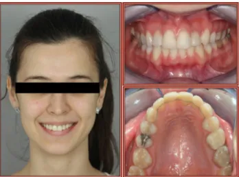
The aim of this study was to demonstrate anterior aesthetic rehabilitation of a patient who had Angle Class I malocclusion with crowding. Discoloration of anterior teeth due to previous treatments, managed with direct composite veneer following vital and non-vital in-office bleaching after orthodontic treatment.
Keywords: anterior crowding, direct composite veneers, laminate veneers, tooth bleaching
ÖZET
Günümüzde bireyler dış görünüşlerine ve yüz estetiklerine geçmiş zamanlara oranla daha fazla önem vermektedir. Sosyal iletişimde kişi, yüzündeki ifade ile algılanır. Gözlerin bulunduğu yüzün orta bölümü ve maksilla ve mandibulanın bulunduğu alt bölümü, ilk dikkat çeken noktalardır ve kişinin sosyal yaşamı içinde tartışmasız önemli bir yer tutmaktadır. Bireylerin gülümsemeleri ön dişleri göz önüne çıkarır ve bu gülümseme yüzün harmonisini bozar ya da geliştirir. Bu nedenle yüz görünümünün önemli bir parçasını oluşturan diş estetiği, ön planda yer almaktadır. Adeziv diş hekimliğinin gelişimi ve varış noktası sonucunda estetik beklentilerin hekim tarafından sağlanması mümkün hale gelmiştir. Diş hekimliğinde estetik amaçlı yapılan tedavilerde esas amaç, fonksiyonun kazandırılmasının yanı sıra, güzel bir gülüş dizaynının da ortaya konmasıdır. Dental girişimler sonrasında, özellikle üst ön bölge dişlerinin görünümü hasta memnuniyeti açısından büyük önem taşımaktadır. Üst çene anterior bölgedeki fonksiyonel ve estetik problemler için birçok tedavi seçeneği bulunmaktadır; fakat ideal bir tedavi için multidisipliner tedavi yaklaşımları önerilmektedir.
Bu olgu sunumunda, Angle Sınıf I maloklüzyon ve çapraşıklığa sahip olan hastanın, üst çene ortodontik tedavisini takiben, tedavi öncesinde mevcut olan restorasyonlar sonucunda meydana gelen renklenme ve form bozukluklarının, devital ve ofis tipi beyazlatma uygulamaları kombine edilerek, direkt kompozit veneer uygulaması ile estetik rehabilitasyonu sunulacaktır.
Anahtar Kelimeler: diş beyazlatma, direkt kompozit veneer, laminate veneer, ön çapraşıklık
© 2016 Published by Istanbul Aydın University, Faculty of Dentistry. All rights reserved.
Aesthetic Management of Anterior Teeth with Direct Composite Veneers and Bleaching After Fixed Orthodontic Treatment
18
INTRODUCTION
In present time people consider more about appearance and facial aesthetics according to the past. The social media determines the desirable appearance, for this reason patients quest to beautify their smiles. The first step in any type of dental therapy is to establish treatment objectives. If the appropriate goals or objectives have not been identified before treatment, it is impossible to achieve the ideal results.1 So, multidisciplinary treatment
approaches are recommended for optimal treatment. Due to the increasing importance of the smile aesthetic which provides confidence-boosting process in social and professional experience, many adult patients prefer orthodontic treatment. Particularly in adult patients, aesthetic solutions should be planned with interdisciplinary strategies as the case requires.2 In fact, the primary goal of
modern orthodontics is to establish the proper occlusal relationship between the maxillary and mandibular arches while maintaining facial esthetics.3 In order to achieve it, the
physician will often have to rely on restorative procedures to reach the optimal esthetic result.4
With advances and interest in adhesive dentistry, bleaching of discolored teeth has become popular. Tooth discoloration is a multifactorial event that can be internal or external origin and classified as intrinsic, extrinsic or both according to the etiology and localization of staining.5,6 Extrinsic
discoloration is caused by chromogenous intake through dietary sources such as tea, coffee, fizzy drinks, wine, tobacco or poor oral hygiene.7 Intrinsic discoloration largely
results of genetic or metabolic causes such as amelogenesis imperfecta, dentinogenesis imperfecta, dentinal dysplasia, fluorosis and tetracycline intake during tooth development
process. Local reasons such as pulp necrosis, pulp tissue remnants after root canal treatment and endodontic filling materials also may occur intrinsic discoloration.5
Intrinsic discoloration involves on enamel, dentin or both of them, while extrinsic discoloration occurs on enamel.8 Therefore,
extrinsic discoloration can be partly removed with mechanical cleaning and/or brushing with toothpaste; internal discoloration can be eliminated only with bleaching.9,10 For
non-vital bleaching, 30% sodium perborate and 35% hydrogen peroxide solution used either in combination or separately. There are two basic bleaching techniques for internal discolored teeth: walking and thermocatalytic bleaching.11 In-office bleaching, different
concentrations of hydrogen peroxide are applied approximately 45 minutes for each practice 2 to 6 times.8 Both of these treatments
are used on non-vital teeth for adapting same color harmony.
Lots of people, consider that the best solution is porcelain crowns for highly discolored teeth. Crowns that made by high aesthetic ceramic materials, have potential to provide long-term aesthetics. On the other hand, the number of physicians who accept the applications of adhesive dentistry are increasing. Composite laminate veneer applications, with a conservative approach to protect the natural tooth structure, are an alternative treatment to porcelain crowns.12 Direct composite veneers
indicated as a complementary treatment option to the bleaching, to ensure aesthetic compliance for old restorations, to replace missing tooth tissue or to mask discolored teeth. The aim of this case-report was to demonstrate an anterior aesthetic rehabilitation of a patient who had Angle Class I malocclusion with crowding.
Engin Fırat ÇAKAN, Fatma YILDIRIM, Ahu TOPKARA
19
Aydın Dental - Year 2 Number 3 - 2016 (17-23)
Discoloration of anterior teeth due to previous root canal treatments and restorations, treated with direct composite veneers following non-vital walking and in-office bleaching after orthodontic treatment.
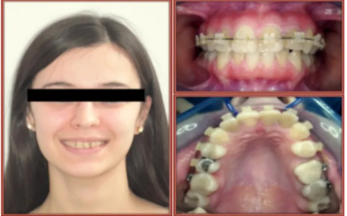
CASE REPORT
A 22-year-old female patient who had Angle Class I malocclusion, crowding and 4 mm midline shift in the maxillary anterior region (Fig 1), was referred to Department of Orthodontics, for existing orthodontic treatment. Patient had started her orthodontic treatment in another clinic and her upper left second premolar teeth was extracted in order to correct upper jaw midline discrepancy. The orthodontist had bonded the ceramic braces just only in the upper teeth (Fig 2). After treatment had started, she had wanted
to continue her treatment in Istanbul Aydin University, Faculty of Dentistry.
Initial case procedures were started when the patient came for the first appointment in faculty clinics. For the assessment of this case; radiographs, photographs and the study cast models were taken. The transverse positions of the maxillary and mandibular midlines were evaluated in postero-anterior cephalogram. There was no skeletal discrepancy between the jaws and it was found that the midline shift was caused from dental asymmetry. Thus, we thought that her previous orthodontist had reached the same conclusion and had chosen the tooth extraction treatment. Before the orthodontic treatment, solving other dental problems is important for oral hygiene recovery. Because it will become so difficult for patient to maintain oral hygiene during the
Figure 1 OPG Before Orthodontic Treatment Figure 2 Case Photos (Before) orthodontic treatment. In this case the physician
whom started the orthodontic treatment, was began without taking into account procedures. Patient had periodontal problems and gingival index scores were measured close to 2 because of poor oral hygiene at first arrival. Furthermore, she had discoloration both as existing root canal treatments and as deformed previous restorations. All under these terms, the treatment plan was determined by a multidisciplinary approach.
As a result of treatment planning, periodontal improvement was defined as to be done first. After periodontal treatment was completed, oral health care education and motivation were given and correction of poor oral hygiene was provided. It was decided to continue treatment with existing braces not to prolong the duration of treatment. Therefore, after consultation with Department of Restorative Dentistry, the renewal of restorative treatments and aesthetic improvement were left to the end
Aesthetic Management of Anterior Teeth with Direct Composite Veneers and Bleaching After Fixed Orthodontic Treatment
20
of the orthodontic treatment. Depending on the patient’s demand, orthodontic treatment has begun only in upper jaw after informed consent form received. To achieve good Class II occlusion at the left side, the extraction space was used to slide and correct the midline. Over a period of 15 months, orthodontic treatment was completed. On the right side Class I molar occlusion and on the left side full cusp Class II molar occlusion with a Class I canine relationship and 2 mm overjet and overbite for both sides were obtained (Fig 3).
Figure 3 OPG After Orthodontic Treatment For the treatment of the patient’s aesthetic expectations, the patient was referred to the Department of Restorative Dentistry following the end of the orthodontic treatment. During this process, Essix retainers were prepared in order to maintain the treatment outcomes. First of all, when the patient visited to the restorative dentistry clinic, discoloration and color mismatch of the teeth had been focused on. In order to remove tooth discoloration due to pre-made root canal treatment, non-vital walking bleaching was planned. Discolored upper left central and both upper lateral incisors were included to the treatment. Residual supplies in the pulp chamber were cleaned and root canal orifices were closed with glass ionomer cement in hermetic manner. Opalesence Endo (Ultradent Productions Inc, UT, USA), 35% hydrogen peroxide was
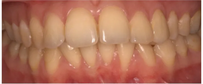
applied at first practice and was changed for 3 times in every 4 days. 4 days after the last appointment, the teeth were bleached substantially. To reach the final colors of the root canal treated teeth, Opalesence Endo was cleaned with physiological saline and has been waited for one week. At the beginning of the following week, the pulp chamber was sealed by calcium hydroxide (Sultan Healthcare, PA, USA) for one week to allow elimination of residual oxygen. During this two-week period, in-office bleaching (Philips Zoom WhiteSpeed, Philips, Holland) was done for 2 times, one week apart in 15-minute sessions, to adapt all the teeth. After reaching the final colors of the teeth in the anterior region, the teeth which has degraded aesthetic features, edge harmony, colors and forms, were restored with Composite Laminate Veneers due to extensive borders of restoration (Fig 4). Existent caries and old restorations has been removed in accordance with minimally invasive treatment procedures. A three-step etch-and-rinse bonding agent (Adper Single Bond 2, 3M/ESPE, St. Paul, MN, USA) and a nanofil composite (Filtek Ultimate, 3M/ ESPE, St. Paul, MN, USA) were used. The composite increments were placed between the tooth and matrix strip by using hand instruments. Subsequent to polymerization, contouring and finishing were performed with microfine finishing diamonds and restorations were polished using abrasive disks (Sof-Lex, 3M/ESPE, St. Paul, MN, USA) ranging from medium to superfine. At the end of all restorative procedures, fixed lingual retainers were bonded to prevent relapse (Fig 4).
Engin Fırat ÇAKAN, Fatma YILDIRIM, Ahu TOPKARA
21
Aydın Dental - Year 2 Number 3 - 2016 (17-23)
Figure 4 Case Photos (After) DISCUSSION
Before the beginning of the orthodontic treatment, required process for oral hygiene maintain should be completed. Substantially when the proper teeth positions and optimal occlusion is achieved, orthodontic treatment can be terminated without the need to any other treatment. Depending on the aesthetic expectations, if there is a requirement for the renewal of previous restorations to rearrange the remaining tooth shapes or colors, restorative dentistry should play a part in the finishing phase of the orthodontic treatment.13
Physician should determine the treatment objectives, as should meet the requirements of the patient. To meet the treatment needs of patients properly, it is necessary to evaluate all aspects of the case. Constant interaction and communication among the team members and the patient at all level of treatment are the keys to the success of the interdisciplinary treatment.14
In dental and facial aesthetics, dental midlines relative to each arches and to the face are very important.15 In diagnosing dental
asymmetries, a through clinical examination and radiographic evaluation are necessary. Each dental arches should be evaluated
separately both clinically and by using dental cast models, to accurately determine the bilateral symmetry of the molar and canine positions. In addition to the clinical and dental model evaluation, differentiation between various types of asymmetries can be aided by the use of postero-anterior cephalograms.16 In
this case by using clinical examination, dental cast model evaluation and postero-anterior cephalometric analyses, it was determined that the 4 mm of midline shift to the right side was dentoalveolar.
Through dental asymmetries are often treated asymmetric extraction sequences and asymmetric mechanics.16 In this case; for
to propose of the treatment of the 4 mm of dentoalveolar midline discrepancy, upper left premolar tooth was extracted and upper left canine was distalized 4 mm to correct the midline and the remaining extraction space was closed by anterior mesialization of molar teeth to provide full Class II molar relationship in the left side.
In order to plan aesthetic rehabilitation of discolored teeth, both porcelain and composite laminate veneers may be preferred. Between both treatment options, technical precision and cost should also be considered as well as aesthetic results to be obtained. Porcelain veneers are more resistant to adhesive and cohesive forces. Therefore, when requested to increase the inciso-gingival sizes of the teeth, porcelain laminate veneers should be preferred.17,18 Although the composite veneers
are aesthetic, they don’t provide exactly all the aesthetic properties of dental structures as much as porcelain. In addition to aesthetic properties, good results are obtained with porcelain veneers in terms of satisfaction of patients.18,19 However, porcelain veneers
require more technical precision and higher costs compared to composite veneers. Due