Romanian Neurosurgery (2014) XXI 4: 477 – 479 477 DOI: 10.2478/romneu-2014-0064
Three level spinal dysraphism: multiple composite Type 1
and Type 2 split cord malformatıon
I. Alatas
1, M. Gundag
2, H. Canaz
1*, E. Emel
31Spina Bifida Center, Florence Nightingale Bilim University, Istanbul, Turkey
2Department of Neurosurgery, Bezmi Alem Vakif Gureba School of Medicine, Istanbul, Turkey 3Department of Neurosurgery, Bakirkoy Psychiatry and Neurology Research and Training
Hospital, Istanbul, Turkey
Abstract: It has reported an uncommon case a 3 year-old girl a composite split cord malformation (SCM) with two different levels of SCM type1 and one level SCM type2, tight filum and sacral dermal sinus. The patient was admitted with a hypertrichosis and hyperpigmented patch. MRI of whole spine and brain was done. SCM type1 at T 7 and L2 levels and SCM typ2 at T11 level were removed then tight filum was cut and dermal sinus was excised at different sites during the same surgery.
Key words: Split Cord Malformation, 3 levels spinal dysraphism.
Introduction
Diastematomyelia literally means spinal cord splitting, while diplomyelia represents cord duplication. In 1992, Pang at all suggested that terms such as diastematomyelia and diplomyelia be abandoned, to make way for a new classification of SCM into two types, based on the state of the dural tube and the nature of the median septum. Type 1 has an osseous or osteocartilagineous midline septum splits the spinal cord into two tubes each containing a hemicord. Type 2 has no osteocartilagineous spur, consist of two hemicords, but they are contained in a single dural sheath, rarely, thin fibrous septa may form. (5)
Composite type is very rare and result from two seperate foci of ectoendodermal adhesions and endomesenchymal tracts leading to development of different SCM types with intervening normal cord in the same patient. Only very few cases of composite SCM have been reported in literature. (6, 8) We present a case of composite type of SCM with two levels SCM Type 1 and single level SCM Type 2, tight filum and sacral dermal sinus.
Case: A 3 year-old girl was admitted hyperpigmented patch and hypertrichosis. There was no history of bladder, bowel and limbs disturbances. Neurological examination was normal. The urodynamic test showed overactive detrusor and increased bladder capacity. MRI revealed the SCM type 1 at level
- 10.2478/romneu-2014-0064 Downloaded from PubFactory at 08/22/2016 09:36:06AM via free access
478 Alatas et al Three level spinal dysraphism
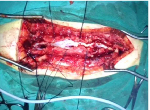
T7 and L2, SCM type2 at T11 level and low-lying conus (Figure 1, 2). A skin incision was made from T5 to L5. Then T7 laminoplasty was done and the dural sheath was incised and the bony spur was removed and dura was unified. Afterthat T11 laminoplasty was performed and the thin fibrous septum was excised. Then L2 laminoplasty was done, the dural sheath was incised and the bony spur was removed and dura was unified. Then L5 laminoplasty was done and dural sheath was incised and the filum terminale was cut and the unification of the dura was done (Figure 3). The control urodinamic test was normal after six month’ surgery.
Figure 1 - MRI sagittal T2 weighted image showing SCM type 1 at level T7 and L2, SCM type 2 at T11 level
Figure 2 - MRI Axial T2 weighted image shows SCM type 1 at T7 , SCM type 2 at T11 and type 1 at L2
Figure 3 - At the operation photograph showing three level SCM
Discussion
Diastematomyelia was introduced by Olliver the disease of the spinal cord and its covering published in 1837. Later Cohen and Sledge emphasised that this term only defines the cleft in the spinl cord or not, as often written, the osseous spur or fibro-cartilaginous tract that separates both hemicords. (1) Diplomyelia was invented by von Recklinghausen, applies to a completely different malformation where one finds a totally formed spinal cord, situated dorsally or ventrally to the original spinal cord, mostly at the lumbo-sacral level. (9) Pang gave a new classification recommending the term SCM for all double spinal cord malformations. He also described the unified theory, explains the embryologic mechanism of development of all variants of SCM. According to the theory, the formation of an “accessory neuroenteric canal" between the yolk sac and amnion through the midline embryonic disc, which is subsequently invested with mesenchyme to form an endomesenchymal tract that splits the notochord and neural plate. The location of
- 10.2478/romneu-2014-0064 Downloaded from PubFactory at 08/22/2016 09:36:06AM via free access
Romanian Neurosurgery (2014) XXI 4: 477 – 479 479 DOI: 10.2478/romneu-2014-0064 this abnormal fistula is variable, but it must be
rostral to the primitive neuroenteric canal, because the primitive pit into which the latter normally opens, ultimately comes to lie opposite the coccyx. In type 1 SCM, the precursor cells within endomesenchymal tract may form a bony spur in the midline, attached to the bone of developing vertebral column, while in type 2 SCM the mesenchymal tract forms a thin fibrous septum in the space between the hemicords. (7)
Accessory neuroenteric canal maybe single or multiple, and the presence of multiple accessory neuroenteric canals results in two or more septa that divide the spinal cord into hemicords. One or both types of SCM at different levels have been reported in the same patients as we have seen in our case. Pang reported about two such cases of composite type of SCM, out of total 39 case studies. (7) Ersahin et al. reported on 4 such cases out of 74 cases of SCM in their study. (2)
Kumar et al labeled the complex spina bifida and feel it is necessary to make minor modification to Pang’s classification to accommodate the pure/combined anomalies together. (3) Kumar and Singh were put forward that meningomyelocele and SCM are seen together 40.8 %. Thus, they emphasized spinal dysrafism together with occult and also open types. (4) Our patient has a composite split cord malformation (SCM) with two different levels of SCM type1 and one level SCM type 2, tight filum and sacral dermal sinus. Our surgery was performed well in a one-stage operation. We did not have any complication during surgery.
Conclusion
Composite type SCM is seen rare. Not only neurological deficits, but also neuro-urological findings should be noted. Our case’s neurological examination was normal, However, The urodynamic test showed overactive detrusor and increased bladder capacity. Control urodinamic test was done after six month surgery and test result was normal. It should be remembered if presence of tethered cord syndrome other congenital spinal malformation can be exist.
Correspondence
Huseyin Canaz
Spina Bifida Center, Florence Nightingale Bilim University, Sisli, Istanbul, Turkey
+905065307853, [email protected]
References
1.Cohen J, Sledge CB Diastematomyelia. An embryological interpretation and report of a case. Am J Dis Child 1960;100: 257-263
2.Ersahin Y, Demirtas E, Mutluer S, Tosun AR, Saydam S. Split cord malformations: Report of three unusual cases. Pediatr Neurosurg. 1996;24:155-9.
3.Kumar R, Bansal KK, Chhabra DK. Occurrence of split cord malformation in meningomyelocele: Complex spina bifida. Pediatr Neurosurg. 2002;36:119-27.
4.Kumar R, Singh V, Singh SN. Split cord malformation in children undergoing neurological intervention in India: A descriptive study. J Pediatr Neurol. 2004;2:21-7. 5. Pang D, Dias MS, Ahab-Barmada M. Split cord malformation: Part I: A unified theory of embryogenesis for double spinal cord malformations. Neuro- surgery 1992; 31:451-480.
6.Pang D Split cord malformation. II. Clinical syndrome. Neurosurgery 1992; 31:481-500.
7.Pang D. Split cord malformation, Proposal for a New Clinicoradiological. Neurosurg Clin N Am. 1995;3:339-52. 8.Vaishya S, Kumarjain P. Split cord malformation: Three unusual cases of composite split cord malformation. Childs Nerv Syst. 2001;17:528-30 9.Von Recklinghausen F Untersuchungen über die spina bifida. Virchows Arch Path Anat 1885;105:243-330
- 10.2478/romneu-2014-0064 Downloaded from PubFactory at 08/22/2016 09:36:06AM via free access