Cukurova Medical Journal
Chondro-osseous Type Metaplastic Carcinoma of Breast
Memenin Kondroosseöz Tipte Metaplastik Karsinomu
Aysin Pourbagher1, Hülya Aslan1, Filiz Aka Bolat2
1
Baskent University Faculty of Medicine, Teaching and Medical Research Center, Department of Radiology,
2
Department of Pathology, ADANA,
Cukurova Medical Journal 2015;40(4):847-849.
DEAR EDITOR,
We read the article ‘A Pathologists Purview of Breast Calcifications’ by Sankaye with great interest1. The author evaluated benign and malignant calcifications of breast with radiological and pathological findings. Metaplastic carcinomas of the breast are high grade uncommon tumors including mixed epithelial and sarcomatoid components2. To our knowledge, a few reports and studies have been reported including the imaging findings of metaplastic carcinoma2,3,4,5. However, a few reports including osseous or chondromatous metaplasia have been reported3,4.
We wish to report one case of grade 3 metaplastic carcinoma including chondro-osseous type mesenchymal components and invasive ductal carcinoma areas in a 75 year old woman. The mammography showed coarse and dense coalescing calcifications which can be easily misdiagnosed as calcifying fibroadenoma.
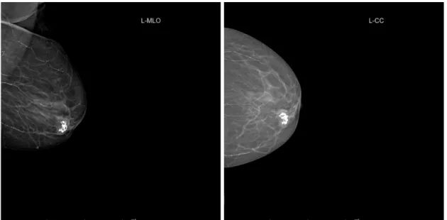
A 75 year old female was admitted to our department with a palpable mass in her right breast two years ago. We performed bilateral mammography. On mammography she had large, irregular, partially circumscribed mass with dens coalescent calcifications in left lower inner quadrant (figures 1A and 1B). The margins of the lesion were partially obscured.
The differential diagnosis is calcifying fibradenoma, primary sarcomas of the breast and invasive ductal carcinoma without metaplasia. Then, we performed ultrasound-guided core biopsy. The final diagnosis was grade 3 metaplastic carcinoma including chondro-osseous type mesenchymal components and invasive ductal carcinoma. She went segmental mastectomy. Patient was alive without any reccurence or metastases.
Metaplastic changes in breast carcinomas are very rare in occurring less than 5% of breast carcinomas 6. Microcalcifications or dense calcifying nidus within the mass has been previously reported in chondroid or osseous metaplasia of the breast carcinomas5,7. The differential diagnosis of the chondro-osseous type metaplastic breast carcinomas are calcifying fibradenoma, primary sarcomas of the breast and invasive ductal carcinoma without metaplasia.
Fibroadenoma is the most challenging diagnosis in this case. It typically shows ‘’popcorn’’ shaped, dense large calcifications on mammography. It can also be seen as pure calcification. This case can be misinterpreted easily as a fibroadenoma and follow-up may be recommended rather than biopsy. Coalescing pattern of the dense calcifications and partially obscured margins of the mass may help us to differentiate it from fibroadenomas.
Editöre Mektup / Letter to the Editor
Pourbagher et al. Cukurova Medical Journal
The patterns of calcifications in the primary invasive ductal carcinoma are fine, linear branching or pleomorphic morphology in contrast to coarse calcifications in our case. It has been shown that calcifications are more frequently seen in metaplastic carcinomas then invasive ductal carcinoma8.The calcifications of the metaplastic carcinomas can not only be in pleomorphic, heterogeneous, linear pattern but also they can be in coarse or amorphous morphology similar to our
case8. Primary sarcomas of the breast are very rare and calcifications are less frequently9.
As a result, chondro-osseous type metaplastic carcinomas can be easily misinterpreted as a fibroadenoma. Coalescing pattern of the dense calcifications and partially obscured margins of the mass may help us to differentiate it from fibroadenomas.
Figures 1A, 1B: Mediolateral oblique and craniocaudal mammograms of the left breast show a partially obscured and circumscribed mass including dense, coarse coalescing calcifications in lower inner quadrant.
Figure 2. Metaplastic carcinoma , chondro-osseous type; invasive ductal carcinoma (long arrows), and osteoid – chondroid differentiation ( short arrows) in metaplastic carcinoma (Hematoxylin and eosinx100).
Cilt/Volume 40 Yıl/Year 2015 Metaplastic Carcinoma of Breast
REFERENCES
1. Sankaye S, Kachewas S. A Pathologists Purview of Breast Calcifications. Cukurova Medical Journal. 2014;39:413-21.
2. Tse GM, Tan PH, Putti TC, Lui PC, Chaiwun B, et al. Metaplastic carcinoma of the breast: a clinicopathological review. J Clin Pathol. 2006;59:1079–83.
3. Shin HJ, Kim HH, Kim SM, Kim DB, Kim MJ, et al. Imaging features of metaplastic carcinoma with chondroid differentiation of the breast. AJR Am J Roentgenol. 2007;188:691-6.
4. Evans HA, Shaughnessy EA, Nikiforov YE. Infiltrating ductal carcinoma of the breast with osseous metaplasia: imaging findings with pathologic correlation. AJR Am J Roentgenol. 1999;172:1420-2.
5. Patterson SK, Tworek JA, Roubidoux MA, Helvie MA, Oberman HA. Metaplastic carcinoma of the breast: mammographic appearance with pathologic correlation. AJR Am J Roentgenol. 1997;169:709–12. 6. Rosen PP, Oberman HA. Invasive carcinoma. In:
Atlas of tumor pathology: tumors of the mammary gland, 3rd series, fasc. 7. Washington, DC: Armed Forces Institute of Pathology, 1993:194-203.
7. Brenner RJ, Turner RR, Schiller V, Arndt RD, Guiliano A. Metaplastic carcinoma of the breast: report of three cases. Cancer. 1998;82:1082–87. 8. Yang WT, Hennessy B, Broglio K, Mills C, Sneige N,
et al. Imaging differences in metaplastic and invasive ductal carcinomas of the breast.AJR Am J Roentgenol. 2007;189:1288-93.
9. Smith TB, Gilcrease MZ, Santiago L, Hunt KK, Yang WT. Imaging features of primary breast sarcoma. AJR Am J Roentgenol. 2012;198:386-93.
Yazışma Adresi / Address for Correspondence: Dr. Hülya Aslan
Başkent University Faculty of Medicine Adana Teaching and Medical Research Center Department of Radiology
ADANA
E-mail: hul_yaaslan@hotmail.com
Geliş tarihi/Received on : 20.03.2015 Kabul tarihi/Accepted on: 15.04.2015