Accepted: 2014.06.17 Published: 2014.11.27
1791
2
2
22
Association between Red Blood Cell Distribution
Width and Acute Pancreatitis
A 1
Keziban Uçar Karabulut
F 2Hüseyin Narcı
E 3
Yıldız Uçar
C 4Mehmet Uyar
Corresponding Author: Keziban Uçar Karabulut, e-mail: dr_kezi@hotmail.com
Source of support: Departmental sources
Background: We aimed to determine whether there are any alterations in red blood cell width in patients diagnosed with acute pancreatitis.
Material/Methods: This was a retrospective study of records of patients treated in emergency units and diagnosed with acute pancreatitis between January 2011 and April 2013. Records were investigated regarding patient age, sex, and causes of acute pancreatitis. Red blood cell width and amylase values obtained from the patients during diag-nosis and after a full recovery from acute pancreatitis were added to the records.
Results: Recorded red blood cell width and amylase values of 104 patients diagnosed with acute pancreatitis were sta-tistically compared according to the time of sample collection. Samples were collected in the post-diagnostic period and after full recovery. Values of red blood cell width and amylase were found to be significantly high-er in samples collected during hospital admission in comparison to those obtained from patients afthigh-er fully re-covering from acute pancreatitis (p<0.05).
Conclusions: An increase in red blood cell width value is a marker of acute pancreatitis; therefore, we suggest that red blood cell width can be used as a tool for the early diagnosis and assessment of disease progression.
MeSH Keywords: Abdomen, Acute • Erythrocyte Volume • Inflammation • Pancreatic alpha-Amylases
Full-text PDF: http://www.medscimonit.com/abstract/index/idArt/891075 Authors’ Contribution: Study Design A Data Collection B Statistical Analysis C Data Interpretation D Manuscript Preparation E Literature Search F Funds Collection G
1 Department of Emergency Medicine, Baskent University Konya Hospital, Konya, Turkey
2 Department of Emergency Medicine, Mersin University, Mersin, Turkey 3 Department of Pulmonary Diseases, Memorial Hospital, Diyarbakır, Turkey 4 Department of Public Health, Necmettin Erbakan University, Konya, Turkey
Background
Acute pancreatitis (AP) occurs due to the auto-digestion of the pancreas by the intrapancreatic activation of digestive enzymes. AP is an acute inflammatory disease of the pancre-as characterized by severe abdominal pain that may be lo-cated in the epigastric region or spread to the back. Local or systemic symptoms may also be associated with the disease [1]. Pancreatic enzymes normally remain inactive. Activation of these enzymes by various etiological factors results in the auto-digestion of pancreatic tissues. Thus, AP leads to inflam-mation of the pancreas and may consequently result in local or systemic complications [2].
Patients are diagnosed with AP with the consideration of a combination of positive findings obtained from a history of pancreatitis, the physical examination of patient, serological markers, and radiological images [3]. Nevertheless, the diag-nosis of AP can be difficult when patients are admitted to the emergency unit, due to the variable clinical properties of AP and factors that may restrict the diagnostic approaches to AP. Moreover, a number of different parameters are still used for the diagnosis and management of AP. Increases in blood and urinary amylase, serum enzymes (including lipase, elastase 1, trypsin and phospholipase 2), C-reactive protein (CRP), inter-leukin 6–8 (IL-6–8) and procalcitonin levels are used to con-firm the diagnosis of AP [4].
Furthermore, there is still no consensus on the diagnosis and (especially) the treatment of AP, which has a heterogeneous distribution ranging from mild interstitial edematous pancre-atitis to necrotizing pancrepancre-atitis with a 20% mortality rate [5]. Therefore, early diagnosis and treatment of AP is crucial. No criterion standard for the diagnosis of AP has been developed. The first approach to the diagnosis of AP is to check the lev-el of serum amylase and lipase; however, the specificity and sensitivity of these enzymes is low. Computerized tomography (CT) is the most reliable tool in the diagnosis of AP and the determination of its severity. The high cost-effectiveness, re-stricted availability of CT, and the potential adverse effects of using contrast chemicals are the disadvantages of CT use [6]. RDW refers to an evaluation of the sizes of the red blood cells (RBCs) circulating in the vasculature, demonstrating the width of distribution of RBCs based on their sizes. It is a component of the complete blood cell (CBC) test. It is calculated by dividing the histogram width of 68.26% of RBCs by the mean corpuscular volume (MCV), then multiplying the result by 100. RDW values are also affected in other of diseases, including celiac disease, pulmonary embolism, and coronary artery disease [7–9]. In ad-dition, elevated levels of RDW were determined in inflammatory and infectious bacteriemia, severe sepsis, and septic shock [10].
This study aimed to determine whether there was a difference in RDW levels and amylase on admission to emergency units, compared to the levels obtained from patients who had ful-ly recovered from AP. Moreover, the role and association of these markers with the early diagnosis of AP was evaluated. Also, we tried to determine whether there were any perma-nent alterations (either increase or decrease) in RDW values in the post-recovery period.
Material and Methods
We included 104 patients diagnosed with AP in the emergen-cy unit of Baskent University Konya Training and Research Hospital between January 2011 and April 2014 were included in this retrospective analysis. Clinical and laboratory examina-tions, in addition to radiological methods, were performed for the diagnosis of AP in patients on admission to the emergen-cy department. Patients were excluded if they had any histo-ry of oncologic pathologies, iron deficiency anaemia, thalas-semia, metabolic disorders, or inflammatory bowel disease. The Ranson criteria were used for the classification of AP se-verity. Patients who died or had severe necrotizing pancreati-tis were also excluded from the study (n=63). Only patients with mild or moderate AP (edematous or interstitial) were in-cluded into the study.
To evaluate the levels of RDW and amylase, blood samples were collected from 104 patients diagnosed with AP on ad-mission.. Patients were then hospitalized in clinics for AP treatment. Standard AP treatment included intravenous flu-id resuscitation, electrolyte replacement, and the use of an-algesics. Nasogastric drainage was applied to protect pa-tients with nausea and vomiting from respiratory aspiration. Patients were given nutritional support [total parenteral nu-trition (TPN)] and antibiotics to protect them from septic com-plications. Patients were also exposed to specific procedures, including endoscopic retrograde cholangiopancreatography (ERCP) and magnetic resonance cholangiopancreatography (MRCP), to reduce mortality rates caused by pancreatic pa-renchymal injury and complications. Patients started to be fed via oral nutrition after about 5–10 days. The patients were ob-served to improve, based on clinical, radiological, and labo-ratory findings. To evaluate the values of RDW and amylase, blood samples were collected from 104 patients who had re-covered from AP.
Lab assessment
For the evaluation of RDW, an electronic blood cell counting device was used (Cell-Dyne 3700, Abbott, Abbott Park, IL, USA). A routine biochemical kit was used to assess amylase values.
Statistically assessment
Collected data were recorded on previously prepared forms. In data analysis, SPSS software was used. Analysis of normal-ization to common distribution was performed. The t test and paired t test (t test for dependent groups) were performed. The numerical values were presented as mean ± standard deviation (SD). A p value less than 0.05 (p<0.05) was accepted as indi-cating a statistically significant difference between 2 groups.
Results
The study included 104 patients hospitalized with the diagno-sis of AP on emergency admission to Baskent University Konya Training and Research Hospital between January 2011 and April 2013. The sex distribution of patients was 51 males (49%) and 53 females (51%). The mean age was 56.5±1.55 years. There were 17 patients (16.3%) over age 70 years. The major etiological fac-tor was determined to be biliary disorders in 82 patients (79%).
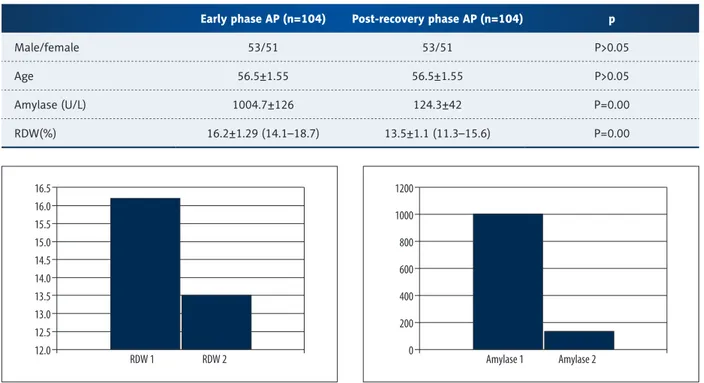
Mean RDW value of AP cases was 16.2±1.29% in the acute period (RDW1) and 13.5±1.1% in the post-recovery period (RDW2) (Table 1) and this difference was statistically signif-icant (p<0.05) (Figure 1). However, no correlation was found between the parameters of RDW1 and RDW2 (Table 2). The mean level of amylase in the acute period of AP was 1004.7±126 U/L and 124.3±42 U/L in the post-recovery peri-od. There was a statistically significant difference between the amylase values of the 2 groups (p<0.05) (Figure 2).
Discussion
AP is an important clinical condition that should be considered in the differential diagnosis of acute abdominal pain in patients examined in emergency departments. The clinical symptoms and findings of patients with AP may differ according to their age or severity of AP attack. The severity of AP may vary from mild edematous condense necrosis to hemorrhage [11,12].
Early phase AP (n=104) Post-recovery phase AP (n=104) p
Male/female 53/51 53/51 P>0.05
Age 56.5±1.55 56.5±1.55 P>0.05
Amylase (U/L) 1004.7±126 124.3±42 P=0.00 RDW(%) 16.2±1.29 (14.1–18.7) 13.5±1.1 (11.3–15.6) P=0.00
Table 1. Demographic properties and RDW/ Amylase values at two time periods;early phase AP and post-recovery AP.
Figure 1. Graphical comparison of RDW values obtained from AP patients at both early phase (RDW1) and recovery (RDW2) period of the disease.
16.5 16.0 15.5 15.0 14.5 14.0 13.5 13.0 12.5 12.0 RDW 1 RDW 2
Figure 2. Graphical comparison of amylase values obtained from AP patients at both early phase (Amylase 1) and recovery (Amylase 2) period of the disease.
1200 1000 800 600 400 200 0 Amylase 1 Amylase 2
parameters Correlation coefficient (r) P value
RDW1-RDW2 –0.069 0.489
However, the diagnosis of AP at emergency departments may be difficult due to the variable clinical properties of the disease and factors such as limited diagnostic tools. Various parame-ters are used for the diagnosis and treatment of AP; hence, no specific biochemical markers have still been determined for the early definite diagnosis and etiological clarification of AP [13]. The clarification of secondary inflammation has been studied in AP patients; increases in blood and urinary amylase, serum li-pase, elastase 1, trypsin and phospholipase A2, C-reactive protein (CRP), interleukin 6–8 and procalcitonin may be observed [11,14]. In this study, we aimed to assess the alterations in amylase levels, as well as RDW values, in patients newly diagnosed with AP and to compare these values to those of individuals who had recovered from AP. A search of the relevant literature reveals that the majority of AP cases have mild pancreatitis (80–85%), and 15–20% have severe pancreatitis. In contrast to previous studies, we included patients with mild (85.1%) and moderate (14.9%) AP and excluded patients with severe AP. Amylase levels increased in the early phase of AP in 3/4 of pa-tients and remained increased for 7–10 days. Therefore, in the conventional approach, checking amylase level for the diagnosis of AP is the criterion standard test. In AP patients, amylase reach-es a peak level at between 12 and 72 hours. The formation of a pseudocyst or abscess should be considered if the level of amy-lase remains high 10 days or longer after an acute attack [15,16]. Although the sensitivity of amylase levels for AP diagnosis is high, the specificity of this test is low. Many clinicians still use an enhanced (3-fold) level of serum amylase and lipase as a diagnostic parameter for AP. The sensitivity of blood amylase level for the diagnosis of AP is 67–100% and the specificity is 85–98% [11,17]. Treacy et al. suggested that the sensitivity and specificity rates for serum amylase were 45% and 97%, respectively; 63% and 85% for urinary lipase; and, 67% and 97% for serum lipase [18].
We found amylase levels to be 3-fold higher in 94 patients (90.4%) in the early phase of AP compared to the recovery period.
RDW is used as a marker to reveal alterations in the number of RBCs. RDW as a parameter is a component of the routine complete blood cell count. The formulation of RDW includes standard deviation of RBC volume, which is divided by the mid-corpuscular volume (MCV) and multiplied by 100 [19]. The value of RDW may be increased due to hemolysis, defi-ciency of iron and folic acids, and in response to blood trans-fusion. In addition, the increased level of RDW may be seen in thrombotic thrombocytopenic purpura (TTP), inflammatory bowel disorders such as Crohn’s disease, and pregnancy [20]. Moreover, RDW is also used as a tool for the differential diag-nosis of microcytic anemia caused by common iron deficien-cy, thalassemia, or hemoglobinopathies [21].
Results of a study conducted to evaluate the association be-tween mortality and RDW in AP patients suggested that RDW is an excellent marker for the assessment of AP prognosis. The patients with severe AP were included in the study [22], and RDW values were found to be significantly increased in these cases, compared to the normal range RDW values [22]. Unlike previous studies, we included only patients with a mild and moderate AP in our study and excluded cases with fatal and severe AP. We also determined elevated RDW values in cases with mild and moderate AP. We found that RDW val-ues are higher in patients with severe AP as well as in pa-tients with early AP. Moreover, we observed a decrease in the elevated level of RDW to normal, without any perma-nent alteration.
Conclusions
AP is an important medical challenge due to its high mortal-ity and morbidmortal-ity. Early determination of disease severmortal-ity is essential to determine the need for intensive care, to start a feasible and adequate treatment, and to determine the effi-ciency of treatment. We suggested that RDW could play an important role in the early diagnosis of AP and evaluation of prognosis of the disease. Expanded clinical and experimental studies are required to verify this.
References:
1. Carroll JK, Herrick B, Gipson T, Lee SP: Acute pancreatitis: diagnosis, prog-nosis, and treatment. Am Fam Physician, 2007, 75: 1513–20
2. Glazer G: Contentious issues in acute pancreatitis. Acute pancreatitis. Experimental and clinical aspects of pathogenesis and management, 1st
ed. London: Bailliare Tindall, 1988; 1–36
3. Vlodov J, Tenner SM: Acute and chronic pancreatitis. Prim Care, 2001; 28: 607–28
4. Koizumi M, Takada T, Kawarada Y et al: JPN Guidelines for the manage-ment of acute pancreatitis: diagnostic criteria for acute pancreatitis. J Hepatobiliary Pancreat Surg, 2006; 13: 25–32
5. Xin M-J, Chen H, Luo B, Sun B-J: Severe Acute Pancreatitis in the elderly: Etiology and clinical characteristics. World J Gastroenterol, 2008; 14(16): 2517–21
6. Balthazar EJ: CT diagnosis and staging of acute pancreatitis. Radiol Clin North Am, 1989; 27: 19–37
7. Harmanci O, Kav T, Sivri B: Red cell distribution width can predict intesti-nal atrophy in selected patients with celiac disease. J Clin Lab Aintesti-nal, 2012; 26: 497–502
8. Hampole CV, Mehrotra AK, Thenappan T et al: Usefulness of red cell dis-tribution width as a prognostic marker in pulmonary hypertension. Am J Cardiol, 2009; 104: 868–72
9. Tonelli M, Sacks F, Arnold M et al., for the Cholesterol and Recurrent Events (CARE) Trial Investigators: Relation Between Red Blood Cell Distribution Width and Cardiovascular Event Rate in People With Coronary Disease. Circulation, 2008; 117: 163–68
10. Ku NS, Kim HW, Oh HJ et al: Red blood cell distribution width is an inde-pendent predictor of mortality in patients with gram-negative bacteremia. Shock, 2012; 38: 123–27
11. Koizumi M, Takada T, Kawarada Y et al: JPN Guidelines for the manage-ment of acute pancreatitis: diagnostic criteria for acute pancreatitis. J Hepatobiliary Pancreat Surg, 2006; 13: 25–32
12. Karaca E, Oktay C: Results The effect of prognostic criteria in patients with acute pancreatitis. Turkey Journal of Emergency Medicine, 2008, 8: 18–25 13. Brunicardi FC, Andersen DK, Billiar TR: Schwartz’s principles of surgery, 8th
edition 2005; 1222–96
14. Carroll JK, Herrick B, Gipson T, Lee SP: Acute pancreatitis: diagnosis, prog-nosis, and treatment. Am Fam Physician, 2007; 75: 1513–20
15. Steinberg W, Tenner S: Acute Panreatitis. N Eng J Med, 1994; 330: 1198–210
16. Wong EC, Butch AW, Rosenblum JL: The Clinical chemistry laboratory and acute pancreatitis. Clin Chem, 1993; 39: 234–43
17. Bollen TL, van Santvoort HC, Besselink MG et al: The Atlanta Classification of acute pancreatitis revisited. Br J Surg, 2008; 95: 6–21
18. Treacy J, Williams A, Bais R et al: Evaluation of amylase and lipase in the diagnosis of acute pancreatitis. ANZ J Surg, 2001; 71: 577–82
19. Fukuta H, Ohte N, Mukai S et al: Elevated plasma levels of B-type natri-uretic Peptide but not C-reactive protein are associated with higher red cell distribution width in patients with coronary artery disease. Int Heart J, 2009; 50(3): 301–2
20. Heymans S, Hirsch E, Anker SD et al: Inflammation as a therapeutic tar-get in heart failure? A scientific statement from the Translational Research Committee of the Heart Failure Association of the European Society of Cardiology. Eur J Heart Fail, 2009; 11: 119–29
21. Öztürk ZA, Ünal A, Yiğiter R et al: Is increased red cell distribution width (RDW) indicating the inflammation in Alzheimer’s disease (AD). Arch Gerontol Geriatr, 2013; 56: 50–54
22. Şenol K, Saylam B, Kocaay F, Tez M: Red cell distribution width as a predic-tor of mortality in acute pancreatitis. Am J Emerg Med, 2013; 31: 687–89