Corresponding Author Filiz Şimsek, MD
Karlıova Sokak, No: 8/17, 06010 Etlik, Ankara, Turkey Phone : +90 312 3623030 / 7293 Fax : +90 312 3620581 E-mail : simsekfi[email protected] Received: Nov 26, 2004 • Accepted: Jan 25, 2005
Oxidant and antioxidant status in beta thalassemia
major patients
Beta talasemi major hastalarinda oksidan ve antioksidan düzeyleri
Filiz Şimşek
1, Gülyüz Öztürk
2, Sabri Kemahlı
1, Deniz Erbaş
3, Alev Hasanoğlu
31 Ankara University, School of Medicine, Department of
Pediatrics
2 İstanbul University, School of Medicine, Department of
Pediatrics
3 Gazi University, School of Medicine, Department of
Physiology
Purpose: It is well documented that disturbances of oxidant-antioxidant balance occur in hemo-globinopathies especially in thalassemia and sickle cell diseases.
Materials and Methods: Oxidant and antioxidant status were studied in 11 regularly transfused thalassemia major patients who were under chelation therapy and their status were compared with 10 sex and age-matched healthy subjects.
Results: Erythrocyte superoxide dismutase (ESOD), which is a preventive antioxidant value, and plasma malonyldialdehyde (MDA) levels, which is the breakdown product of lipid peroxidation were found to be higher in thalassemia major patients. Serum vitamin E levels were lower in pa-tients with thalassemia major than healthy children.
Conclusion: Oxidative damage especially due to iron overload and depletion of antioxidant sta-tus play an important role in pathogenesis of thalassemias. Increased oxidative damage in thalas-semias may be due to the depletion of lipid soluble antioxidants such as vitamin E.
Key words: beta-thalassemia major, lipid peroxidation, antioxidants
Amaç: Oksidan ve antioksidan dengedeki bozukluklar talasemi ve orak hücreli anemi gibi hemog-lobinopatilerde saptanmıştır. Çalışmada düzenli olarak transfuzyon yapılan ve şelasyon tedavisi alan 11 beta talasemi major hastasında oksidan ve antioksidan özellikler çalışılmıştır.
Gereç ve yöntem: Hastaların laboratuar değerleri cinsiyet ve yaşları çalışma grubuna uygun 10 sağlıklı çocuk ile karşılaştırılmıştır.
Bulgular: Koruyucu bir antioksidan olan eritrosit superoksid dismutazı (ESOD) ve lipit peroksidas-yonunun yıkım ürünü olan plazma malonildialdehid (MDA) düzeyleri çalışma grubunda sağlıklı çocuklara göre yüksek bulunmuştur. Serum vitamin E düzeyleri ise sağlıklı çocuklara gore düşük bulunmuştur.
Sonuç: Talasemilerin patogenezinde demir yüklenmesi sonucu oluşan artmış oksidatif yıkım önemlidir. Ayrıca vitamin E gibi yağda eriyen vitaminlerin yetersizliği sonucu olarak antioksidan durumun bozulması oksidatif yıkımda artışa neden olur.
Anahtar sözcükler: beta talasemi major, lipit peroksidasyonu, antioksidanlar
S
everal studies in which it is found that increased level of lipid peroxidationand decreased level of antioxidants play important roles in the pathogen-esis of anemias indicated that erythrocytes might be expected to be highly susceptible to peroxidation (1,2).
The degree of lipid peroxidation in the organism can be evaluated by malo-nyldialdehyde (MDA), which is the breakdown product of lipid peroxidation (1). Antioxidants, which are working against the oxidative damage within the cell, consist of preventive and chain breaking mechanisms. Superoxide dismutase (SOD) is a preventive antioxidant whereas vitamin E is a chain breaking anti-oxidant (1,2).
It is well documented that disturbances of oxidant-antioxidant balance occur in hemoglobinopathies, especially in thalassemia and sickle cell diseases (3). In beta thalassemias there are several causes of oxidative damage. Anemia, which is seen in beta-thalassemia, is caused as a result of ineffective erythropoiesis and
premature hemolysis of erythrocytes in the peripheral cir-culation (4). In beta-thalassemia syndromes, decreased or impaired biosynthesis of beta-globin leads to accumulation of unpaired alpha globin chains. Excess presence of the al-pha-globin chains is the primary reason for the cellular oxi-dative damage in thalassemias (4,5). And also iron overload as a result of both high plasma iron and high intracellular nonhemoglobin iron in beta-thalassemias leads to an en-hanced generation of reactive oxygen species and oxidative stress (6). Due to increased consumption low plasma levels of tocopherol, a chain breaking antioxidant, may induce lipid peroxidation within the red blood cells and conse-quently hemolysis (7,8). Thus the efficacy of antioxidant therapy especially treatment by vitamin E was evaluated in several studies previously.
The aim of this study is to investigate the oxidant-an-tioxidant status in regularly transfused beta-thalassemia major patients, and evaluate the necessity of the vitamin E treatment in patients with beta-thalassemia.
Mateials and methods
Present study was conducted in Gazi University School of Medicine and Ankara University School of Medicine. 3 male and 8 female patients with beta-thalassemia major whose mean age was 7 ± 2.18 years, were selected randomly and included in the study. Informed assents were provided from all of the patients.
All of the patients were examined regularly once or twice a month in Paediatric Haematology Departments of these universities. They regularly received erythrocyte transfusions every month. Transfusion characteristics, and duration of transfusion were similar in all patients. Defer-rioxamine was administered to each of the patients (by pump, five days a week, 50mg/kg/d, 12 hours infusion). None of the patients was treated with vitamin E.
Blood samples were obtained after at least 48 h from the last deferrioxamine infusion and just before transfusion from the patients.
5 male and 5 female healthy children whose mean age was 7.75 ± 1.39 years, were selected as the control group. None of these ten healthy subjects had history of anemia, abnormal complete blood counts and abnormal hemoglo-bin electrophoresis results. They were born in-term, and during sample collection it was ensured that children had neither infection nor any acute or chronic disease state.
Clinical laboratory examinations including complete blood count, periferal blood analysis, reticulocyte count, serum ferritin levels, serum vitamin E levels, erythrocyte superoxide dismutase (ESOD) and plasma malonyldialde-hyde (MDA) levels were obtained.
Serum ferritin levels were determined by using an en-zyme-linked colorimetric immunoassay method. Plasma MDA levels were determined by using the thiobarbituric acid reaction substance (TBARS] methods (9), and ESOD levels were determined by the method described by Win-terbourn (10). Serum vitamin E levels were measured spec-throphotometrically by the method described by Rindi (11).
All data were compared by Mann-Whitney U test be-tween patient group and control group in SPSS 10.0 for Windows. The values within the tables were given as mean ± standard deviation.
Results
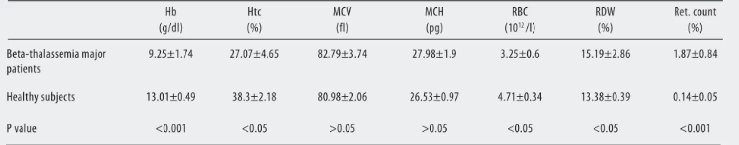
The hemoglobin and the hematocrit values and the red blood cell counts (RBC) were found to be lower in the pa-tients with thalassemia when compared with healthy chil-dren. Red cell distribution width (RDW) and reticulocyte counts were higher in patient group. All of these findings were found to be statistically significant. Haematological parameters of patient and control groups are shown in Table 1.
Table 1. Complete blood count results of the patients and the healthy subjects. Hb (g/dl) Htc (%) MCV (fl) MCH (pg) RBC (1012 /l) RDW (%) Ret. count (%) Beta-thalassemia major patients 9.25±1.74 27.07±4.65 82.79±3.74 27.98±1.9 3.25±0.6 15.19±2.86 1.87±0.84 Healthy subjects 13.01±0.49 38.3±2.18 80.98±2.06 26.53±0.97 4.71±0.34 13.38±0.39 0.14±0.05 P value <0.001 <0.05 >0.05 >0.05 <0.05 <0.05 <0.001
*Hb, haemoglobin; Htc,hematocrit; MCV, mean corpuscular volume; MCH, mean corpuscular hemoglobin; RBC; red blood cell count; RDW, red cell distribution width (RDW); Ret. count, reticulocyte count.
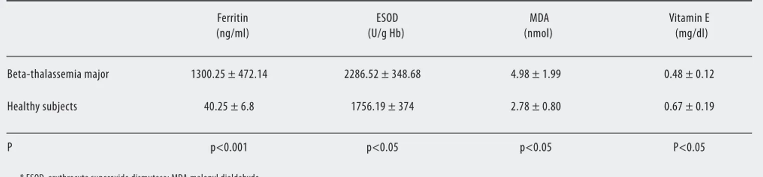
In the patient group, serum ferritin, ESOD, and plas-ma MDA levels were determined to be higher (Table 2). Also we found that serum vitamin E levels were lower than healthy children. This finding was found significant sta-tistically.
Serum ferritin levels, ESOD levels, vitamin E and plas-ma MDA levels and serum vitamin E levels are also shown in Figure 1, Figure 2, and Figure 3.
Discussion
Previous studies have demonstrated that a variety of morphological, biochemical, and metabolic disturbances of the thalassemic red cell with shortened life span (3). There is extensive evidence of in vivo oxidative damage as well as enhanced sensitivity to exogenous oxidant stress in red cells of beta-thalassemia (3). It has been postulated that the biochemical and metabolic changes of beta-thalassemic red blood cells (RBC) are associated with a constant oxida-tive stress within the cell caused by precipitation of excess
alpha-globin chains, iron decompartimentalization, and release of free iron (2,3)
Increased plasma malonyldialdehyde (MDA) level, which is measured by the thiobarbituric acid reaction substance (TBARS) methods, was found in beta-thalasse-mia patients (12,13). MDA is a good indicator of oxida-tive damage. In one of the previous studies, free and total MDA was found to be higher in regularly transfused thal-assemia major patients than in the thalthal-assemia intermedia patients (14). In beta-thalassemia intermedia patients in-creased concentration of lipid peroxidation products (such as MDA) was found in another study (15). Similarly in our study the increased plasma MDA levels were found. As a result of continuous blood transfusions, our patients might be subjected to peroxidative tissue injury by the second-ary iron overload. These finding might support the idea of iron overload in beta-thalassemia leads to an enhanced generation of reactive oxygen species and oxidative stress.
Erythrocytes are protected from oxidative stress by in-tracellular enzymes such as superoxide dismutase and sev-eral other constituents such as vitamin E (1). ESOD is a preventive antioxidant. Increased ESOD activities were found in the patients with beta-thalassemia by the inves-tigators previously (12,16). Both beta-thalassemia and ac-companying iron overload lead in vivo lipid peroxidation and the compensatory increase in the antioxidant enzyme levels of SOD and glutathione peroxidase (GPx) (12). The significant increased catalytic activities of SOD and GPx in beta-thalassemic erythrocytes were found when compared with healty subjects and beta-thalassemic carriers (17). In a study from Jakarta, highly significant decrease in antioxi-dants was found in a group of transfusion-dependent
thal-Figure 1. The comparison of plasma MDA values in patients and control group, p<0.05
Figure 2. The comparison of ESOD activities in patients and control group, p<0.05
Figure 3. The comparison of vitamin E levels in patients and control group, p<0.05
assemia major patients, and this picture was even worsened in long-term transfused patients. In this study insufficient chelation after transfusions was suggested the cause of de-creased antioxidant levels (18). In another study, inde-creased MnSOD and Cu/ZnSOD levels, which might be induced by mediators of oxidative stress, was found in Turkish thalassemia patients, (19). In our study, increased ESOD activity finding was probably due to an increase in the pro-portion of younger red blood cells, and the compensatory mechanism after increased oxidant stress. Our patients re-ceived regular blood transfusions and chelation therapies. In homozygous beta-thalassemia, low serum levels of alpha-tocopherol, which is a lipid soluble antioxidant, have been found (2). The patients in our study didn’t receive any vitamin E supplementation, and decreased vitamin E levels were found these patients. Vitamin E deficiency in thalassemias is due to its increased consumption as a result of the oxidative stress, and imposes both to the red blood cells and the other tissues by haemochromatosis (7). Pre-vious studies have shown that iron-induced liver damage in thalassemia may play a role in the depletion of lipid-soluble antioxidants (6). While no significant changes oc-curred in hemoglobin levels and transfusion requirements, parenteral administration of vitamin E appeared to be ef-fective to attenuate the oxidative damage of the erythro-cytes in homozygous beta-thalassemia (2). Similarly, the oral vitamin E treatment improves the antioxidant/oxidant
balance within the plasma, LDL particles, and red blood cells, and counteracts with lipid peroxidation processes in beta-thalassemia intermedia patients (13). Since oxygen free radicals are involved in the pathogenesis of increased destruction of thalassemic red blood cells, our results may be assumed that the use of antioxidants may have a protec-tive effect by improving the red blood cells survival. The recovery of antioxidant status may be helpful for decreas-ing oxidative damage.
In conclusion, while ESOD (a preventive antioxidant) values, and plasma MDA (the breakdown product of lipid peroxidation) levels were found to be higher in beta-thal-assemic patients than healthy children, serum vitamin E levels were higher in healthy children. Iron overload in beta-thalassemia leads to an enhanced generation of reac-tive oxygen species and oxidareac-tive stress. Increased oxida-tive damage in thalassemias may be due to the depletion of lipid soluble antioxidants such as vitamin E. Our results suggest that the measurement of the peroxidation prod-ucts, together with the evaluation of the antioxidants may be the simple measurement of iron toxicity in thalassemia. The administration of selective antioxidants such as vita-min E with an appropriate diet or appropriate treatment might represent a promising way of counteracting with the oxidative damage and its deleterious effects on the progres-sion of the disease.
References
1. Chiu D, Kuypers F, Lubin B: Lipid peroxidation in human red cells. Semin Hematol 1989; 26:257-276.
2. Clemens MR: Antioxidant therapy in hematological disorders. In Antioxidants in Therapy and Preventive Medicine I. Emerit et al (ed), Plenum Press, New York 1990:423-433.
3. Kattamis C, Kattamis AC. Oxidative stress disturbances in erythrocytes of β-thalassemia. Pediat Hematol Oncol 2001;18:85-88.
4. McDonagh KT, Nienhuis AW: The thalassemias. In Nathan DG,Oski FA, ed. Hematology of Infancy and Childhood, 4th ed. WB Saunders, Philadelphia 1993: 783-879.
5. Scott MD, Van den Berg JJM, Repka T, et al. Effect of excess β-hemoglobin chains on cellular and membrane oxidation in model β-thalassemic erythrocytes. J Clin Invest 1993; 91:1706-1712. 6. Livrea MA, Tesoriere L, Pintaudi AM, et al. Oxidative stress and
antioxidant status in beta-thalassemia major: iron overload and depletion of lipid soluble antioxidants. Blood 1996; 88:3608-3614.
Table 2. Ferritin levels, erythrocyte superoxide dismutase activities, malonyldialdehyde and vitamin E values of the patients and the healthy subjects. Ferritin (ng/ml) ESOD (U/g Hb) MDA (nmol) Vitamin E (mg/dl) Beta-thalassemia major 1300.25 ± 472.14 2286.52 ± 348.68 4.98 ± 1.99 0.48 ± 0.12 Healthy subjects 40.25 ± 6.8 1756.19 ± 374 2.78 ± 0.80 0.67 ± 0.19 P p<0.001 p<0.05 p<0.05 P<0.05
* ESOD, erythrocyte superoxide dismutase; MDA,malonyl dialdehyde * Values were given as mean ± standard deviation.
7. Rachmilewitz EA, Kornberg A, Acker M. Vitamin E deficiency due to increased consumption in β-thalassemia and in Gaucher’s disease. Ann NY Acad Sci 1982;393:336-347.
8. Miniero R, Canducci E, Ghigo D, et al. Vitamin E in beta-thalassemia. Acta Vitaminol Enzymol 1982; 4:21-25. 9. Kurtel H, Granger DN, Tso P, et al: Vulnerability of intestinal
interstitial fluid to oxidant stress. Am J Physiol 1992;263: G573-578.
10. Winterbourn CC, Hawkins RE, Brian M, et al. The estimation of red cell superoxide dismutase activity. J Lab Clin Med 1975;85: 337-341.
11. Rindi G. A rapid colorimetric method for determination of tocopherol and tocopheryl acetate in plasma. Int Rev Vit Res 1958; 28:225.
12. Meral A, Tuncel P, Surmen-Gur E, et al. Lipid peroxidation and and antioxidant status in beta-thalassemia. Pediatr Hematol Oncol 2000; 17:687-693.
13. Tesoriero L, D’Arpa D, Butera D,et al. Oral supplements of vitamin E improve measures of oxidative stress in plasma and reduce oxidative damage to LDL and erythrocytes in beta-thalassemia intermedia patients. Free Radical Res 2001;34(5): 529-540.
14. Cighetti G, Duca L, Bortone L,et al. Oxidative status and malonyldialdehyde in beta-thalassemia patients. Eur J Clin Invest 2002;32 Suppl 1:55-60.
15. Cappelini MD, Tavazzi D, Duca L, et al. G. Non-transferrin-bound iron, iron related oxidative stress and lipid preoxidation in beta-thalassemia intermedia. Transfusion Science 2000;23:245-246.
16. Yenchitsomanus P, Wasi P: Increased erythrocyte superoxide dismutase activities in ß-thalassaemia/haemoglobin H diseases. J Clin Pathol 1983;36:329-333.
17. Chakraborty D, Bhattacharyya M. Antioxidant defence status of red blood cells of patients with β-thalassemia and Eβ-thalassemia. Clinica Chimica Acta 2001;305:123-129.
18. Laksmitawati DR, Handayani S, Udyaningsih-Freisleben SK, et al. Iron status and oxidative stress in beta-thalassemia patients in Jakarta. Biofactors 2003;19:53-62.
19. Tekin M, Akar N, Egin Y, et al. Serum superoxide dismutase levels of beta thalassemia patients and effects of high dosage of intravenous desferrioxamine treatment on superoxide dismutase levels. Pediatr Hematol Oncol 1998;15:371-374.