Case Reports
158
A novel oral anticoagulant,
dabigatran, in acute renal infarction
Cihan Altın, Onur Sakallıoğlu*, Esin Gezmiş**, Haldun Müderrisoğlu*** Departments of Cardiology, *Nephrology, **Radiology, Faculty of Medicine, Başkent University; İzmir-Turkey***Department of Cardiology, Faculty of Medicine, Başkent University; Ankara-Turkey
Introduction
Acute renal infarction (ARI), which is a rare cause of flank pain, results from interruption in the blood supply of renal tissue. The sever-ity of its clinic depends on the width of the affected part of the kidney. It is often impossible to find the underlying cause (1, 2). Thromboembolic states related to cardiac diseases, such as atrial fibrillation, rheumatic mitral stenosis, endocarditis, and left ventricular wall aneurysms, are the major causes, whereas occlusion of the renal artery following endovascular aortic or renal intervention, renovascular diseases, malignant hypertension, paradoxical cardiac emboli associated with atrial septal defects, polyarteritis nodosa, and cocaine have also been reported as rare causes in the etiology (3-5). In the literature, the inci-dence of ARI lies within a range of 0.007%-1.4% (1-5).
Herein, we present a patient with ARI and its treatment with a novel oral anticoagulant, dabigatran.
Case Report
A 55-year-old male patient was admitted to our emergency depart-ment with right flank tenderness preceding 4-6 hours. He had no dysuria, hematuria, fever, nausea, or vomiting. Except for the right flank tenderness and 3/6 systolic murmur at the cardiac apex, his physical examination and hemodynamic parameters were normal. His
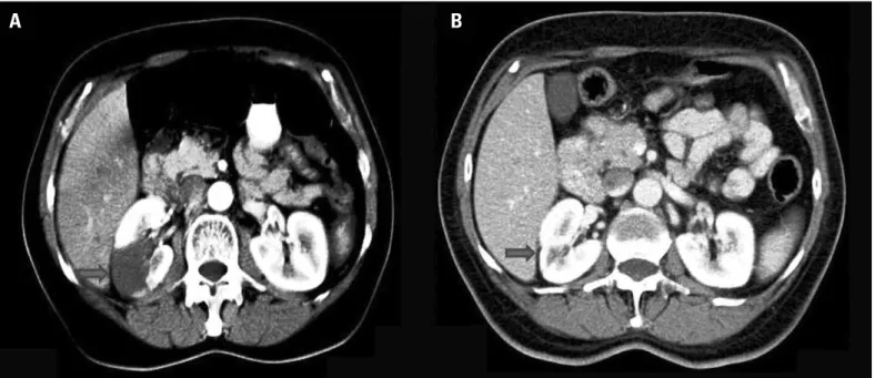
medical history was remarkable for non-ischemic dilated cardiomyopa-thy, which was diagnosed by coronary angiography (CAG) and cardiac catheterization in another clinic 2 days ago, and the drugs in use were carvedilol, ramipril, furosemide, and acetylsalicylic acid. The abdominal x-ray, urinary ultrasonography (US), and laboratory analysis were all normal. Only mild microscopic hematuria was detected. The patient was clinically diagnosed with renal colic, treated with analgesics, and then discharged home for further ambulatory urologic investigation. A few hours later, the patient returned to the emergency department with severe right flank pain. On contrast-enhanced computed tomography (CECT), performed for persistent flank pain, to rule out nephrolithiasis and CAG complications, such as retroperitoneal hematoma, a hypodense, non-enhancing, wedge-shaped area in the right kidney, which was compatible with renal infarction, was seen (Fig. 1A). Globally systolic dysfunction (ejection fraction: 35%), mild-moderate mitral insufficiency, dilated left chamber without thrombi or vegetation, and multiple self-terminating atrial fibrillation (AF) episodes compatible with paroxysmal AF were detected on transthoracic echocardiography and 24-hour Holter monitoring, respectively. CHA2DS2-VASc and HAS-BLED scores were calculated as 3 and 2, respectively. Dabigatran (150 mg, b.i.d.), analgesics, and hydration were initiated. In 5 days, he recovered and was discharged home on dabigatran. On the control CECT scan per-formed 1 month later, reperfusion and contrast enhancement were seen at the previously hypodense renal infarct area (Fig. 1B).
Discussion
The clinical diagnosis of ARI is frequently missed or delayed because of its nonspecific presentation, which can mimic more com-mon conditions, such as nephrolithiasis, pyelonephritis, and appendici-tis. X-ray and US, the primary imaging modalities for flank pain, have low sensitivity; so, CT is mostly performed for the differential diagnosis in patients with persistent severe flank pain. Magnetic resonance imaging with gadolinium and radioisotope scans are alternatives to CT in the
Figure 1. A, B. Contrast-enhanced abdominal computed tomography images at the same level. (A) The hypodense, wedge-shaped infarct area in the right kidney (arrow). (B) One month later, reperfusion and contrast enhancement are seen at the previously hypodense infarct area (arrow). A slight decrease in cortical thickness is detected
diagnosis of ARI. Inflammatory symptoms and parameters can be help-ful to rule out infection (lobar nephronia/pyelonephritis) (6, 7).
There are no clinical research data or therapeutic guidelines for renal artery embolism; however, hydration, systemic anticoagulation, percutaneous endovascular therapy, local thrombolysis, and surgical embolectomia are recommended treatment options in limited reports. Conservative therapy is favorable in unilateral ARI (8). Common concepts for anticoagulation therapy in ARI are intravenous heparin, followed by oral warfarin. Unlike other cases in the literature, we preferred oral dabigatran (150 mg, b.i.d.) for anticoagulation. Although there is no spe-cific suggestion about the usage of dabigatran in ARI, the American College of Cardiology and American Heart Association have recom-mended that dabigatran is useful as an alternative to warfarin for the prevention of stroke and systemic embolism in patients with paroxysmal to permanent AF and risk factors for stroke or systemic embolization who do not have a prosthetic heart valve or hemodynamically significant valve disease, severe renal failure (creatine clearance <15 mL/min), or advanced liver disease (impaired baseline clotting function) (9, 10).
In patients with a high risk of systemic thromboembolism and pre-senting with flank tenderness, ARI should be kept in mind to prevent a delayed or missed diagnosis. Clinicians should be aware of the clinical importance of paroxysmal AF, which has similar risks for stroke and systemic embolism as permanent AF. Also, 24-hour Holter monitoring can be useful to detect underlying causes of ARI. In our case, we har-vested an excellent clinical outcome with the proper diagnostic and treatment methods.
Conclusion
Dabigatran can be preferred as an oral anticoagulant in the conser-vative therapy of systemic embolisms, like ARI. Clinical usage of dabiga-tran in such rare cases will help clinicians in their daily practice.
Acknowledgment: The authors are thankful to Dr. Kutlay Naci Tutucu from the Department of Nephrology, Başkent University, Faculty of Medicine, for his valuable contribution in the differential diagnosis of the presented case.
References
1. Bourgault M, Grimbert P, Verret C, Pourrat J, Herody M, Halimi JM, et al. Acute renal infarction: a case series. Clin J Am Soc Nephrol 2013; 8: 392-8.
[CrossRef]
2. Bolderman R, Oyen R, Verrijcken A, Knockaert D, Vanderschueren S. Idiopathic renal infarction. Am J Med 2006; 119: 356: e9-12.
3. Paris B, Bobrie G, Rossignol P, Le Coz S, Chedid A, Plouin PF. Blood pressure and renal outcomes in patients with kidney infarction and hypertension. J Hypertens 2006; 24: 1649-54. [CrossRef]
4. Antopolsky M, Simanovsky N, Stalnikowicz R, Salameh S, Hiller N. Renal infarction in the ED: 10-year experience and review of the literature. Am J Emerg Med 2012; 30: 1055-60. [CrossRef]
5. Mahamid M, Francis A, Abid A, Awawde M, Abu-Elhija O. Embolic renal infarc-tion mimicking renal colic. Int J Nephrol Renovasc Dis 2014; 7: 157-9. [CrossRef]
6. Jeffrey RB, Federle MP. CT and ultrasonography of acute renal abnormali-ties. Radiol Clin North Am 1983; 21: 515-25.
7. Kim SH, Park JH, Han JK, Han MC, Kim S, Lee JS. Infarction of the kidney: role of contrast enhanced MRI. J Comput Assist Tomogr 1992; 16: 924-8. [CrossRef]
8. Moyer JD, Rao CN, Widrich WC, Olsson CA. Conservative management of renal artery embolus. J Urol 1974; 109: 138-43.
9. Eikelboom JW, Wallentin L, Connolly SJ, Ezekowitz M, Healey JS, Oldgren J, et al. Risk of bleeding with 2 doses of dabigatran compared with warfarin in older and younger patients with atrial fibrillation: an analysis of the ran-domized evaluation of long-term anticoagulant therapy (RE-LY) trial. Circulation 2011; 123: 2363-72. [CrossRef]
10. Wann LS, Curtis AB, Ellenbogen KA, Estes NA, Ezekowitz MD, Jackman WM, et al. American College of Cardiology Foundation/American Heart Association Task Force. 2011 ACCF/AHA/HRS focused update on the management of patients with atrial fibrillation (update on Dabigatran): a report of the American College of Cardiology Foundation/American Heart Association Task Force on practice guidelines. Circulation 2011; 123: 1144-50. [CrossRef]
Address for Correspondence: Dr. Cihan Altın, 6471/5 Sok., No:7, Yalı Mahallesi
Bostanlı, Karşıyaka/İzmir-Türkiye Phone: +90 232 241 10 00 E-mail: [email protected] Available Online Date: 21.01.2015
©Copyright 2015 by Turkish Society of Cardiology - Available online at www.anakarder.com DOI:10.5152/akd.2015.5837
Case Reports