1 Departmant of General Surgery, Dicle University Medical Faculty, Diyarbakir, Turkey 2 Departmant of General Surgery, Diyarbakır State Hospital, Diyarbakir, Turkey 3 Departmant of Pathology, Dicle University Medical Faculty, Diyarbakir, Turkey 4 Departmant of Radiology, Dicle University Medical Faculty, Diyarbakir, Turkey
Yazışma Adresi /Correspondence: Sadullah Girgin,
Dicle Üniversitesi Tıp Fakültesi, Genel Cerrahi Anabilim Dalı, Diyarbakır, Türkiye Email: [email protected] Geliş Tarihi / Received: 16.01.2015, Kabul Tarihi / Accepted: 26.02.2015
ORIGINAL ARTICLE / ÖZGÜN ARAŞTIRMA
Granulomatous mastitis: A retrospective review of 49 patients
Granülamatöz mastit: 49 hastanın retrospektif incelenmesi
Sadullah Girgin1, Ömer Uslukaya1, Edip Erdal Yılmaz2, Uğur Fırat3, Hatice Gümüş4,
Murat Kapan1, Metehan Gümüş1
ÖZET
Amaç: İdiopatik granulomatöz mastit, nadir görülen, be-nign, kronik inflamatuar bir hastalık olup meme kanserini taklit eder. Bu çalışmada idiopatik mastitli 49 hastanın te-davisini ve kliniğini sunmayı amaçladık.
Yöntemler: Hastane otomasyon sisteminde 2007-2012 yılları arasında histolojik olarak granulomatöz tanısı ko-nulmuş hastalar incelendi. Hastalara ait klinik bulgular, demografik veriler ve tedavi yöntemleri kaydedildi. Bulgular: Ortalama yaş 37.71±7.1 idi. Tedavi gruplar arasında etiyolojik ve demografik açıdan istatistiksel fark yoktu. Nüks oranları grup 1, grup 2 ve grup 3’te sırasıyla %8,3, %14,8 %20 olup, gruplar arasında istatistiksel an-lamlı farklılık yoktu.
Sonuç: Steroid ve cerrahi tedavinin idiopatik granuloma-töz mastitli hastalarda uygulanabileceğini, steroid tedavi-sinin ilk basamak tedavi olması gerektiği kanaatindeyiz. Bununla birlikte tedavi algoritması belirlenebilmesi için prospektif, randomize klinik çalışmalara ihtiyaç vardır. Anahtar kelimeler: İdyopatik, granülamatöz mastit, te-davi.
ABSTRACT
Objective: Idiopathic granulomatous mastitis, a rare, be-nign, chronic inflammatory condition of the breast, which usually mimics breast carcinoma. In this study, we report on the management of 49 cases of idiopathic granuloma-tous mastitis and their clinical presentation.
Methods: The breast and anatomical databases of one center were reviewed from 2007 to 2012 to identify pa-tients with histological diagnosis of idiopathic granuloma-tous mastitis. Clinical and demographic characteristics were retrieved and treatment modalities were were re-corded.
Results: The mean patient age was 37.71±7.1 years. There were no differences between the management groups about etiological and demographic factors. The rate of recurrence in group 1, group 2, and group 3 was 8.3%, 14.8% and 20%, respectively; there were no statis-tically significant difference among the groups.
Conclusion: Steroids and surgical methods codified in idiopathic granulomatous mastitis may be preferred. We recommend steroid therapy as first-line treatment. How-ever, prospective, randomized clinical trials are needed to determine the treatment algorithm.
Key words: Idiopathic, granulomatous, mastitis, man-agement
INTRODUCTION
Idiopathic granulomatous mastitis (IGM), also known as granulomatous lobular mastitis, is a rare benign inflammatory disease of the breast. It has a poorly understood etiology and unpredictable dura-tion, and there is no consensus on optimal treatment [1-4]. Breast surgeons are usually adept at manag-ing women with breast cancer and keep abreast of
the evidence (Figure 1). Benign breast disorders, however, form a significant majority of the condi-tions presented to the breast surgeon and there is often a paucity of literature and evidence to manage these potentially more difficult diseases. Idiopathic granulomatous mastitis is rare and is a great mim-icker of other breast conditions including breast carcinoma [5]. If not managed cautiously and thor-oughly, treatment of IGM may require mastectomy.
Figure 1. A photograph of the patient’s left breast . Clini-cal appearance of the patient’s breast including skin ab-scesses and fistulae. Photo obtained during her hospital admission
Although surgical excision and steroid therapy are the most commonly used treatments, different management options may be implemented. These include conservative approaches with close surveil-lance, wide excision, abscess drainage, mastectomy, and immunosuppressive therapy [6-9]. However, there is no consensus on the optimal treatment of patients with IGM [10-11]. Furthermore, reported rates of recurrence with IGM are as high as 50% [12]. In this study, we report on the management of 49 cases of IGM and their clinical presentation.
METHODS
This study included 49 women who met the re-quired histological criteria of IGM and were treated between 2007 and 2012 in Dicle University Medical School Hospital. The design of the study has been approved by Dicle University Faculty of Medicine ethics committee and thus meets the standards of the Declaration of Helsinki. Due to retrospective nature of the study, the ethics committee did not re-quire a written consent from the participants. Dur-ing this period, each case of idiopathic granuloma-tous mastitis was diagnosed from among the 1,587 patients treated for breast pathologies. Diagnostic and therapeutic management of all 49 patients was undertaken by the same team.
We recorded a variety of etiologic factors, in-cluding: smoking; use of oral contraceptives; local trauma; time to presentation since last delivery or breast feeding; family history of breast cancer;
tu-berculosis contact history; history of autoimmune disease; presenting symptoms; clinical findings (in-cluding size of lesion, the presence of an abscess, or sinus formation); radiographic, histological, and microbiological results; treatment modalities; and follow-up recurrence data.
All patients underwent a clinical breast exami-nation and ultrasonography (USG) examiexami-nation, while patients older than 40 years of age also under-went mammography. On suspicion of malignancy, magnetic resonance imaging (MRI) was performed for further investigation. Definitive diagnosis was obtained by core needle biopsy (14-gauge) or surgi-cal excision or from biopsy specimens taken from the abscess wall during surgical drainage. The obtained specimens were fixed overnight in 10% formalin, and the residual tissues were cultured for bacteria. Paraffin-embedded specimens were stained with hematoxylin and eosin, Ziehl-Neelsen, and the pe-riodic acid-Schiff reaction with or without diastase digestion. Other possible causes of granulomatous inflammatory lesions of the breast were excluded by serological tests, histological analysis, and tests of the affected tissue for aerobic and anaerobic bacte-ria, mycobactebacte-ria, and fungi.
Group 1 had twelve patients that were only given medical treatment in the form of steroid therapy at 32 mg/day depending on the dimensions of the lesion the severity of symptoms, and the pa-tient’s general health and personal treatment pref-erences. Group 2 (27 patients) underwent surgical excision. In group 3 (10 patients), breast abscesses were drained as an initial treatment step for those patients that had them. In addition, group 3 patients also received antibiotic therapy (as did any patient with positive microbiological culture) and steroid therapy at 32 mg/day after the regression of inflam-matory process.
Patients who had receiving steroid treatment and the dosage of steroid were monitored month-ly until remission. Once remission was achieved, monitoring and dosage control was adjusted every 3 month for the first year; after the first year, they were controlled on a yearly basis. All patients were assessed on follow-up by physical examination, USG, and laboratory tests (including C reactive protein, ESR, white blood cell count, and alkaline phosphatase).
Remission was conceptualized as a complete absence of disease activity, with no signs or symp-toms of active disease. Practically, the patients would need to meet the following criteria to be con-sidered in remission:
• Clinical criteria: No tender or swollen mass on physical exam.
• Laboratory criteria: Normal C reactive pro-tein, ESR, white blood cell count, and alkaline phosphatase.
• Radiological: Absence of ultrasound finding • Duration: At least three weeks of meeting the above criteria.
Statistical analysis
Statistical analysis was performed by SPSS for Windows 13.0 (SPSS Inc., Chicago, IL, USA). Data were expressed as the mean ± SD (standard devia-tion) values for continuous values. Chi-square test was used for categorical variables. Groups were compared by using the non-parametric Kruskal-Wallis test. The Mann-Whitney U test was used for continuous data that did not meet a normal distribu-tion. P-value of less than 0.05 was considered sig-nificant.
RESULTS
All patients were female and the median age at the time of diagnosis was 37.71±7.1 years (24-57 years). Patients’ mean ages in group 1, group 2, and group 3 were 35.42±6.37, 39±7.94 and 36.90±4.97 years old, respectively, without significant statisti-cal difference between the groups (p=0.350). Most of the patients (42 of the 49) were at a reproductive age. All patients were less than five years removed from a previous pregnancy (average 26 months) and had a history of breast feeding. Average duration of breast feeding was 17 months (with a range of 4-34 months). Eight patients (16.3%) reported use of tobacco products and nine (18.4%) reported prior use of oral contraceptive pills. None of the patients had local trauma, family history of breast cancer, tuberculosis contact history, history of autoimmune disease, history of diabetes mellitus, or alpha-1 an-titrypsin deficiency. The mean duration of symp-toms was nine weeks (with a range of 20 days to 9 months) before presentation. Twenty-three patients (47%) had already been treated with antibiotics be-fore presentation to our department.
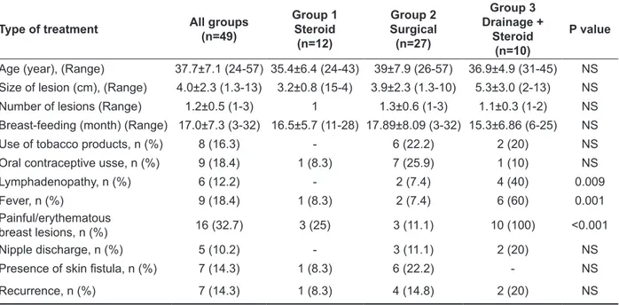
Table 1. Summary of the characteristics of patients with idiopathic granulomatous mastitis
Type of treatment All groups(n=49) Group 1Steroid (n=12) Group 2 Surgical (n=27) Group 3 Drainage + Steroid (n=10) P value Age (year), (Range) 37.7±7.1 (24-57) 35.4±6.4 (24-43) 39±7.9 (26-57) 36.9±4.9 (31-45) NS Size of lesion (cm), (Range) 4.0±2.3 (1.3-13) 3.2±0.8 (15-4) 3.9±2.3 (1.3-10) 5.3±3.0 (2-13) NS Number of lesions (Range) 1.2±0.5 (1-3) 1 1.3±0.6 (1-3) 1.1±0.3 (1-2) NS Breast-feeding (month) (Range) 17.0±7.3 (3-32) 16.5±5.7 (11-28) 17.89±8.09 (3-32) 15.3±6.86 (6-25) NS
Use of tobacco products, n (%) 8 (16.3) - 6 (22.2) 2 (20) NS
Oral contraceptive usse, n (%) 9 (18.4) 1 (8.3) 7 (25.9) 1 (10) NS
Lymphadenopathy, n (%) 6 (12.2) - 2 (7.4) 4 (40) 0.009
Fever, n (%) 9 (18.4) 1 (8.3) 2 (7.4) 6 (60) 0.001
Painful/erythematous
breast lesions, n (%) 16 (32.7) 3 (25) 3 (11.1) 10 (100) <0.001
Nipple discharge, n (%) 5 (10.2) - 3 (11.1) 2 (20) NS
Presence of skin fistula, n (%) 7 (14.3) 1 (8.3) 6 (22.2) - NS
Recurrence, n (%) 7 (14.3) 1 (8.3) 4 (14.8) 2 (20) NS
NS= not significant (p>0.05)
The left breast was affected in 29 patients
Pain-ful and/or erythematous breast lesions, fever and lymphadenopathy symptoms were significantly higher patients from group 3 (p<0.0001, p=0.001 and p<0.009 respectively). There were no differ-ences between the groups about etiological and de-mographic factors. The rate of recurrence in group 1, group 2, and group 3 was 8.3%, 14.8% and 20%, respectively; there were no statistically significant difference among the groups. Remission occurred by surgical excision at three patients with relapses in group 1 and group 3; and by steroid treatment at four patients with relapses in group 2. All patients in group 1 had only one lesion and there was no tobacco use, nipple discharge, or lymphadenopathy (Table 1). Steroid treatment period were 7.52±1.6 months at group 2 and were 7±1.7 months at group 3 as there were no statistically significant difference between the groups (p>0.05). Complications due to steroid treatment were observed in only two pa-tients who developed a moon face and acne. Mean follow up for all groups was 48 months.
DISCUSSION
IGM, a rare benign breast disease, was first de-scribed in 1972 by Kessler and Wolloch [13]. It is a benign inflammatory breast disease that often clini-cally simulates carcinoma. Few articles exist in the literature, and most of those are case reports and in-clude only small series of patients [14]. Our series of 49 patients is one of the largest single-institution series reported to date in Turkey. Similar to other published series, the age at presentation was be-tween 24 and 57 years; however, patients as young as 11 and as old as 83 years have been reported [14-15].
IGM is characterized by the presence of epithe-lioid and multinucleated giant cell granulomas limi-ted to the mammary lobules with micro-abscesses in the absence of obvious etiology [16]. Local or systemic irritants as well as undetected infective ca-uses may result in damage to the ductal epithelium. Luminal fat and protein rich secretion into the lobu-lar connective tissue produces a localized granulo-matous response [17]. This association has recently been challenged, because serologic and bacterial tests are usually negative in patients presenting with IGM [8]. Also, some authors have suggested that the prior use of oral contraceptives, a history of bre-ast feeding, and pregnancy are linked to IGM. Many
women with IGM breast-fed their children and the majority of those that breast-fed were pregnant or lactating in the five years prior to the onset of IGM symptoms [18]. High serum prolactin levels, as well as distention of the acini and ducts may result in a rupture of these structures, inducing a granuloma-tous response [19,20]. In our series, nine patients (18.4%) had previously used oral contraceptive pills and eight patients (16.3%) had a history of using tobacco products.
Idiopathic granulomatous mastitis usually pre-sents with a unilateral, firm, discrete, tender mass, ranging from 0.5 to 10 cm in size. Overlying skin is often inflamed, though it can sometimes present with a non-tender mass or pain alone. Nipple retra-ction and discharge, sinus formation, and axillary lymphadenopathy may be seen [17]. In the literatu-re, the most common presentation symptoms have been reported as a mass in the breast (48-100%), sinus or abscess (16-52%), and inflammation or ery-thema (11%) [6,17,20]. In this study, all patients had unilateral breast mass. However, lymphadenopathy, pain, erythematous breast lesions, and fever seen most frequently in patients who had breast abscess. Mammography, ultrasonography, and MRI are used primarily to rule out malignancy and confirm a diagnosis of IGM. The mammographic findings of IGM are not well documented and often mimic breast carcinoma [1,2]. The USG appearance of IGM is non-specific, non-homogeneous, irregular hypoechoic lesion with focal posterior shadowing [21]. The appearance of multiple clustered, often contiguous, hypoechoic tubular lesions, associated with a large hypoechoic mass has been considered to be suggestive of the condition, but these features are not always found [22]. MRI shows a focal ho-mogeneous enhancing mass with irregular borders and parenchymal distortion, but it is difficult to dif-ferentiate GM from breast cancer based on such MRI findings [3]. The role of MRI in the charac-terization of inflammatory processes of the breast is non-specific, but it is still useful in patients who have no significant pathology on mammography or USG [1,2,17]. MRI is also reported to provide no additional information for the differentiation of mastitis from carcinoma. This process has high sen-sitivity, but low specificity for most breast lesions [14].
Other causes of granulomatous lesions in the breast that require different treatments must be ex-cluded. These include infective causes such as tu-berculosis or brucellosis, parasitic infections such as filariasis, fungal infections such as actinomy-cosis, and systemic diseases such as sarcoidosis, Wegener’s granulomatosis, or giant-cell arteritis [13,23]. Foreign-body reaction, fat necrosis, and ductal ectasia must also be excluded [2,13]. Since clinical and imaging diagnoses are often difficult and inconclusive, histopathologic evaluation plays a crucial role in diagnosing IGM. Diagnosis can be challenging and cytological features can be difficult to distinguish from those of carcinoma and other granulomatous disease of the breast, such as specif-ic infections (mycobacterium tuberculosis, fungus, parasites), duct ectasia, periductal mastitis, plasma-cell mastitis, sarcoidosis, and vasculitis [11,24]. Tuberculosis must be considered in the differential diagnosis, especially since it is more prevalent in an endemic region like ours, southeastern Turkey. In the published literature, most diagnoses of IGM are based upon ruling out tuberculosis by histochemical staining and clinical evaluation [6,25]. Chest X-ray, tuberculin skin test, Quantiferon assay, determina-tion of erythrocyte sedimentadetermina-tion rate, and PCR may also aid in diagnosis [26].
The optimal treatment approach for IGM has not yet been established; research is limited by an insufficient number of diagnoses. Available treat-ment options include close follow-up, antibiotic therapy, surgical excision, steroids, and immuno-suppressive drugs [9,25,27,28]. The management of patients with IGM varies broadly from conservative approaches to mastectomy [1,14]. Lai et al. [15] re-ported spontaneous resolution in four of eight cases of IGM with a mean interval of 14.5 months for complete resolution. Wide surgical excision is tra-ditional and the most commonly performed surgi-cal intervention [4]. Although limited excision may be possible, complete excision of the breast mass or involved inflammatory tissue provides more satis-factory results in terms of preventing further com-plications and recurrence [1,9]. Breast abscesses must initially be drained. The role of antibiotics in the treatment of IGM is of little value [12]. Antimi-crobial therapy should be administered in cases of positive culture results for the biopsy samples. In
the case of abscess or sinus formation, 2 weeks em-piric antibiotic treatment is also recommended [27].
DeHertogh et al. in 1980 were the first who in-vestigated the efficacy of different treatments and they concluded that corticosteroid is an appropriate option for the treatment of the disease. Furthermore, Sakurai et al. sought this pharmaceutical approach and they authenticated that corticosteroid could be efficient in 87% of patients without any relapse. The proceeded results were validated by Hisuing et al. while their research implied that low doses of corticosteroids were efficient; though, more inves-tigations in this field were recommended by them. Moreover, in 2010, it was authenticated that corti-costeroids provide appropriate treatment, whereas first option should be allocated to surgery in the case of relapse, fistula, and abscess [21]. Pandey et al.14 described their prospective study of 49 women with a mean age of 35 years and found that 80% were of Hispanic origin (OR = 3.00, 91% CI 1.42-6.24, P = 0.0032), 80% presented with a painful mass and 90% were prescribed steroids with an 80% com-plete resolution rate. It should be kept in mind that steroids may exacerbate infectious disease of the breast; therefore, it is essential to exclude an infec-tious etiology before giving steroids. Furthermore, the side effects of steroid therapy can include glu-cose intolerance, acne and cushingoid features [5].
Based on our findings, oral corticosteroids and surgery have both been used as treatment options. Because recurrence ratios were the same. The surgi-cal approach should be considered in the following circumstances: the presence of contraindications or complications of corticosteroid therapy; failure to achieve the desired benefits in long-term treatment; or the patient requests surgery. We advise steroid treatment after surgical or radiological drainage with antibiotherapy to the patients which have cavi-ties or abscess formation. In the study, recurrences were seen at two of the patients with abscess forma-tion and they were treated with extensive surgical excision. We mostly do not advise extensive surgi-cal excision primarily to the patients which have large cavities or abscess formation. The recurrences at four patients who had surgical treatment before were treated with steroids and the recurrences at two patients who had steroid treatment before were treated with steroids surgical excision successfully.
So we think that these treatments can be alternative-ly used with the patients who have recurrences. In our study, the recurrence at one patient who had ste-roid treatment before were treated surgically and the recurrence at four patients who had treatment surgi-cally before were treated with steroids successfully.
This study shows that both of surgical and ste-roid treatments can be used for granulomatous mas-titis. But as there is no statistically significant differ-ence about the recurrdiffer-ence ratios, it can be concluded that steroid treatment should be used primarily and surgical treatment should be used at the patients with recurrences, when the side effects and also re-sistance are occurred with steroids.
There is still not a commonly accepted optimal treatment plan for IGM. Wide local excision and/or corticosteroid administration has been reported for the first line treatment of IGM. The wide spectrum of clinical signs, symptoms, and course of IGM make it difficult to choose and standardize a favor-able treatment. Which one of the treatment modali-ties is favorable? When should wide local excision or corticosteroids be preferred? Is it best to admin-ister the same dose of corticosteroids for the same duration for all patients? What should be done if patient is reluctant to accept corticosteroid therapy? Clearly, there is a need for prospective randomized trials comparing conservative approach, wide local excision, and corticosteroid therapy to optimize and standardize the diagnosis and treatment of IGM. Nevertheless, it may be possible to compose a man-datory treatment algorithm for IGM.
REFERENCES
1. DeHertogh DA, Rossof AH, Harris AA, Economou SG. Pred-nisone management of granulomatous mastitis. N Engl J Med 1980;303:799-800.
2. Fletcher A, Magrath IM, Riddell RH, Talbot IC. Granulomatous mas-titis: a report of seven cases. J Clin Pathol 1982;35:941-945. 3. Going JJ, Anderson TJ, Wilkinson S, Chetty U.
Granulomatouslobu-lar mastitis J Clin Pathol 1987;40:535-540.
4. Jorgensen MB, Nielsen DM. Diagnosis and treatment of granuloma-tous mastitis. Am J Med 1992;93:97-101.
5. Skandarajah A, Marley L. Idiopathic granulomatous mastitis:a medi-cal or surgimedi-cal disease of the breast? ANZ J Surg 2014; 24:234-239. doi: 10.1111/ans.12929.
6. Afridi SP, Memon A, Shafiq-ur-Rahman, Memon A. Granulomatous mastitis: a case series. J Coll Physicians Surg Pak 2010;20:365-368.
7. Sellitto A, Santoriello A, De Fanis U, et al. Granulomatous lobular mastitis: another manifestation of systemic lupus erythematosus? Breast J 2013;19:331-332.
8. Gurleyik G, Aktekin A, Aker F, et al. Medical and surgical treatment of idiopathic granulomatous lobular mastitis: a benign inflam-matory disease mimicking invasive carcinoma. J Breast Cancer 2012;15:119-123.
9. Bani-Hani KE, Yaghan RJ, Matalka II, Shatnawi NJ. Idiopathic granulomatous mastitis: Time to avoid unnecessary mastectomies. Breast J 2004;10:318-322.
10. Kok KY, Telisinghe PU. Granulomatous mastitis: presentation, treatment and outcome in 43 patients. Surgeon 2010;8:197-201. 11. Sakurai K, Fujisaki S, Enomoto K, et al. Evaluation of follow-up
strategies for corticosteroid therapy of idiopathic granulomatous mastitis. Surg Today 2011;41:333-337.
12. Altintoprak F, Kivilcim T, Ozkan OV. Aetiology of idiopathic granulomatous mastitis. World J Clin Cases 2014;2:852-858. doi: 10.12998/wjcc.v2.i12.852.
13. Erhan Y, Veral A, Kara E, et al. A clinicopathologic study of a rare clinical entity mimicking breast carcinoma: idiopathic granuloma-tous mastitis. Breast. 2000;9:52-56.
14. Asoglu O, Ozmen V, Karanlik H, et al. Feasibility of surgical management in patients with granulomatous mastitis. Breast J 2005;11:108-114.
15. Lai ECH, Chan WC, Ma TK, et al. The role of conservative treat-ment in idiopathic granulomatous mastitis. Breast J 2005;11:454-456.
16. Casteren CV, Jacquemyn Y, Himpe E, Verslegers I. Idiopathic ganu-lomatous mastitis. BMJ Case Rep 2012. doi: 10.1136/bcr-2012-006187.
17. Ocal K, Dag A, Turkmenoglu O, et al. Granulomatous masti-tis: clinical, pathological features, and management. Breast J. 2010;16:176-182.
18. Gautier N, Lalonde L, Tran-Thanh D, et al. Chronic granuloma-tous mastitis: Imaging, pathology and management. Eur J Radiol. 2013;82:e165-175.
19. Ozel L, Unal A, Unal E, et al. Granulomatous mastitis: is it autoim-mune disease? diagnostic and therapeutic dilemmas. Surg Today 2012;42:729-733.
20. Kiyak G, Dumlu EG, Kilinc I, et al. Management of idiopathic granulomatous mastitis: dilemmas in diagnosis and treatment. BMC Surg 2014;14:66. doi: 10.1186/1471-2482-14-66.
21. Salehi M, Salehi H, Moafi M, et al. Comparison of the effect of surgical and medical therapy for the treatment of idiopathic granu-lomatous mastitis. J Res Med Sci. Mar 2014;19:5-8.
22. Memis A, Bilgen I, Ustun EE Ozdemir N, et al. Granulomatous mastitis: imaging findings with histopathologic correlation. Clin Radiol 2002;57:1001-1006.
23. Wilson JP, Massoll N, Marshall J, et al. Idiopathic granuloma-tous mastitis: in search of a therapeutic paradigm. Am Surg. 2007;73:798-802.
24. Heer R, Shrimankar J, Griffith CDM. Granulomatous mastitis can mimic breast cancer on clinical, radiological or cytological exami-nation: a cautionary tale. Breast 2003;12:283-286.
25. Guven HE, Pak I, Oral S. Granulomatous mastitis: surgical out-comes. J Coll Physicians J Coll Physicians Surg Pak 2006;16:431-433.
26. Joseph KA, Luu X, Mor A. Granulomatous Mastitis: A New York Public Hospital Experience. Ann Surg Oncol 2014. doi:10.1245/ s10434-014-3895-z.
27. Al-Khaffaf B, Knox F, Bundred NJ. Idiopathic granulomatous mas-titis: a 25-year experience. J Am Coll Surg 2008;206:269-273. 28. Pereira FA, Mudgil AV, Macias ES, Karsif K. Idiopathic
granuloma-tous lobular mastitis. Int J Dermatol 2012;51:142-151.
29. Pandey TS, Mackinnon JC, Bressler L, et al. Idiopathic granulo-matous mastitis - a prospective study of 49 women and treatment outcomes with steroid therapy. Breast J 2014;20:258-266.