Original Article
INTRODUCTION
Tympanic membrane perforations (TMPs) usually result from infection and trauma. Most acute TMPs heal within 7–10 days [1]. Over-all, 10%–20% of TMPs do not heal within 3 months and reach a clinically chronic state [2, 3]. The traditional method of repairing chron-ic TMPs is myringoplasty, in whchron-ich TM is reconstructed using graft materials such as temporalis muscle fascia, cartilage, and fat tis-sue. For cost effectiveness, avoiding the risks associated with general anesthesia, limiting the intra-operative time, and preventing the requirement of care and sick leaves after surgery, simpler therapeutic strategies are being investigated [2]. Various biomaterials and molecules are being studied for the evaluation of their unknown safety and efficacy profiles with respect to the healing process of tympanic membranes (TMs) and in animal models before clinical trials [1,4]. In the present study, we aimed to assess the efficacy of the application of platelet-rich plasma (PRP) in the healing process of acute TMPs.
MATERIALS and METHODS Animal Study
Twelve (3 females, 9 males) healthy New Zealand rabbits with an average weight of 3200 g were used in the study. All animals were treated in accordance with Principles of Laboratory Animal Care formulated by the American National Society for Medical Research.
Platelet-Rich Plasma Application for Acute Tympanic
Membrane Perforations
OBJECTIVE: To assess the efficacy of the application of platelet-rich plasma (PRP) in the healing process of acute tympanic membrane perforations
(TMPs).
MATERIALS and METHODS: Acute TMPs were made in both the ears of 12 New Zealand rabbits. Plasma gel was applied at the right tympanic
membrane (TM) of the same animal until the perforations were closed. The left TM was left untreated. On days 1, 4, 7, 10, 13, 16, 21, 28, and 35, the TMs were monitored to check the closure of perforations. The days of perforation closure for the 2 groups were compared using the paired t-test. The animals were sacrificed 2 months after making the perforations. Seven histopathological parameters were reviewed by 2 blinded patholo-gists: acute inflammation, chronic inflammation, edema in the lamina propria, congestion in the lamina propria, sclerosis, fibroblastic reaction, and an increase in the thickness of the squamous epithelial layer. The presence or absence of each histological parameter in both groups was compared using the Pearson Chi–square test.
RESULTS: The average number of days for closure in the plasma gel group was 12 (range 8–18 days) and that in the control group was 17.7 (range
8–31 days). The difference was statistically significant (p=0.0145). There was no sclerosis or fibroblastic reaction in any of the specimens. No sta-tistically significant difference was seen between the 2 groups with respect to acute inflammation, chronic inflammation, edema in the lamina propria, congestion in the lamina propria, and an increase in thickness of the squamous epithelial layer (p>0.05).
CONCLUSION: Platelet-rich plasma fastens TMP closure; in long term, the eventual outcome is both microscopically and macroscopically same
for the control as well as study groups in a rabbit traumatic TMP model. We believe that this study will encourage the clinical use of PRP for acute TMPs and trigger clinical studies in this field.
KEYWORDS: Tympanic membrane perforation, acute, trauma, platelet rich plasma
Selmin Karataylı Özgürsoy, M. Emin Tunçkaşık, Fatma Tunçkaşık, Egemen Akıncıoğlu,
Handan Doğan, Sinan Kocatürk
Department of Otolaryngology, Ufuk University School of Medicine, Ankara, Turkey (SKÖ, MET, FT, SK) Department of Pathology, Ufuk University School of Medicine, Ankara, Turkey (EA, HD)
Cite this article as: Karataylı Özgürsoy S, Tunçkaşık ME, Tunçkaşık F, Akıncıoğlu E, Doğan H, Kocatürk S. Platelet-Rich Plasma Application for Acute
Approval for conducting this study was obtained from the Ankara University Ethical Committee for Animal Studies (2015-2-24). Medical conditions of the animals were monitored by the veterinary and laboratory technicians to avoid any systemic disease that could interfere with the results of our study. None of the animals had otitis media or externa during the experiment. The animals were anesthe-tized using xylazine (5 mg/kg i.m.) and ketamine (45 mg/kg i.m.), and carprofen (1.5 mg/kg) was subcutaneously applied as an analgesic. All the surgical procedures were conducted and photographed using a 0-degree endoscope (Storz totendoscope 0 degree, Storz flowki-ght source Tuttlingen, Germany), camera (Dr. Camscope DCSM 102 & 103E, Sometech, Korea), and recorder (MediCap USB 200, PA, USA). Right ears constituted the experimental group because the plasma gel of the same animals was applied to these ears; left ears were left untreated and constituted the control group.
Preparation of PRP
Under the effect of general anesthesia, 4 cc blood was drawn from the central ear artery of the rabbits into a sterile tube preloaded with 0.6 cc of anticoagulant citrate dextrose solution. The blood was cen-trifuged at 1500 rpm in a centrifugal chamber for 15 min to separate it into 3 layers: red blood cells at the bottom, white cells at the inter-mediate level, and plasma at the top. Upper white part was drawn to an injection syringe to readily apply on TMP.
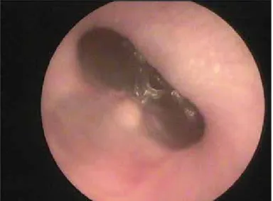
Acute TMPs were made in both ears of all animals using a cone-shaped needle in the posteroinferior part of the pars tensa (Figure 1). Plasma gel was prepared from the blood of each rabbit on the day of perforation and applied to the right TM of the same animal (Figure 2). The left TM was left untreated. On days 1, 4, 7, 10, 13, 16, 21, 28, and 35, the TMs were monitored to check the closure of the perforations. Plas-ma gel obtained from the same aniPlas-mal was applied to its right TM on every observation day until the perforations were closed, as observed using an endoscope. The left TM was left untreated and was observed using an endoscope on the same days until no perforation was ob-served. The results were expressed with respect to whether the perfo-rations were closed or not. Because the animals were not monitored daily, perforation closure observed on a particular observation day was assumed to have occurred at the middle of the time interval from the previous observation day. The days of perforation closure for 2 groups were compared using the paired t-test (SPSS, IBM, NYC, USA).
The animals were sacrificed 2 months after making the perforation; their skulls were cut into half and fixed using 10% formaldehyde solu-tion for approximately 1 week. Before decalcificasolu-tion, each bone was reduced until only the mastoid bone, external auditory canal, tympan-ic membrane, middle ear cavity, and petrous apex remained (Figure 3). The minimized specimens were decalcified using TBD-1 Rapid De-calcifier (Shandon, Thermo Fisher Scientific Inc. NYSE:TMO, Waltham, MA, USA) solution for approximately 1 week. After decalcification, the tympanic membranes were dissected out as a whole. On macroscopic examination, all tympanic membranes were intact. Specimens were washed under running tap water for half an hour. After routine tissue processing (Leica ASP300S, Wetzlar, Germany), the tissue samples were embedded in paraffin (Leica EG1150H, Wetzlar, Germany), cut into slic-es of 5-µm thicknslic-ess (Leica RM2245, Wetzlar, Germany), and stained using hematoxylin–eosin (Leica Autostainer XL, Wetzlar, Germany).
Two pathologists, blinded to the nature of the specimens, performed the histological analysis. Seven histopathological parameters were reviewed: acute inflammation, chronic inflammation, edema in the lamina propria, congestion in the lamina propria, sclerosis, fibroblastic reaction, and an increase in the thickness of squamous epithelial lay-er. Category of microscopic parameters were dichotomized as present (“1”) or absent (“0”) in statistical analyses. The presence or absence of each histological parameter in both groups was compared using the Pearson Chi–square test (SPSS, IBM.; NYC, USA).
Figure 1. Tympanic membrane perforations in the posteroinferior part of the
pars tensa.
RESULTS
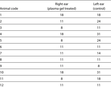
None of the ears showed reperforation or otorrhea; thus, we did not have to exclude any of the animals from the study. The number of days required for TM closure in each ear is provided in Table 1. The average number of days of closure for the plasma gel group was 12 (range 8–18 days) and that for the control group was 17.7 (range 8–31 days). The difference was statistically significant (p=0.0145).
Based on the findings of the histopathological examination, there was no sclerosis or fibroblastic reaction in any of the specimens. When the 2 groups were compared for other 5 parameters, no sta-tistically significant difference was observed with respect to acute inflammation, chronic inflammation, edema in the lamina propria, congestion in the lamina propria, and an increase in thickness of the squamous epithelial layer (p>0.05). Histological views of the healed TMs belonging to each group have been provided as Figures 4 (right ear) and 5 (left ear).
DISCUSSION
Although acute TMPs are reported to heal within 7–10 days [1], 10%– 20% of the patients have persistent TMPs that become chronic. The mechanism underlying the presence of persistent perforations is unknown. Numerous factors are considered to impede the sponta-neous healing mechanism of acute TMP. In their review, Wang et al. [4] classified the hypotheses into structural, histological, infectious, and growth-related mechanisms. In this context, the prevention of an acute TMP from becoming chronic is at least as important as try-ing to cure chronic TMPs to avoid the risks associated with general anesthesia, for cost effectiveness, and for limiting the time required for surgery.
Major criticisms regarding the use of acute TMP models in animal studies are as follows: TMPs mostly heal spontaneously and acute TMPs are not analogous to chronic TMPs, wherein acute repair mech-anisms are deactivated [5]. However, not all acute TMPs heal sponta-neously, and today, there are no specific symptoms for a clinician to predict as to which TMP will heal with no intervention and which will
Figure 3. Inside view of a half skull reduced until only the mastoid bone,
exter-nal auditory caexter-nal, tympanic membrane, middle ear cavity, and petrous apex remained.
Figure 4. Histopathological view of a right tympanic membrane stained
us-ing hematoxylin–eosin under ×400 magnification: the healed (thicker) part is marked with a rectangle.
Figure 5. Histopathological view of a left tympanic membrane stained using
hematoxylin–eosin under ×400 magnification: the healed (thicker) part is marked with a rectangle.
Table 1. Days required for the closure of tympanic membrane perforations
in each group
Right ear Left ear Animal code (plasma gel treated) (control)
1 18 18 2 11 24 3 8 11 4 18 31 5 8 24 6 11 11 7 11 14 8 11 11 9 11 8 10 18 31 11 8 18 12 11 11
not. Thus, the authors believe that there is still a need to explore bio-materials that will help acute TMPs heal faster.
In the last decade, there have been investigations on new materials such as gelfoam patch, EGF, PGF, TGFb1, PDGF, autocollagen, egg shell membrane, estrogen, and plasminogen in human and animal studies for both acute and chronic TMPs [2, 6-11]. Although the efficacy of PRP has been investigated in wound healing [12-15], its effects on TMP heal-ing have not yet been investigated in acute TMPs. In the literature, the only study on the effect of PRP on TMP closure is that reported by El-Anwar et al. [16], wherein they studied 64 patients with chronic TMPs who had undergone postauricular myringoplasty with underlay graft using conchal perichondrial graft. They reported that postoperatively at 6 months, the success rate (graft taking) in the case group with the use of autologous PRP (100%) was significantly higher than that in the control group without the use of autologous PRP (81.25%).
Platelet-rich plasma is a concentration of autologous platelets which release polypeptides, including PDGF, TGFb1, VEGF, and EGF, each of which have different roles in wound healing, immune reactions, an-giogenesis [12], and bone regeneration [17, 18]. There have been human studies to report that PRP has a positive effect on wound healing [12-15]. In animal studies, histological examinations revealed enhanced fibroblastic and endothelial cell formation during wound healing, in-creased neovascularization [12, 19-22], and enhanced granulation tissue formation [12, 19, 21, 22].
The role of various immunological molecules on TMP closure has been investigated. The effect of local TGFb1 application has been studied on acute TMP models in rats, and repeated application of TGFb1 seems to accelerate the healing process of TMPs [7]. Early im-mune reaction after acute TMP was reported in a study on rats [23], wherein T cells were demonstrated to peak on day 3 and B cells on day 6 after the perforation. It was concluded that products inducing the healing process secreted by these cells should be identified to promote the closure of TMPs. Because PRP contains many growth factors with different roles in the immune mechanism, our results to show statistically faster closure of acute TMPs with local PRP applica-tion, supporting the existing knowledge.
Regarding our histopathological examination, the reason for not ob-serving any significant difference in any of the parameters between the 2 groups could be the time at which we sacrificed the animals. It has been shown in the literature that healing process in TMP is com-plete by day 14 in rat models [24], and most of the histological studies on TMPs in animal models in the literature are limited to a 14-day fol-low-up [24, 25]. Shen et al. [2] showed that in 9 days, a single local injec-tion of plasminogen resulted in TMP closure with a continuous but rather thick outer keratinocyte layer; however, three plasminogen in-jections led to a completely healed TM with a thin keratinizing squa-mous epithelium covering a connective tissue layer. We observed that in long term (2 months), there is no significant difference in scar indicators such as the thickness of TM and fibrosis between control and experimental groups.
Thus, PRP fastens TMP closure in ways we do not exactly know yet; however, in long term, eventual outcomes are both microscopically and macroscopically same in the study and control groups using a
rabbit traumatic TMP model. Clinical studies are needed to evaluate if PRP increases the closure rate of acute TMPs with various etiologies under variable underlying conditions.
In the present study, we demonstrated that PRP is an effective autol-ogous material for the healing process of acute TMPs in a rabbit mod-el. In particular, in acute TMPs such as trauma, local PRP application on TMP may be a rapid, easy, cheap, noninvasive, and safe procedure that can be repeatedly applied in outpatient settings. We believe that this study will encourage the clinical use of PRP in acute TMPs and trigger clinical studies in this field.
Ethics Committee Approval: Ethics committee approval was received for this
study from the ethics committee of Ankara University Ethical Committee for Animal Studies (2015-2-24).
Informed Consent: N/A.
Peer-review: Externally peer-reviewed.
Author Contributions: Concept - S.K.Ö.; Design - S.K.Ö., M.E.T., F.T.;
Supervi-sion - S.K.Ö., S.K.; Resources - S.K.Ö., S.K., F.T., M.E.T.; Materials - H.D., E.A., S.K.Ö.; Data Collection and/or Processing - H.D., E.A., S.K.Ö.; Analysis and/or Interpre-tation - H.D., E.A., S.K.Ö.; Literature Search - S.K.Ö., E.A., M.E.T., H.D.; Writing Manuscript - S.K.Ö., E.A.; Critical Review - S.K.Ö., S.K.; Other - S.K.
Conflict of Interest: No conflict of interest was declared by the authors. Financial Disclosure: The authors declared that this study has received no
financial support.
REFERENCES
1. Teh BM, Marano RJ, Shen Y, Friedland PL, Dilley RJ, Atlas MD. Tissue en-gineering of the tympanic membrane. Tissue Eng Part B Rev 2013; 19: 116-32. [CrossRef]
2. Shen Y, Guo Y, Wilczynska M, Li J, Hellström S, Ny T. Plasminogen initiates and potentiates the healing of acute and chronic tympanic membrane perforations in mice. J Transl Med 2014; 12: 5. [CrossRef]
3. Lindeman P, Edström S, Granström G, Jacobsson S, von Sydow C, Westin T, et al. Acute traumatic tympanic membrane perforations. Cover or ob-serve? Arch Otolaryngol Head Neck Surg 1987; 113: 1285-7. [CrossRef]
4. Wang AY, Shen Y, Wang JT, Friedland PL, Atlas MD, Dilley RJ. Animal mod-els of chronic tympanic membrane perforation: a ‘time-out’ to review evidence and standardize design. Int J Pediatr Otorhinolaryngol 2014; 78: 2048-55. [CrossRef]
5. Truy E, Disant F, Morgon A. Chronic tympanic membrane perforation: an animal model. Am J Otol 1995; 16: 222-5.
6. Lou ZC, Tang YM, Chen HY, Xiao J. The perforation margin phenotypes and clinical outcome of traumatic tympanic membrane perforation with a Gelfoam patch: our experience from a retrospective study of seven-ty-four patients. Clin Otolaryngol 2015; 40: 389-92. [CrossRef]
7. Kaftan H, Herzog M, Miehe B, Hosemann W. Topical application of trans-forming growth factor-beta1 in acute traumatic tympanic membrane perforations: an experimental study in rats. Wound Repair Regen 2006; 14: 453-6. [CrossRef]
8. Lou Z, Yang J, Tang Y, Fu Y. Topical application of epidermal growth factor with no scaffold material on the healing of human traumatic tympanic membrane perforations. Clin Otolaryngol 2016; doi: 10.1111/coa.12627. [Epub ahead of print] [CrossRef]
9. Röösli C, von Büren T, Gassmann NB, Huber AM. The impact of plate-let-derived growth factor on closure of chronic tympanic membrane perforations: a randomized, double-blind, placebo-controlled study. Otol Neurotol 2011; 32: 1224-9. [CrossRef]
10. Jun HJ, Oh KH, Yoo J, Han WG, Chang J, Jung HH, et al. A new patch ma-terial for tympanic membrane perforation by trauma: the membrane of a hen egg shell. Acta Otolaryngol 2014; 134: 250-4. [CrossRef]
11. Barati B, Abtahi SH, Hashemi SM, Okhovat SA, Poorqasemian M, Tabrizi AG. The effect of topical estrogen on healing of chronic tympanic mem-brane perforations and hearing threshold. J Res Med Sci 2013; 18: 99-102.
12. Sommeling CE, Heyneman A, Hoeksema H, Verbelen J, Stillaert FB, Mon-strey S. The use of platelet-rich plasma in plastic surgery: a systematic review. J Plast Reconstr Aesthet Surg 2013; 66: 301-11. [CrossRef]
13. Kazakos K, Lyras DN, Verettas D, Tilkeridis K, Tryfonidis M. The use of au-tologous PRP gel as an aid in the management of acute trauma wounds. Injury 2009; 40: 801-5. [CrossRef]
14. Saad Setta H, Elshahat A, Elsherbiny K, Massoud K, Safe I. Platelet-rich plasma versus platelet-poor plasma in the management of chronic di-abetic foot ulcers: a comparative study. Int Wound J 2011; 8: 307-12.
[CrossRef]
15. Knighton DR, Ciresi K, Fiegel VD, Schumerth S, Butler E, Cerra F. Stimula-tion of repair in chronic, nonhealing, cutaneous ulcers using platelet-de-rived wound healing formula. Surg Gynecol Obstet 1990; 170: 56-60. 16. El-Anwar MW, El-Ahl MA, Zidan AA, Yacoup MA. Topical use of
autolo-gous platelet rich plasma in myringoplasty. Auris Nasus Larynx 2015; 42: 365-8. [CrossRef]
17. Chang SH, Hsu YM, Wang YJ, Tsao YP, Tung KY, Wang TY. Fabrication of pre-determined shape of bone segment with collagen-hydroxyapatite scaffold and autogenous platelet-rich plasma. J Mater Sci Mater Med 2009; 20: 23-31. [CrossRef]
18. Marx RE, Carlson ER, Eichstaedt RM, Schimmele SR, Strauss JE, Georgeff KR. Platelet-rich plasma: Growth factor enhancement for bone grafts.Oral Surg Oral Med Oral Pathol Oral Radiol Endod 1998; 85: 638-46. [CrossRef]
19. Pietramaggiori G, Kaipainen A, Czeczuga JM, Wagner CT, Orgill DP. Freeze-dried platelet-rich plasma shows beneficial healing properties in chronic wounds. Wound Repair Regen 2006; 14: 573-80. [CrossRef]
20. Pietramaggiori G, Scherer SS, Mathews JC, Alperovich M, Yang HJ, Neu-walder J, et al. Healing modulation induced by freeze-dried platelet-rich plasma and micronized allogenic dermis in a diabetic wound model. Wound Repair Regen 2008; 16: 218-25. [CrossRef]
21. Pietramaggiori G, Scherer SS, Mathews JC, Gennaoui T, Lancerotto L, Ragno G, et al. Quiescent platelets stimulate angiogenesis and diabetic wound repair. J Surg Res 2010; 160: 169-77. [CrossRef]
22. Takikawa M, Nakamura S, Nakamura S, Nambu M, Ishihara M, Fujita M, et al. Enhancement of vascularization and granulation tissue formation by growth factors in human platelet-rich plasma-containing fragmin/ protamine microparticles. J Biomed Mater Res B Appl Biomater 2011; 97: 373-80. [CrossRef]
23. Tahar Aissa J, Hultcrantz M. Acute tympanic membrane perforations and the early immunological response in rats. Acta Otolaryngol 2009; 129: 1192-7. [CrossRef]
24. Araújo MM, Murashima AA, Alves VM, Jamur MC, Hyppolito MA. Sponta-neous healing of the tympanic membrane after traumatic perforation in rats. Braz J Otorhinolaryngol 2014; 80: 330-8. [CrossRef]
25. Santa Maria PL, Redmond SL, Atlas MD, Ghassemifar R. Histology of the healing tympanic membrane following perforation in rats. Laryngo-scope 2010; 120: 2061-70. [CrossRef]