26
Bir çocukta serebellumun disembriyoplastik nöroepiteliyal
tümörü
Dysembryoplastic neuroepithelial tumor of cerebellum in a
child
Doğan Köse1, Erdal Kalkan2, Yahya Paksoy3, Hatice Toy4, Nurcan Üçüncü Ergün5, Yavuz Köksal1
1
Selçuk Üniversitesi Tıp Fakültesi, Pediatri Ana Bilim Dalı, Konya
2
Necmettin Erbakan Üniversitesi Tıp Fakültesi, Beyin Cerrahisi Ana Bilim Dalı, Konya
3
Medipol Üniversitesi Tıp Fakültesi, Radyoloji Ana Bilim Dalı, İstanbul
4
Necmettin Erbakan Üniversitesi Tıp Fakültesi, Patoloji Ana Bilim Dalı, Konya
5
İzmit Kadın Hastalıkları Ve Çocuk Hastanesi, Pediatri Bölümü, İzmit
Olgu sunumu/Case report
Özet
Disembriyoplastik nöroepitelyial tümörler (DNT) tipik olarak supratentorial yerleşen benign neoplazmalardır.
Bilinç ve postural tonus kaybı şikayetleri ile bir lokal hastaneye başvuran 12 yaşındaki erkek hasta saptanan serebellar kitlesi nedeniyle hastanemize refere edildi. Hikayesinden benzer şikayetlerinin 3 ve 10 yaşlarında da tekrar ettiği ancak o dönemde pediatric kardiyolog ve pediatric nörolog tarafından yapılan incelemelerde herhangi bir anomaliye
rastlanmadığı öğrenildi. Hastanın fizik muayenesi ve rutin laboratuvar tetkikleri normaldi.
Serebellumda saptanan kitle total olarak çıkarıldı. Alınan dokunun histopatolojik değerlendirmesi ile hastaya DNT tanısı konuldu. Hasta halen, 5 yıldır, herhangi bir nüks belirtisi olmadan ayaktan takip edilmektedir.
Bu yazıda serebellum yerleşimli DNT’ye sahip bir çocuk vakanın literatürle paylaşılması
amaçlanmıştır.
Anahtar Kelimeler: beyin neoplazmları, selim çocuk, serebellum
Türkçe kısa makale başlığı: Serebellumun disembriyoplastik nöroepiteliyal tümörü
Abstract
Dysembryoplastic neuroepithelial tumors are benign neoplasms with typical supratentorial location. 12 year old patient, who applied to a local hospital with complaints of loss of consciousness and postural tonus, was referred to our hospital with cerebellar mass diagnosis. According to the anamnesis, similar complaints also appeared when the patient was 3 and 10 years old, however no anomaly was found in the examinations done by pediatric cardiologist and pediatric neurologist in that period. Physical
examination and routine laboratory examinations of the patient were normal. The mass identified in the cerebellum was totally removed. The patient was diagnosed as dysembryoplastic neuroepithelial tumor by histopathological evaluation of the tissue. The patient has still been under outpatient follow-up and in complete remission without any evidence of recurrence for 5 years.
In this report a child patient having dysembryoplastic neuroepithelial tumor with cerebellar location is aimed to be shared with the literature.
Key words: brain neoplasms, benign, child, cerebellum
İngilizce kısa makale başlığı: Dysembryoplastic neuroepithelial tumor of cerebellum
İletişim (Correspondence):
Dr. Doğan Köse / Selçuk Üniversitesi Tıp Fakültesi, Pediatri Ana Bilim Dalı, Konya Tel: 03322244512/ E-mail: [email protected]
27
Introduction
Dysembryoplastic neuroepithelial tumor (DNT) an infrequently seen condition and a benign neoplasm, was first defined by Daumas-Duport et. al. in 1988 (1).
The disease, whose incidence was determined as 1.2% for below 20 year old, 0.24% for above 20 years old (2), affects male patients more than female patients (3). Familial types are also reported for DNTs and approximately 30% of them are related to neighbor cortical dysplastic changes (4). These changes lead to the thought of a possible developmental defect occurring in second trimester of utero in the etiology (3).
Tumors typically have a supratentorial location (1). Multifocal (midbrain, diencephalon, subependymal regions with bilateral mesial temporal lobe occurence) (6) and extracortical located (in the anterior horn of the lateral ventricle) (5) types of these tumors, which essentially locate in cortical or deep grey matter as a single lesion, are also reported. In this document, we aim to share with the literature a child patient with cerebellum retention, which is an infrequent region for location of DNT.
Case report
12 year old patient, who applied to a local hospital with complaints of loss of consciousness and postural tonus, was referred to our hospital with cerebellar mass diagnosis. According to the anamnesis, similar complaints also appeared when the patient was 3 and 10 years old, however no anomaly was found in the examinations done by pediatric cardiologist and pediatric neurologist in that period. The physical examination was normal. Complete blood count and biochemistry analysis were within normal limits. In the magnetic resonance imaging (MRI), a mass with lobule contour having an approximate size of 1,5x1x0,9 cm, showing hypo-intense surrounding on T1 series, mild hyper-intense on T2 series, isointense at center was seen in the left cerebellar hemisphere. There was no edema region around the mass and the lesion was restricted by the cortical grey substance. It had a multicystic (Bubbly) appearance (Figure 1a, b). Contrast enhanced T1 weighted image showed superficial band-like contrast enhacement (Figure 1c). The mass was removed totally. In the histopathologic examination of the removed tissue, a tumoral
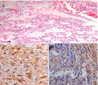
structure composed of oval/spherical cells having normo/hyperchromatic seed and pink/transparent cytoplasm in the microcystic level was seen. This structure was including nodules consisted of neuroglial elements and was being painted strongly positive with synaptophysin, and negative with glial fibrillary acidic protein (GFAP) immunohistochemically (Figure 2). Pathology combined with immunohistochemistry findings was diagnostic of DNT (grade I). The patient has still been under outpatient follow-up and in complete remission without any evidence of recurrence for 5 years.
Fig. 1a, b: T2 weighted oblique MRI images show a mass in left cerebellar hemisphere causing remodeling in the inner calvarial table. The lesion is only limited to cortical gray matter. There was no white matter involvement. It shows multicystic (Bubbly) appearance. c: Contrast enhanced T1 weighted image shows superficial band-like contrast enhancement
Discussion
Nearly 40% of these tumors are cystic, however they can also be in nodular, multi-nodular and diffuse forms. These neoplasms, which are considered in grade I mixed neoroglial tumor group according to classification of World Health Organization for brain tumors, are caused by glial and neuronal elements (oligodendrocyte-like cells) similar to
Köse ve ark. Kocaeli Tıp Dergisi 2014; 3:26-30 Dysembryoplastic neuroepithelial tumor of cerebellum Medical Journal of Kocaeli 2014; 3:26-30
28 Fig. 2a: Microcystic background of the tumour
(Hematoxylin-eosin, original magnification, X200) b: Oligodendroglial-like
cells showing strongly positivity for synaptophysin
(Synaptophysin, original magnification, X100) c:
Oligodendroglial-like cells showing negativity for GFAP (GFAP, original magnification, X200)
gliomas. It might not be possible to
immunohistochemically eliminate
oligodendrogliomas with light microscope. Presence of a myxoid matrix is another interesting characteristic (4). In the histopathologic examination of our case, a tumoral structure composed of oval/spherical cells having normo/hyperchromatic seed and pink/transparent cytoplasm in the microcystic level was seen. This structure was including nodules consisted of neuroglial elements and was being painted strongly positive with synaptophysin, and negative with GFAP immunohistochemically.
Tumors are most frequently seen in mesial or lateral temporal lobe (62%) and frontal lobe (31%), however there are also tumors which were reported to locate in parietal (3), occipital (5,7), cerebellar (7,8,9-13), insular cortex, brain stem and striatum (5). Locating in posterior fossa is very rare and a cerebellar tumor was reported with DNT diagnosis for the first time in 1994 (8). In our case, tumor was located in the left cerebellar cortex and according to our knowledge this case is the second child case out of total eight DNT cases reported to locate in cerebellar (Table 1). These cases are thought to represent neurocytic neoplasms caused by cerebellum external granular layer (1). These tumors grow very slowly and remain stable for years, even Daumas-Duport et. al. showed that
the disease remained stable for 4.5 years on average before the operation by serial imagings (14). Clinically, the patients typically apply for complex partial attacks (3). These attacks generally resistant to medication and secondary generalization can be seen. Even a partial resection is applied, activities of the attacks generally cease after the operation (1), however residual tumor is a risk factor for recurrence of the attacks. If complex congenital anomalies are not accompanying, unlike to other brain tumors, neurological deficit is not common (3). Infrequently, neurological deterioration can occur with bleeding (4). The patients, whose tumors locate in cerebellum, are young adults and they can have minor symptoms such as headache, vertigo or tinnitus without development of any attack for several years (15). For our case, an attack activity, which was defined as the loss of conscious and postural lotus, occurred. However, it was learned from the anamnesis that the same incident had recurred two times previously (on 3 and 10 year-old) and the examination results had been normal during that period. The fact that those attacks occurred even though there was no tumoral structure that could be identified and there were no attacks in the cases reported previously made us think that the attacks could be due to a dysplasia accompanying to the tumor rather than the tumor itself.
The imaging studies show a mass with solid and cortical location and having a cystic component. Calcification is not common. Multiple “pseudocyctic” regions with different sizes, which were seen as hyper-intense on T2 weighted images and hypo-intense on T1 weighted images, were identified (16). These structures can be surrounded by a high signal separating them from the glioneuronal elements in the FLAIR cross-sections. Peritumoral edema, middle line shift or contrast retention are not typical (17), however there are cases reporting contrast retention (12,13). Erosion which was seen in the adjacent bone structures supported this diagnosis (15). In our case, compatible with the literature, the mass was seen with hypo
Köse ve ark. Kocaeli Tıp Dergisi 2014; 3:26-30 Dysembryoplastic neuroepithelial tumor of cerebellum Medical Journal of Kocaeli 2014; 3:26-30
29 Table 1: Demographical features of previously published cases
which settled in the cerebellum
surrounding and isointense center on the T1 series and the mass was seen mildly hyper-intense on T2 series. There was no edema around it and it had a multicystic appearance. Even it was not typical, a contrast retention having a superficial band-like form was diagnosed similar to the previously reported three cases (7,12,13).
Complete surgical removal is generally preferred in the treatment, since this approach controls the attacks without using anticonvulsants and prevents the malign transformation of tumors (18). Since a normal life is expected for these patients there is no need for chemotherapy or radiotherapy after the operation. Recurrence and malign transformation is very infrequent. Most of the recurrent tumors were shown to occur in the patients who were applied subtotal or partial recession at the beginning (19) Transformation to astrocytomas with a high degree can be due to inappropriate post-operative radiotherapy. Today, there is no treatment guide for recurrent tumors, however, generally surgical recession is applied. A
patient, who showed transformation to astrocytomas (grade III) and recurrence, was applied temozolomide and the patient was reported to show a very good post-operative prognosis (20). In our case, the tumor removed totally and the patient has been leading his life without any recurrence or symptom on the fifth year of the follow-up.
In conclusion, cerebellum is one of the infrequently seen location region for DNTs. However, since, DNT with cerebellar location is a relatively newly defined entity, retrospective screening of the previous cases may change this frequency. In addition to this, long-term follow-up for these cases is appropriate in terms of recurrence and malign transformation.
References
1.Daumas-Duport C, Scheithauer BW, Chodkiewicz JP, et al. Dysembryoplastic neuroepithelial tumor: a surgically curable tumor of young patients with intractable partial seizures. Report of thirty-nine cases. Neurosurgery 1988; 23:545-56.
2.Rosemberg S, Vieira GS. Dysembryoplastic neuroepithelial tumor. An epidemiological study from a single institution. Arq Neuropsiquiatr 1998; 56:232-6.
3.Daumas-Duport C, Pietsch T, Lantos PL. Dysembryoplastic neuroepithelial tumour. In: Kleihues P, Cavenee WK eds. Pathology and genetics of tumours of the nervous system. 2nd ed. Lyon: IARC, 2000; 103-6.
4.Thom M, Gomez-Anson B, Revesz T, et al. Spontaneous intralesional haemorrhage in dysembryoplastic neuroepithelial tumours: a series of five cases. J Neurol Neurosurg Psychiatry 1999; 67:97-101.
5.Onguru O, Deveci S, Sirin S, et al. Dysembryoplastic neuroepithelial tumor in the left lateral ventricle. Minim Invasive Neurosurg 2003; 46:306-9.
6.Whittle IR, Dow GR, Lammie GA, et al. Dsyembryoplastic neuroepithelial tumour with discrete bilateral multifocality: further evidence for a germinal origin. Br J Neurosurg 1999; 13:508-11. 7.Kuchelmeister K, Demirel T, Schlörer E, et al. Dysembryoplastic neuroepithelial tumour of the cerebellum. Acta Neuropathol 1995; 89:385-90.
Köse ve ark. Kocaeli Tıp Dergisi 2014; 3:26-30 Dysembryoplastic neuroepithelial tumor of cerebellum Medical Journal of Kocaeli 2014; 3:26-30
30 8.Leung SY, Gwi E, Ng HK, et al. Dysembryoplastic
neuroepithelial tumor. A tumor with small neuronal cells resembling oligodendroglioma. Am J Surg Pathol 1994; 18:604-14.
9.Yasha TC, Mohanty A, Radhesh S, et al. Infratentorial dysembryoplastic neuroepithelial tumor associated with Arnold-Chiari malformation. Clin Neuropathol 1998; 17:305-10.
10.Fujimoto K, Ohnishi H, Tsujimoto M, et al. Dysembryoplastic neuroepithelial tumor of the cerebellum and brainstem. Case report. J Neurosurg 2000; 93:487-9.
11.Litrico S, Desjardins T, Dran G, et al. Infratentorial localisation of a dysembryoplastic neuroepithelial tumor. A case report. Neurchirurgie 2004; 50:47-52.
12.Tailor JK, Kim AH, Folkerth RD, et al. The development of ring-shaped contrast enhancement in a case of cerebellar dysembryoplastic neuroepithelial tumor: case report. Neurosurgery 2008; 63:609-10.
13.Vaquero J, Saldaña C, Coca S, et al. Complex form variant of dysembryoplastic neuroepithelial tumor of the cerebellum. Case Rep Pathol, 2012: 718651. doi: 10.1155/2012/718651.
14.O'Brien DF, Farrell M, Delanty N, et al. The Children's Cancer and Leukaemia Group guidelines for the diagnosis and management of
dysembryoplastic neuroepithelial tumours. Br J Neurosurg 2007; 21:539-49.
15.Sarrazin JL, Hélie O, Lévêque C, et al. Tumeurs de la fosse cérébrale postérieure de l’adulte. Encycl Med Chir, Radiodiagnostic-Neuroradiologie-Appareil Locomoteur 1999; 10:16.
16.Campos AR, Clusmann H, von Lehe M, et al. Simple and complex dysembryoplastic neuroepithelial tumors (DNT) variants: clinical profile, MRI, and histopathology. Neuroradiology 2009; 51:433-43.
17.Yu AH, Chen L, Li YJ, et al. Dysembryoplastic neuroepithelial tumors: magnetic resonance imaging and magnetic resonance spectroscopy evaluation. Chin Med J 2009; 122:2433-7.
18.Chan CH, Bittar RG, Davis GA, et al. Long-term seizure outcome following surgery for dysembryoplastic neuroepithelial tumor. J Neurosurg 2006; 104:62-9.
19.Schittenhelm J, Mittelbronn M, Wolff M, et al. Multifocal dysembryoplastic neuroepithelial tumor with signs of atypia after regrowth. Neuropathology 2007; 27:383-9.
20.Ray WZ, Blackburn SL, Casavilca-Zambrano S, et al. Clinicopathologic features of recurrent dysembryoplastic neuroepithelial tumor and rare malignant transformation: a report of 5 cases and review of the literature. J Neurooncol 2009; 94:283-92.
Köse ve ark. Kocaeli Tıp Dergisi 2014; 3:26-30 Dysembryoplastic neuroepithelial tumor of cerebellum Medical Journal of Kocaeli 2014; 3:26-30