Abstract. –OBJECTIVE:Disulfiram (DSF) ex-erts its therapeutic effects through oxidative, proteasome, and nuclear factor kappa beta
(NF-κκB) pathways. The study was planned to test the
impact of DSF on growing of endometriotic im-plants in rats with experimentally induced en-dometriosis.
PATIENTS AND METHODS: Thirty rats were labeled as the control (n = 8), sham (n = 6), GnRH-agonist (n = 8) and the DSF (n = 8) groups. The rats in the group 3 exposed to sin-gle dose leuprolide acetate. The rats in group 4 were treated with DSF for 21 days. The serum activity of oxidant and antioxidant markers, total oxidant status (TOS), total antioxidant status (TAS), interleukin-1ββ, and tumor necrosis
factor-αα (TNF-factor-αα) were determined. Implants were
processed for NF-κκB, PCNA, and CD34 im-munostaining.
RESULTS:The serum concentration of malon-dialdehyde in the DSF group was significantly higher than those in other groups. The concen-tration of TAS, TNF-αα, and interleukin-1ββ in the DSF group considerably decreased compared to control group. Following treatment with DSF while the percentage of Grade 1 and 2 implants increased the percentage of Grade 3 and 4 im-plants decreased. The imim-plants disappeared to-tally in two cases in the DSF group and one case in the GnRH-agonist group. The mean H-Scores of implant NF-κκB and PCNA in DSF treated ani-mals were found to significantly lower than those of the control group.
CONCLUSIONS:By decreasing NF-κκB expres-sion, angiogenesis, and cell proliferation DSF prevents the growth of endometriotic implants.
Key Words:
Endometriosis, Disulfiram, NF-κB, Oxidative stress.
Disulfiram, as a candidate NF-
κκB and
proteasome inhibitor, prevents endometriotic
implant growing in a rat model of endometriosis
O. CELIK
1, A. ERSAHIN
2, M. ACET
3, N. CELIK
4, Y. BAYKUS
5,
R. DENIZ
5, E. OZEROL
6, I. OZEROL
71Private Clinic Obstetrics and Gynecology, Usak, Turkey
2Department of Obstetrics and Gynecology, Bahcesehir University, School of Medicine, Istanbul, Turkey 3Department of Obstetrics and Gynecology, Istanbul Medipol University Hospital, Istanbul, Turkey 4Department of Biochemistry, Behçet Uz Children’s Hospital, Izmir, Turkey
5Department of Obstetrics and Gynecology, Kafkas University Faculty of Medicine, Kars, Turkey 6Department of Medical Biochemistry, Medical Faculty, Inonu University, Malatya, Turkey 7Department of Medical Microbiology, Medical Faculty, Inonu University, Malatya, Turkey
Introduction
Endometriosis is a chronic inflammatory dis-ease affecting 30-50% of infertile women and women with chronic pelvic pain during repro-ductive period1,2. Adhesion, invasion, and prolif-eration of the endometrial cells shedding to peri-toneal cavity through retrograde menstruation is well-known hypothesis regarding the occurrence of peritoneal endometriosis3. The primary aim of the current medical treatment with hormonal preparations is to inhibit implant growth and pelvic pain, and also improve fertility chance and quality of life1-3. Nevertheless, long-term treat-ment is usually needed due to the high recurrence rate after discontinuation of the medical treat-ment. Nevertheless, high side effects occurence of both steroids and GnRH agonist treatment do not allow long-term management of subjects suf-fering endometriosis. Therefore, new and more effective medical agents that allows long-term treatment with minimal side effect are needed.
Different treatment agents targeting possible pathways underlying the formation of endometri-otic implants were used in many experimental studies. Tumor necrosis factor α-blockers, NF-κB inhibitors, statins, immunomodulators, apop-totic agents, antiangiogenic molecules, antioxi-dants, anti-inflammatory drugs, flavonoids, his-tone deacetylase inhibitors are some of them4). Due to lack of clear evidence regarding their impacts on endometriosis treatment the use of all these agents in human endometriosis is of concern.
The dithiocarbamate (DC) drug, disulfiram (DSF) is a NF-κB inhibitor5,6). It can affect both oxidative and proteasome pathways in healthy and pathological tissues7-9. Conventional effect of DSF on proteasome pathway is inhibition7-9. NF-κB activity has been demonstrated in eutopic and ectopic endometrial tissues10,11. NF-κB as-sists the transcription of a variety of immune and inflammatory molecules that involved in the de-velopment of endometriotic implants10,11. NF-κB also contributes the regulation of cell prolifera-tion, apoptotic events, adhesion, invasion, and angiogenetic processes in many cell types12,13. Concordantly, DSF has been shown to reduce an-giogenesis14,15 and induce apoptotic cell death in healthy and cancerous tissues16.
The pathophysiological pathways involved in the development of the endometriosis also oper-ate in normal tissues. Hence, there are many concerns about the possible side effects of the current medical agents that targeting these path-ways in human endometriosis. Although DSF has not been used for the treatment of en-dometriosis in animals and humans it has cur-rently been used for the treatment of bacterial and fungal infections and to break off alcohol abuse in humans with relatively mild side effect8,17. Therefore, DSF is a valuable molecule for investigating its effects in the endometriosis treatment. In a previous study18, the positive ef-fect of the first proteasome-inhibitor PS-341 and DC on endometriotic implants were demon-strated by our team. This one encouraged us to use of DSF in the treatment of endometriosis. In the current study, we planned to test the effects of DSF as a candidate NF-κB and proteasome inhibitor on endometriotic implants of rats with experimentally induced endometriosis. Since DSF is a lipophilic and pro-oxidative drug the balance between oxidant and antioxidant sys-tems during implant development was taken in-to account.
Materials and Methods
Experimental AnimalsThis study was carried out in the Experimental Research Laboratory of the Inonu University Faculty of Medicine, complying with the ap-proval of the ethic committee, the guidelines for care and use of experimental animals. Thirty-five adult female Wistar rats each weighing between 250 and 300 g were included. Daily vaginal
smears of the rats were taken to establish the es-trous cycle of each animal. Rats observed for at least two successive 4-day estrous cycles. Methods
Endometriosis was induced surgically by us-ing the method described by previously by our team and others18,19. Detailed information could be find elsewhere18. Briefly, A 0.5 × 0.5 × 0.1 cm piece excised by micro scissors from the uterine horn was attached on the right side peritoneal wall close to an artery. This surgical method had been defined as auto transplantation technique. The second laparotomy was performed after 3 weeks in estrous phase to determine the attach-ment and viability of implants. The vesicle-like appearance at the sutured peritoneum was evalu-ated and animals were graded according to aver-age vesicle diameter (D) as: Grade 1 (for cases in which the implant had disappeared or, if it was visible, never became a cyst), Grade 2 (D < 2 mm), Grade 3 (2 mm < D > 4.5 mm) or Grade 4 (D > 4.5 mm). Five out of 35 experimental rats were not developed any signs of vesicles and therefore these animals were excluded. The re-maining 30 rats were put into four groups. The groups were labeled as control (n = 8), sham con-trol (n = 6), GnRH-agonist (GnRH-a; n = 8), and the DSF (n = 8) groups. For the sham group, the only suture was attached to the peritoneum. The rats in group 4 were treated with DSF (100 mg/kg body weight per rat, intraperitoneally) for 21 days. The rats in group 3 were exposed to subcutaneous single dose leuprolide acetate de-pot formulation (1 mg/kg body weight per rat, Lucrin; Abbott, Cedex, Istanbul, Turkey). This dose was determined based on a previous study in which 1 mg/kg leuprolide acetate was found to optimal for female rats20). Since the DSF was di-luted with an isotonic saline solution containing 1% carboxymethylcellulose, an identical amount of this solution was used in control group (0.1 ml/day per rat, ip) for 21 days. Due to DSF ex-hibited similar absorption rates when it was giv-en orally or intraperitoneally in the currgiv-ent study peritoneal administration was chosen. Following administration DSF rapidly metabolizes to the di-ethyldithiocarbamate-glucuronide and inorganic sulfate. Previously, the micromolar concentration of DSF was shown to inhibit the proteasome in the human embryo kidney cell line9. Likewise, NF-κB inhibiting effect of DSF analogue PDTC occurs with the dose of 100 mg/kg in rats. There-fore, in the DSF group we used minimum 100
mg/kg (0.3 µmol/kg) DSF in order to inhibit im-plant NF-κB pathway. These doses were also cho-sen because a recent study indicates that micromo-lar concentrations (0.16 µmol/L) of DSF inhibited proteasome activity in cell-based screening assay9. It has not been stated in any comment by the man-ufacturer whether DSF has any impact on the es-trous cycle we accepted the DSF does not alter cy-cle characteristic of rats. Following the confirma-tion of estrous a third laparotomy was performed. The sizes of the implants were measured again with the same method by the same researchers who were blinded to the groups. Due to the small size of the endometriotic implants blood samples were used for measuring oxidative stress markers and cytokines. The endometriotic implants were excised and processed for immunohistochemical studies.
Biochemical Analysis
All biochemical determinations were per-formed on serum obtained after centrifugation using spectrophotometric methods. Samples were stored at -80°C until assay. The biochemist was blinded to the blood samples.
SOD and GSH-Px Measurement
Total SOD activity was determined according to the method of Sun et al21. GSH-Px activity was measured by the method of Paglia and Valentine22. The activity of SOD and GSH-Px was expressed as U/L.
GSH and PON1 Measurement
Reduced glutathione (GSH) content was deter-mined according to Ellman23. The results were expressed as µmol/L. Paraoxonase-1 (PON1) ac-tivity was measured using the commercially available kit (Relassay, Rel Assay Diagnostics, Gaziantep, Turkey) and results expressed as U/L. ADA and XO Measurement
Adenosine deaminase (ADA) activity was es-timated spectrophotometrically by the method of Giuisti24. Results were expressed as units per liter (U/L). Xanthine oxide (XO) activity was mea-sured spectrophotometrically by the formation of uric acid from xanthine. Results were expressed in units per liter plasma (U/L).
MPO and MDA Measurement
Myeloperoxidase (MPO) activity was deter-mined using a 4-aminoantipyrine/phenol solution as the substrate for MPO-mediated oxidation by
H2O2, and changes in absorbance at 510 nm were recorded25. Data were presented as U/L. Malon-dialdehyde (MDA) level was determined by the method of Esterbauer and Cheeseman26which is based on the reaction with thiobarbituric acid at 90-100°C. The results were expressed according to a standard graphic, which was prepared from a standard solution.
PC Measurement
The protein carbonyl (PC) content was deter-mined spectrophotometrically by the method based on the reaction of the carbonyl group with 2,4-dinitropheniylhydrazine to form 2,4-dinitro-phenylhydrazone27. Results were expressed as µmol/dL.
IL-1ββ and TNF-αα Measurement
Interleukin-1 beta (IL-1β) levels were deter-mined with an enzyme-linked immunosorbent assay (Rat IL-1β Platinum ELISA – Catalog Number BMS630, eBioscience, Inc. Vienna, Austria); which could measure IL-1β in serum with a detection limit is 4 pg/ml. The intra- and interassay coefficients of variation (CV) were < 10% and < 10%, respectively. The rat TNF-α ELISA analysis on the serum was carried out in duplicate using commercially available ELISA kit (Rat TNF-α Platinum ELISA, BMS622, eBioscience, Vienna, Austria). The intra and in-terassay CV were < 5% and < 10% respectively. The TNF-α concentration was measured from the absorbance of each well was read at 450 nm using with an autoanalyser (Alisei system, SEAC Radim Group, Caserta, Italy). The results were presented as ng/ml.
Total Antioxidant Status Assay (TAS) Total antioxidant assay (TAS) was determined with an enzyme-linked immunosorbent assay (Antioxidant Assay Kit ELISA – Item No. 709001, Cayman Chemical Co. Ann Arbor, MI, USA) which could measure the total antioxidant capacity of cell lysates. The intra- and interassay CV were < 3.4% and < 3% respectively.
Total Oxidant Status Assay (TOS)
The total oxidant status (TOS) was deter-mined using a novel automated measurement method as described previously28. The assay is calibrated with hydrogen peroxide and the re-sults are expressed in terms of micromolar hy-drogen peroxide equivalents per liter (µmol H2O2Equiv/L).
Implant Immunohistochemistry
Formalin-fixed endometriotic implants were embedded in paraffin cut into 5 mm thick sec-tions and stained with hematoxylin and eosin. The sections were also stained for NF-κB/65 (Rel A) Ab-1, proliferating cell nuclear antigen (PCNA), and CD34 immunohistochemistry. NF-κκB/p65 (Rel A) Ab-1
Four micrometer paraffin sections were de-waxed in xylene, rehydrated in ethanol and then incubated for 10 min in 3% hydrogen peroxide to block endogenous peroxidase. After washing in phosphate buffer saline (PBS), the sections incu-bated 8 min at ultra V block. The immunoreac-tion was performed for 60 min with ready to use NF-κB/p65 Ab-1 antibody (NeoMarkers, Labvi-sion co., Fremont, CA, USA). After washing in PBS, slides were incubated with horseradish per-oxidase kit. The third trimester human placenta served as the positive control.
PCNA
Monoclonal anti-PCNA clone PC10 from mouse ascites fluid was diluted to 1/1000 and ap-plied to 5-mm paraffin sections deparaffinized in xylene using the labeled streptavidin–biotin method (Clone PC10; Sigma-Aldrich, St. Louis, MO, USA). Human tonsil tissue served as the positive control. Negative controls (primary anti-body omitted) were routinely performed on adja-cent serial sections.
CD-34
Endothelial cells were stained using mouse monoclonal antibody against CD34 antigen, a gly-coprotein expressed on the luminal surface of en-dothelial cells (ready to use, clone QBEnd/10; Novocastra, Newcastle, UK). Rat peritoneal vessel served as an internal positive control for CD34. Timm’s Staining for Copper
Timm’s copper stain is a silver technique where copper sulfide is converted to silver sul-fide which is then visualized as black deposits in the light microscopic sections29. Tissues from liver cirrhosis were used as positive control.
Histological slides were evaluated for NF-κB p65, PCNA, and CD34 immunoreactivity under light microscopy. The H-Score method was used to score the degree of histological change of stro-mal and glandular epithelial cells. This semi-quan-titative method consists of the percentages of posi-tively stained cells multiplied by a weighted
inten-sity of staining: H-Score = ∑Pi (i+1), where Pi is the percentage of stained cells in each intensity category (0–100%), and i is the intensity indicat-ing weak (i = 1), moderate (i = 2), or strong stain-ing (i = 3)30. Due to weak Timm’s staining for copper in treatment and control groups H-score method could not be implemented.
Statistical Analysis
Data distribution was tested using the Kol-mogorov-Smirnov test. Comparison among the groups was performed using the Kruskal-Wallis analysis of variance and post-hoc Mann-Whitney U tests for continuous variables. Data was pre-sented as mean and standard deviation (SD) for continuous variables. p < 0.05 was accepted as significant.
Results
Concentration of Oxidative Stress Markers, TAS and TOS
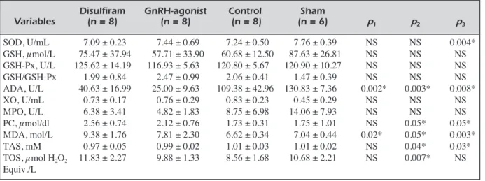
Insignificant difference in serum SOD activity was detected between the group treated with DSF, GnRH-agonist, and the control groups (p = 0.15 and p = 0.67 respectively). SOD activity in the DSF group was significantly different from than the sham group (p = 0.004). Despite high levels of reduced-GSH were detected in the DSF group this did not reach the statistical signifi-cance when compared to the GnRH, control, and sham groups (p = 0.16, p = 0.61, and p = 0.20 re-spectively). Likewise, the mean serum activity of GSH-Px in the DSF group was not different from the GnRH-a, control, and sham groups (p = 0.19, p = 0.61, and p = 0.75 respectively) (Table I). Additionally, the ratio of reduced GSH to GSH-Px was not different in the DSF group compared to other three groups (p = 0.37, p = 1, and p = 0.12 respectively). However, the concentration of serum ADA considerably decreased in the DSF group when compared to the control and sham groups (p = 0.032 and p = 0.008 respectively). Further, significant difference was found regard-ing serum ADA levels between DSF and the GnRH-a group (p = 0.002). Serum concentra-tions of XO were the same all groups.
The concentration of serum MDA, end product of lipid peroxidation, was significantly different in the DSF group than the control and sham control groups (p = 0.05 and p = 0.003, respectively). The concentration of serum MPO was not different among the groups. Further, the serum
concentra-tion of MDA was significantly higher in the DSF group than the GnRH-a group (p = 0.05). Despite the similar PC levels in the DSF and GnRH-a groups the mean serum PC levels of DSF group were significantly different than that of the control and sham animals (p < 0.05).
The concentration of TOS in the DSF group was significantly different from the control group (p = 0.007). The concentration of TAS in the DSF group was significantly lower when com-pared to the control and the sham groups (p = 0.04, p = 0.03 respectively).
TNF-αα and IL-1ββ Concentrations
The concentration of TNF-α considerably de-creased in the group treated with DSF when compared to the control group (p = 0.006). How-ever, there were no significant differences re-garding mean serum concentration of TNF-α be-tween the DSF and GnRH-a groups (p = 0.21 and p = 0.89 respectively).
The concentration of IL-1β in the DSF group was significantly lower than the control and the sham group (p = 0.007 and p = 0.02 respective-ly). However, in the DSF group, the concentra-tion of IL-1β was not different from the GnRH-a group (p = 0.66). The serum concentration of PON was the same all groups (Table II).
Grade of Endometriotic Implants
After the treatment with DSF and GnRH-a, while the percentage of Grade 1 and 2 vesicles increased the percentage of Grade-3 and -4 vesi-cles decreased. The endometrial foci disappeared totally in two of the cases in the DSF group and one case in the GnRH-a group.
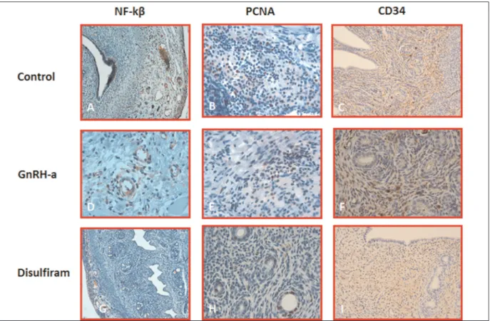
H-Score of NF-κκB, PCNA and CD34 Immunohistochemical analysis confirmed the ELISA findings in each group. The increased NF-κB p65 immunoreactivity was predominantly localized to the cytoplasm of luminal and glan-Disulfiram GnRH-agonist Control Sham
Variables (n = 8) (n = 8) (n = 8) (n = 6) p1 p2 p3 SOD, U/mL 7.09 ± 0.23 7.44 ± 0.69 7.24 ± 0.50 7.76 ± 0.39 NS NS 0.004* GSH, µmol/L 75.47 ± 37.94 57.71 ± 33.90 60.68 ± 12.50 87.63 ± 26.81 NS NS NS GSH-Px, U/L 125.62 ± 14.19 116.93 ± 5.63 120.80 ± 5.67 120.90 ± 10.27 NS NS NS GSH/GSH-Px 1.99 ± 0.84 2.47 ± 0.99 2.06 ± 0.41 1.47 ± 0.39 NS NS NS ADA, U/L 40.63 ± 16.99 25.00 ± 9.63 109.38 ± 42.96 130.83 ± 7.36 0.002* 0.003* 0.008* XO, U/mL 0.73 ± 0.17 0.76 ± 0.29 0.83 ± 0.23 0.45 ± 0.29 NS NS NS MPO, U/L 6.38 ± 3.41 4.82 ± 1.83 8.75 ± 6.98 14.06 ± 7.93 NS NS NS PC, µmol/dl 2.56 ± 0.74 2.12 ± 0.76 1.73 ± 0.31 1.75 ± 1.01 NS 0.05* 0.05* MDA, mol/L 9.38 ± 1.76 7.81 ± 2.30 6.62 ± 0.34 7.04 ± 0.44 0.02* 0.05* 0.003* TAS, mM 0.97 ± 0.05 0.99 ± 0.02 1.01 ± 0.03 1.01 ± 0.02 NS 0.04* 0.03* TOS, µmol H2O2 11.83 ± 2.27 9.88 ± 1.33 8.56 ± 1.68 10.68 ± 2.21 NS 0.007* NS Equiv./L
Table I.Comparison of TAS, TOS and oxidative stress markers among the groups.
The values are presented as mean and standard deviation. p1-value was presented for comparison between the DSF group with
GnRH-a group. p2-value was presented for comparison between the DSF group with the control group, and p3-was presented
for comparison between the DSF group with sham control group. *p-value was significant at < 0.05.
Variables Disulfiram GnRH agonist Control Sham p1 p2 p3
PON, U/L 17.88 ± 4.99 16.44 ± 4.08 26.71 ± 24.01 22.92 ± 6.72 NS NS NS IL-1β 0.08 ± 0.02 0.15 ± 0.07 0.29 ± 0.36 0.21 ± 0.28 NS 0.007* 0.02* TNF-α 0.06 ± 0.02 0.09 ± 0.03 0.11 ± 0.07 0.07 ± 0.02 NS 0.006* NS
Table II.Comparison of PON, IL-1β and TNF-α levels among the groups.
The values are presented as mean and standard deviation. p1-value was presented for comparison between the DSF group with
GnRH-a group. p2-value was presented for comparison between the DSF group with the control group, and p3-was presented
dular epithelial cells in the DSF group (Figures 1, 2). The mean H-Score of NF-κB p65 expres-sion in the endometriotic implants of disulfiram group was significantly lower than those of the control group (30.0 ± 27.38 vs. 98.13 ± 75.21, p = 0.03). The mean H-Scores of PCNA and CD34 in the DSF group were also lower than those of controls (30.63 ± 25.42 vs 112.50 ± 64.97 and 27.38 ± 18.41 vs. 128.75 ± 81.84, p = 0.02 and p = 0.007 respectively). In the GnRH-agonist group, the mean H-Score of NF-κB p65 expres-sion was not significantly different from the DSF (68.13 ± 61.29 vs. 30.63 ± 25.42, p = 0.43) and the control group (p = 0.23). The mean H-Score of PCNA in the GnRH-a group was statistically different from those of the control (31.88 ± 23.29 vs. 112.50 ± 64.97, p = 0.02) but not from the DSF group (p = 0.87). In GnRH-a group, while the mean H-Score of CD34 was not different from those of the control group (93.13 ± 69.94 vs. 128.75 ± 81.84, p = 0.34) it was significantly lower than the DSF group (p = 0.02). The com-parisons of mean H-Score among the groups were presented in Figure 3.
Discussion
In the present work, we demonstrated a signif-icant reduction in the size of the endometriotic implant after DSF treatment. In line with this, both CD34 and PCNA immunoreactivity of im-plants also decreased that reflecting the reduced cell proliferation and angiogenesis in the animals were exposed to intraperitoneal DSF. The anti-endometriotic activity of DSF might depend on its impact on NF-κB pathway. Concordantly, studies demonstrated that DSF inhibited the NF-κB pathway both directly and indirectly through proteasome inhibitory action and/or antioxidative mechanisms8,9,31. We showed significantly de-creased NF-κB-65 expression after DSF treat-ment. Therefore, reduced cell proliferation and angiogenesis in the DSF treated animals may be through the reduced NF-κB activity within the implants.
Both the growth and survival of implants de-pend on inflammatory pathways including NF-κB. The classical NF-κB pathway is induced by TNF-α and IL-1β31-33. The regulation of NF-κB
Figure 1.Representative photographs of the endometriotic implants obtained from control, GnRH-a and Disulfiram groups
after treatment. In DSF (G-I, X10, X40, X10) group, H-Score for NF-κB/65 (Rel A), PCNA and CD34 were significantly re-duced as compared to control (A-C, X10, X40, X10) and GnRH-a (D-F, X40, X40, X40) groups
involves positive feedback stimulation through the NF-κB-mediated synthesis of IL-1β and TNF-α34. Concordantly we clearly showed that DSF attenuated the TNF-α and IL-1β expression in the endometriotic implants. The decreased NF-κB expression within the implants can be due to the decreased cytokine levels or vice ver-sa. Whatever the mechanism, we can strongly suggest that DSF inhibits the progression of im-plant development by reducing the expression of inflammatory and growth-promoting cytokines.
Despite high serum TOS activity detection of low TAS activity let us think that treatment with DSF can lead to the emergence of oxidative stress within the implants. The possible role of oxidative stress on implant development has been reported by several clinical and experimtal studies. In cases with advanced stage en-dometriosis copper, ceruloplasmin and oxidative stress markers were found to be associated with
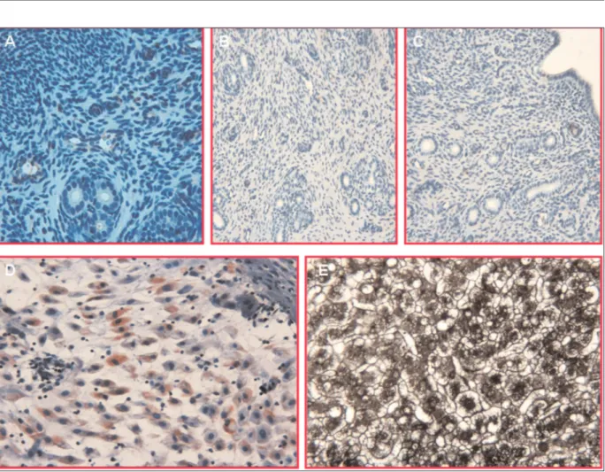
the development of endometriosis35. Likewise, increased miR-210 expression in the hypoxic conditions enhances survival rates of en-dometriotic cells36. As supportive, in the present study, both the markers of reactive oxygen species (ROS) mediated protein oxidation and markers oxidative damage of membrane lipids were significantly elevated in the animal on DSF. Our findings disagree with the previous studies indicating the antioxidative effect of DSF as a possible mechanism for indirect inhi-bition of NF-κB7,8. Nevertheless, oxidative stress-induced NF-κB activation was noted to be cell-specific events and mostly facilitatory rather than causal37. Therefore, in the endometriotic im-plants, DSF does not seem to inhibit the NF-κB through antioxidant mechanisms. As supportive, we found a similar level of PON 1, a marker of antioxidant protection against lipid peroxidation, and decreased levels of ADA, a potent inhibitor Figure 2.Negative control reactions, in which the NF-κB (A, X20), CD34 (B, X10) and PCNA (C, X10) antibody have been
omitted; no specific staining has been observed. (D, X20) Positive control for NF-κB (term human placental tissue) and cop-per (liver cirrhosis due to Wilson disease) (E, X20). A cop-peritoneal rat vessel provides a positive internal control for CD34.
of oxy-radical injury of neutrophils, in the DSF treated group. Moreover, low serum levels of an-tioxidant SOD and similar levels of reduced GSH, GSH-Px, and GSH/GSH-Px ratio in all groups support our idea that DSF does not inhibit the NF-κB activation through antioxidant mecha-nisms.
Dithiocarbamates (DCs) are known to exhibit both pro-oxidant and antioxidant effects in both cell-free and biological systems38-42. DC oxidizes reduced glutathione through a glutathione perox-idase-like activity39,40. They also inhibit glu-tathione S-transferases41. Low levels of DC in-duce oxidative stress leading to necrosis and cell death42. Likewise, some oxidants have been re-ported to cause a similar dose-dependent cyto-toxicity42,43. Therefore, in addition to NF-κB in-hibitory action, DSF also leads to oxidative stress and cell death in the endometriotic implants.
This research has some limitations such as rel-atively small size of groups and lack of the mea-surement of tissue oxidant status. The evaluation of the oxidative tissue parameters would
certain-ly be more valuable to discuss the effects of DSF on either local or systemic redox status in the en-dometrial implants. However, in the current ex-perimental study, we could not split up the im-plants due to their small size.
Conclusions
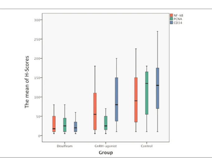
We showed for the first time that DSF exerts a toxic pro-oxidant effect on endometriotic im-plants. It also inhibits angiogenesis, cell prolifer-ation, and inflammatory events in the implants. This anti-endometriotic activity may depend on the ability of the DSF (1) to inhibit NF-κB path-way and /or (2) stimulate oxidative stress. Over-all, the present paper demonstrated that DSF re-duced the serum levels of IL-1β, TNF-α, and TAS activity. DSF also increased the serum MDA, PC, and TOS activity, whereas attenuated the NF-κB expression, angiogenesis, and cell proliferation suggesting a possible molecular mechanism of DSF for controlling the implant Figure 3.The mean of H-scores for NF-κB, PCNA, and CD34 in each treatment and control groups.
growing. DSF did not induce toxicity in kidney and liver cells and is well tolerated clinically. Considering the relatively mild side effects and the large clinical experience of the clinical drug studies using DSF in treatment of endometriosis and/or endometrioma appears to be warranted. –––––––––––––––––-––––
Conflict of Interest
The Authors declare that there are no conflicts of interest.
References
1) ESKENAZI B, WARNER ML. Epidemiology of
en-dometriosis. Obstet Gynecol Clin North Am 1997; 24: 235-258.
2) GOLDSTEIN DP, DECHOLNOKY C, EMANS SJ, LEVENTHAL
JM. Laparoscopy in the diagnosis and
manage-ment of pelvic pain in adolescents. J Reprod Med 1980; 24: 251-256.
3) BURNEY RO, GIUDICELC. Pathogenesis and
patho-physiology of endometriosis. Fertil Steril 2012; 98: 511-519.
4) SOARESSR, MARTÍNEZ-VAREA A, HIDALGO-MORAJJ, PEL
-LICER A. Pharmacologic therapies in
endometrio-sis: a systematic review. Fertil Steril 2012; 98: 529-555.
5) LIU GY, FRANK N, BARTSCHH, LIN JK. Induction of
apoptosis by thiuramdisulfides, the reactive metabolites of dithiocarbamates, through coordina-tive modulation of NF Kappa B, c-fos/c-jun, and p53 proteins. Mol Carcinog 1998; 22: 235-246. 6) WANGW, MCLEODHL, CASSIDYJ.
Disulfiram-mediat-ed inhibition of NF-kappaB activity enhances cy-totoxicity of 5-fluorouracil in human colorectal cancer cell lines. Int J Cancer 2003; 104: 504-511.
7) CEN D, BRAYTON D, SHAHANDEH B, MEYSKENS FL, FARMER PJ. Disulfiram facilitates intracellular Cu
uptake and induces apoptosis in human melanoma cells. J Med Chem 2004; 47: 6914-6920.
8) SCHRECKR, MEIERB, MÄNNELDN, DRÖGEW, BAEUERLE
PA. Dithiocarbamates as potent inhibitors of
nu-clear factor kapa B activation in intact cells. J Exp Med 1992; 175: 1181-1194.
9) LÖVBORG H, OBERG F, RICKARDSON L, GULLBO J, NY
-GREN P, LARSSON R. Inhibition of proteasome
activi-ty, nuclear factor-Kappa B translocation and cell survival by the antialcoholism drug disulfiram. Int J Cancer 2006; 118: 1577-1580.
10) GONZÁLEZ-RAMOSR, DONNEZJ, DEFRÈRES LECLERCQI,
SQUIFFLET J, LOUSSE JC, VAN-LANGENDONCKT A.
Nu-clear factor-kappa B is constitutively activated in peritoneal endometriosis. Mol Hum Reprod 2007; 13: 503-509.
11) GUOSW. Nuclear factor-kappab (NF-kappaB): an
unsuspected major culprit in the pathogenesis of
endometriosis that is still at large? Gynecol Ob-stet Invest 2007; 63: 71-97.
12) PERKINS ND. Integrating cell-signalling pathways
with NF-κB and IKK function. Nat Rev Mol Cell Biol 2007; 8: 49-62.
13) VIATOURP, MERVILLE MP, BOURSV, CHARIOTA.
Phos-phorylation of NF-kappaB and I kappaB proteins: implications in cancer and inflammation. Trends Biochem Sci 2005; 30: 43-52.
14) MARIKOVSKY M, ZIV V, NEVO N, HARRIS-CERRUTI C, MAHLERO. Cu/Zn superoxide dismutase plays
im-portant role in immune response. J Immunol 2003; 170: 2993-3001.
15) SHIANSG, KAOYR, WUFY, WUCW. Inhibition of
in-vasion and angiogenesis by zinc-chelating agent disulfiram. Mol Pharmacol 2003; 64: 1076-1084. 16) CHEND, CUIQC, YANG H, DOU QP. Disulfiram, a
clinically used antialcoholism drug and copper-binding agent, induces apoptotic cell death in breast cancer cultures and xenografts via inhibi-tion of the proteasome activity. Cancer Res 2006; 66: 10425- 10433.
17) MALAGUARNERAL, PILASTROMR, DIMARCOR, SCIFOC, RENIS M, MAZZARINO MC, MESSINA A. Cell death in
human acute myelogenous leukemic cells in-duced by pyrrolidinedithiocarbamate. Apoptosis 2003; 8: 539-545.
18) CELIKO, HASCALIKS, ELTERK, TAGLUKME, GURATESB, AYDIN NE. Combating endometriosis by blocking
proteasome and nuclear factor-kappaB path-ways. Hum Reprod 2008; 23: 2458-2465. 19) VERNON MW, WILSON EA. Studies on the surgical
induction of endometriosis in the rat. Fertil Steril 1985; 44: 684-894.
20) ALTINTASD, KOKCU A, TOSUNM, CETINKAYAMB, KAN
-DEMIRB. Comparison of the effects of cetrorelix, a
GnRH antagonist, and leuprolide, a GnRH ago-nist, on experimental endometriosis. J Obstet Gy-naecol Res 2008; 34: 1014-1019.
21) SUNY, OBERLEYLW, LIY. A simple method for
clini-cal assay of superoxide dismutase. Clin Chem 1988; 34: 497-500.
22) PAGLIADE, VALENTINEWN. Studies on the
quantita-tive and qualitaquantita-tive characterization of erythrocyte glutathione peroxidase. J Lab Clin Med 1967; 70: 158-169.
23) EL L M A N GL. Tissue sulfhydryl groups. Arch
Biochem Biophys 1959; 82: 70-77.
24) GIUSTI G. Adenosine deaminase. In: Bergmeyer
MV, editor. Methods of enzymatic analysis. 2nd ed. New York, USA: Academic Press, 1974; pp. 1092-1098.
25) WEIH, FRENKELK. Relationship of oxidative events
and DNA oxidation in sencar mice to in vivo moting activity of phorbol ester-type tumor pro-moters. Carcinogenesis 1993; 14: 1195-1201. 26) ESTERBAUER H, CHEESEMAN KH. Determination of
aldehydic lipid peroxidation products: malonalde-hyde and 4-hydroxynonenal. Methods Enzymol 1990; 186: 407-421.
27) LEVINERL, GARLANDD, OLIVERCN, AMICIA, CLIMENT
I, LENZ AG, AHN BW, SHALTIELS, STADTMANER.
De-termination of carbonyl content in oxidatively modified proteins. Methods Enzymol 1990; 186: 464-478.
28) ERELO. A new automated colorimetric method for
measuring total oxidant status. Clin Biochem 2005; 38: 1103-1111.
29) TIMM F. Histochemical demonstration of copper in
the brain. Z Zellforch Microsk Anat Histochem 1961; 2: 332-341.
30) BUDWIT-NOVOTNYDA, MCCARTYKS, COXEB, SOPERJT, MUTCHDG, CREASMANWT, FLOWERSJL, MCCARTY KS.
Immunohistochemical analyses of estrogen re-ceptor in endometrial adenocarcinoma using a monoclonal antibody. Cancer Res 1986; 46: 5419-5425.
31) LAIRDSM, TUCKERMANEM, CORKBA, LI TC.
Expres-sion of nuclear factor κB in human endometrium; role in the control of interleukin 6 and leukaemia inhibitory factor production. Mol Hum Reprod 2000; 6: 34-40.
32) LINDSTRÖMTM, BENNETTPR. The role of nuclear
fac-tor kappa B in human labour. Reproduction 2005; 130: 569-581.
33) SAKAMOTO Y, HARADA T, HORIE S, IBA Y, TANIGUCHIF, YOSHIDA S, IWABE T, TERAKAWA N. Tumor necrosis
factor-a-induced interleukin-8 (IL-8) expression in endometriotic stromal cells, probably through nu-clear factor-kB activation: gonadotropin-releasing hormone agonist treatment reduced IL-8 expres-sion. J Clin Endocrinol Metab 2003; 88: 730-735. 34) KARIN M. Nuclear factor- kB in cancer
develop-ment and progression. Nature 2006; 441: 431-436.
35) TURGUTA, OZLERA, GORUKNY, TUNC SY, EVLIYAOGLU
O, GÜL T. Copper, ceruloplasmin and oxidative
stress in patients with advanced-stage en-dometriosis. Eur Rev Med Pharmacol Sci 2013; 17: 1472-1478.
36) XUTX, ZHAOSZ, DONGM, YUXR. Hypoxia
respon-sive miR-210 promotes cell survival and au-tophagy of endometriotic cells in hypoxia. Eur Rev Med Pharmacol Sci 2016; 20: 399-406. 37) BOWIEA, O'NEILL LA. Oxidative stress and nuclear
factor-kappa B activation: a reassessment of the evidence in the light of recent discoveries. Biochem Pharmacol 2000; 59: 13-23.
38) WHO. Environmental Health Criteria. World Health Organization Vammala, Finland, 1980. 39) KUMAR KS, SANCHOAM, WEISS JF. A novel
interac-tion of diethyldithiocarbamate with the glu-tathione/glutathione peroxidase system. Int J Ra-diat Oncol Biol Phys 1986; 12: 1463-1467. 40) HOSNIM, MESKININ, PRIGENTAF, ANKERG, JOULAINC,
ELHABIBR, LAGARDEM.Diethyldithiocarbamate
(di-tiocarb sodium) effect on arachidonic acid metab-olism in human mononuclear cells. Glutathione peroxidase-like activity. Biochem Pharmacol 1992; 43: 1319-1329.
41) DIERCKXPJ.In vitro interaction of dithiocarb with rat
liver glutathione S-transferases. Pharmacol Res Commun 1984; 16: 135-143.
42) NOBEL CI, KIMLANDM, LIND B, ORRENIUS S, SLATER
AF. Dithiocarbamates induce apoptosis in
thy-mocytes by raising the intracellular level of re-dox-active copper. J Biol Chem 1995; 270: 26202-26208.
43) DYPBUKTJM, ANKARCRONAM, BURKITTM, SJÖHOLMA,
STRÖM K, ORRENIUS S, NICOTERAP. Different
prooxi-dant levels stimulate growth, trigger apoptosis, or produce necrosis of insulin-secreting RINm5F cells. The role of intracellular polyamines. Biol Chem 1994; 269: 30553-30560.