O R I G I N A L A R T I C L E
Clinicopathologic characteristics, treatment outcomes, and
prognostic factors of primary thoracic soft tissue sarcoma:
A multicenter study of the Anatolian Society of Medical
Oncology (ASMO)
Olcun Umit Unal1, Ilhan Oztop2, Nurgul Yasar3, Zuhat Urakci4, Tahsin Ozatli5, Oktay Bozkurt6,
Alper Sevinc7, Yusuf Gunaydin8, Burcu Yapar Taskoylu9, Erkan Arpaci10, Arife Ulas11, Hilmi Kodaz12,
Onder Tonyali8, Nilufer Avci13, Asude Aksoy14& Ahmet Ugur Yilmaz15
1 Division of Medical Oncology, Department of Internal Medicine, Ataturk University Medical Faculty, Erzurum, Turkey 2 Division of Medical Oncology, Department of Internal Medicine, Dokuz Eylul University Medical Faculty, Izmir, Turkey 3 Department of Medical Oncology, Dr. Lutfi Kirdar Kartal Education and Research Hospital, Istanbul, Turkey 4 Department of Medical Oncology, Dicle University Faculty of Medicine, Diyarbakir, Turkey
5 Department of Medical Oncology, Dr. Abdurrahman Yurtaslan Training and Research Hospital, Ankara, Turkey 6 Department of Medical Oncology, Erciyes University Faculty of Medicine, Kayseri, Turkey
7 Department of Medical Oncology, Gaziantep University Faculty of Medicine, Gaziantep, Turkey 8 Department of Medical Oncology, Gazi University Faculty of Medicine, Ankara, Turkey 9 Department of Medical Oncology, Pamukkale University Faculty of Medicine, Denizli, Turkey 10 Department of Medical Oncology, Sakarya Education and Research Hospital, Sakarya, Turkey 11 Department of Medical Oncology, Ali Sonmez Oncology Hospital, Bursa, Turkey
12 Department of Medical Oncology, Trakya University Faculty of Medicine, Edirne, Turkey 13 Department of Medical Oncology, Balikesir Goverment Hospital, Balikesir, Turkey 14 Department of Medical Oncology, Firat University Faculty of Medicine, Elazig, Turkey 15 Department of Medical Oncology, Izmir University Faculty of Medicine, Izmir, Turkey
Keywords
Primary thoracic soft tissue sarcoma; prognostic factors; treatment.
Correspondence
Olcun Umit Unal, Division Of Medical Oncology, Department of Internal Medicine, Ataturk University Medical Faculty, Erzurum 25050, Turkey. Tel:+90 04422317883 Fax:+90 04422361301 Email: [email protected] Received: 9 June 2014; Accepted: 23 June 2014. doi: 10.1111/1759-7714.12150 Thoracic Cancer 6 (2015) 85–90
Abstract
Background: Soft tissue sarcomas (STSs) are rare malignant tumors of embryo-genic mesoderm origin. Primary thoracic STSs account for a small percentage of all STSs and limited published information is available. This study aimed to identify the prognostic factors for thoracic STSs and evaluate the disease’s clinical outcomes. Methods: The medical records of 109 patients with thoracic STSs who were treated between 2003 and 2013 were retrospectively reviewed. Patients’ survival rates were analyzed and potential prognostic factors evaluated.
Results: The median follow-up period was 29 months (range: 1–121 months). STSs were most frequently localized on the chest wall (n= 42; 38.5%) and lungs (n = 42; 38.5%). The most common histological types were malignant fibrous histiocytoma (n= 23; 21.1%), liposarcoma (n = 17; 15.6%), and leiomyosarcoma (n = 16; 14.7%). The median survival time of all patients was 40.3 months (95% confidence interval, 14.22–66.37 months), with one and five-year survival rates of 93.4% and 63.5%, respectively. Univariate analysis of all groups revealed that metastatic stage, unresectability, tumor diameter of>10 cm, tumor location other than the chest wall, and grade 3 diseases were predictable of poor survival. However, only grade 3 dis-eases and tumor location other than the chest wall were confirmed by multivariate analysis as poor prognostic factors.
Conclusions: Primary thoracic STSs are rarely seen malignant tumors. Our results indicated that patients with low-grade tumors and those localized on the chest wall often experienced better survival outcomes.
Introduction
Soft tissue sarcomas (STSs) are rare malignant tumors of embryogenic mesoderm origin.1They account for less than
1% of all human malignancies.1Approximately 60% of STSs
are localized on the extremities with the remaining com-monly found in the gastrointestinal, retroperitoneal, head, and neck regions.2Thoracic STSs account for a small
percent-age (3–8%) of STSs3,4with tumors often found in the lungs,
mediastinum, pleura, pericardium, heart, and chest walls.5
STS is a heterogenous disease with more than 60 histologi-cal types.6Malignant fibrous histiocytomas and liposarcomas
are the most commonly observed STSs of the extremities, whereas the frequency of thoracic STS histological types varies depending on tumor location,2such as fibrosarcomas
and leiomyosarcomas in the lungs, angiosarcomas in the heart, and malignant fibrous histiocytomas on the chest wall.7–10
Multimodality therapy, including surgery, radiotherapy, and chemotherapy, is commonly used for the treatment of STSs.2,11–13Of these, surgery is the most important
compo-nent. Clinical parameters such as patients’ age, Eastern Coop-erative Oncology Group (ECOG) performance status, tumor histology, and tumor grade are often considered when other treatment modalities are elected. Although the surgical treat-ment outcomes of thoracic STSs have been analyzed in several published studies, such analyses are not available for any other treatment modalities.
Furthermore, although the prognostic factors for overall STS patient survival have been thoroughly analyzed,11,14only
one study thus far has specifically examined those in primary thoracic STSs.15 Otherwise, most published reports have
focused on surgical or radiological studies of thoracic STS retrospective case series or case reports.16–18
The present study aimed to determine the clinico-pathological features, survival rates, and prognostic factors of patients with primary thoracic STSs treated at major hospi-tals in Turkey.
Materials and methods
Study designThe present study was designed as a retrospective analysis of a primary thoracic STS patient cohort.
Methods
The Executive Committee of Anatolian Society of Medical Oncology approved this study. We collected data from patients with primary STSs treated at 15 different medical oncology centers in Turkey. Data collection began in Febru-ary 2012. Information on patients’ age, gender, tumor size,
location, and the presence of primary tumor and metastatic site were retrieved from their medical records. The 2002 World Health Organization criteria were used for histopatho-logical diagnosis of all patients,19 and the Fédération
Nationale des Centres de Lutte Contre le Cancer system was used for tumor grading.20In addition, treatment modalities
(cytotoxic agents, chemotherapy regimen, surgery, and radio-therapy), clinical outcomes, time to disease progression, mor-tality rates, and the length of follow-up periods were recorded until the last visit in October 2013.
The inclusion criteria were as follows: (i) STS histology; (ii) the primary tumor site being within the thoracic region, including the chest wall, lungs, pleura, mediastinum, pericar-dium, and heart; and (iii) patient’s age of >18 years. The exclusion criteria included: (i) skeletal sarcoma (excluding extra-skeletal Ewing sarcoma and extra-skeletal chondrosar-coma); (ii) desmoid tumor; and (iii) dermatofibrosarcoma protuberans.
Statistical analysis
Data were analyzed using the Statistical Package for the Social Sciences (SPSS) version 15.0 for Windows (SPSS Inc., Chicago, IL). Overall survival (OS) was calculated from the surgery time or the time of diagnosis to death or the last day of the follow-up period. The Kaplan-Meier method was used for OS analysis. For the comparison of survival rate differ-ences, a log-rank (Mantel-Cox) test was used for univariate analysis and Cox-proportional hazards model for multivari-ate analysis. A P-value of<0.05 was considered statistically significant. The following factors were evaluated in the prog-nostic factor analysis: gender (male or female); age (<50 and ≥50 years), stage (local, locally advanced, and metastatic), tumor diameter (0–5 cm, 5–10 cm, or ≥10 cm), grade (1 and 3), resection (complete, incomplete resection, and unresectable), ECOG performance status (0, 1, and 2), tumor location (the chest wall, lungs, or other), adjuvant radio-therapy (applied or not), and adjuvant chemoradio-therapy (applied or not).
Results
Global patient characteristics and treatment alternatives
This study enrolled 109 patients with a male/female ratio of 1:3. Patient characteristics are summarized in Table 1. The primary tumor was commonly located in the mediastinum (n= 16; 14.7%), pleura (n = 5; 4.6%), intracardiac region (n = 3; 2.8%), and pericardium (n= 1; 0.9%). Malignant fibrous histiocytoma was the most frequently observed histological type. When separate groups were considered, malignant fibrous histiocytoma was often found on the chest wall
(n= 16; 38%), leiomyosarcoma in the lungs (n = 12; 28.5%), liposarcoma in the mediastinum (n= 4; 25%), unclassified sarcoma in the pleura (n= 2; 40%), and angiosarcoma in the intracardiac region (n= 2; 66.6%). Synovial sarcoma was detected in the pericardium of one patient. Ewing sarcomas were observed in the lungs (n= 9) and mediastinum (n = 2). Additionally, chondrosarcomas were detected in the lungs of two patients.
Seventy-five patients underwent tumor resection, mostly complete resection with only 11% incomplete resection (R1 resection). The characteristics of these surgical patients are listed in Table 1. Adjuvant chemotherapy (n = 46) or radiotherapy (n= 39) was provided to all surgical patients. The adjuvant chemotherapy regimens included ifosfamide-doxorubicin (n = 32),
vincristine-adriamycin-cyclopho-sphamide (VAC) and ifosfamide-etoposide (IE) combination (n= 7), cisplatin and doxorubicin combination (n = 4), and VAC (n= 3).
Palliative chemotherapy was provided to 58 patients with metastatic diseases at baseline or progression. The administered regimens included IE combination, gemcitabine-docetaxel combination, cisplatin-etoposide combination, cyclophosphamide, vincristine, doxorubicin, and dacarbazine (CYVADIC), and paclitaxel.
Survival outcomes
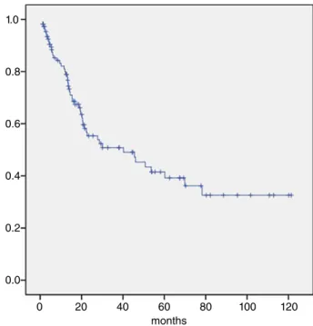
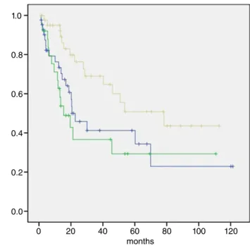
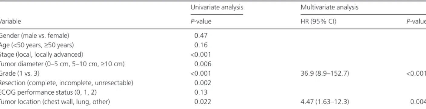
The median follow-up period was 29 months (range, 1–121 months). At the time of the present analysis, 51 patients had died. The median OS of all patients was 40.3 months (95% confidence interval [CI], 14.2–66.3 months) (Fig 1) with one and five-year survival rates of 93.4% and 63.5%, respectively. The median OS of patients undergoing resection was 53.6 months (95% CI, 16–91.3 months) with one and five-year survival rates of 91.5% and 46.5%, respectively (Fig 2). Patients with tumors located on the chest wall tended to expe-rience a better OS (median, 78.2 months) than those with dis-eases in the lungs (median, 20.6 months) and other locations (median, 15.4 months) (P= 0.022) (Fig 3).
Analysis of potential prognostics
The factors included in the univariate and multivariate analy-ses of surgical patients’ survival rates are listed in Table 2. Table 1 Patient characteristics
All patients (n= 109) Surgery group (n= 75) Median age 46 (18–85) 46 (19–85) Gender Male 62 (56.9%) 43 (57.3%) Female 47 (43.1%) 32 (42.7%) Primary tumor site Chest wall 42 (38.5%) 36 (48%) Lung 42 (38.5%) 25 (33.3%) Others† 25 (23%) 14 (18.7%) Histological type Malignant fibrous histiocytoma 23 (21.2%) 20 (26.7%) Liposarcoma 17 (15.6%) 12 (16%) Leiomyosarcoma 16 (14.7%) 10 (13.3%) Unclassified 14 (12.8%) 10 (13.3%) Ewing sarcoma 11 (10.1%) 6 (8%) Synovial sarcoma 10 (9.1%) 6 (8%) Fibrosarcoma 6 (5.5%) 4 (5.3%) Rare types 12 (11%)‡ 7 (9.4%) Tumor diameter <5 cm 20 (18.3%) 17 (22.7%) 5–10 cm 45 (41.3%) 37 (49.3%) ≥10 cm 19 (17.4%) 18 (24%) Unknown 25 (23%) 3 (4%) Grade 1 33 (30.3%) 26 (34.7%) 2 4 (3.7%) 1 (1.3%) 3 54 (49.5%) 37 (49.3%) Unknown 18 (16.5%) 11 (14.7%) Stage at diagnosis Local 57 (56.1%) 54 (72%) Locally advanced 29 (22.8%) 21 (28%) Metastatic 23 (21.1%)
Surgery Complete resection 63 (57.8%) 63 (84%)
Incomplete resection§ 12 (11%) 12 (16%)
Unresectable 34 (31.2%)
ECOG 0 27 (24.8%) 23 (30.6%)
1 58 (53.2%) 41 (53.3%)
2 24 (22%) 11 (16.1%)
ECOG, Eastern Cooperative Oncology Group performance status. †Other, mediastinal, pleural, pericardial, or cardiac locations; ‡Rhabdo-myosarcoma (3), angiosarcoma (2), hemangiopericytoma (2), chondro-sarcoma (2), spindle cell (2), alveolar soft chondro-sarcoma (1), epiteloid sarcoma (1), giant cell sarcoma (1); §R1 resection.
1.0 0.8 0.6 0.4 0.2 0.0 0 20 40 60 months 80 100 120
Figure 1 Mean survival of all groups. , Survival Function; , Censored.
Univariate analysis revealed that the absence of adjuvant che-motherapy, tumor diameter of>10 cm, tumor location other than the chest wall, and the presence of a grade 3 tumor were poor prognostic factors. These four factors were included in the subsequent multivariate analysis, which confirmed grade 3 tumors and tumor location other than the chest wall as poor prognostic factors. Similarly, the univariate and multivariate analyses of all patients’ survival rates are summarized in Table 3. The univariate analysis identified metastatic stage, unresectability, tumor diameter of>10 cm, tumor location other than the chest wall, and the presence of a grade 3 tumor as poor prognostic factors, whereas the multivariate analysis subsequently confirmed the prognostic value of grade 3 tumors and tumor locations other than the chest wall.
Discussion
The present study aimed to define the clinical characteristics, treatment alternatives, and prognostic factors for thoracic STSs using data from multiple institutes in Turkey. The inci-dences of both STSs and thoracic STSs peaked at 50 years of age with a male dominancy. In our series, the median patient age was 46 years, and there were slightly more male than female patients, which is comparable to other published STS and thoracic STS series.2,15,21
Malignant fibrous histiocytoma is the most common STS21
and thoracic STS type, but other histological types can also be observed at other thoracic locations.10In our series,
angiosar-comas were frequently detected in the cardiac region, whereas
Table 2 Univariate and multivariate analyses of surgical patients’ survival rates (n= 75)
Variable
Univariate analysis Multivariate analysis
P-value HR (95% CI) P-value
Gender (male vs. female) 0.98
Age (<50 years, ≥50 years) 0.39
Stage (local, locally advanced) 0.67
Tumor diameter (0–5 cm, 5–10 cm,≥10 cm) 0.003
Grade (1 vs. 3) <0.001 27.6 (6.8–112.2) <0.001
Resection (complete, incomplete) 0.09
Tumor location (chest wall, lung, other) 0.02 4.63 (1.57–13.7) 0.006
Adjuvant radiotherapy (present, absent) 0.38
Adjuvant chemotherapy (present, absent) 0.01
CI, confidence interval; HR, hazard ratio. 1.0 0.8 0.6 0.4 0.2 0.0 0 20 40 60 months 80 100 120
Figure 2 Mean survival of resected patients. , Survival Function; , Censored. 1.0 0.8 0.6 0.4 0.2 0.0 0 20 40 60 months 80 100 120
Figure 3 Mean survival of primary site. , Lung; , Other; , Chest Wall; , Lung-censored; , Other-censored; , Chest Wall-censored.
liposarcomas were observed more often in the mediastinum. Our findings are in agreement with those previously reported on these two locations.9,22Leiomyosarcoma and fibrosarcoma
are commonly reported for lung STSs,7,8which was also the
case in our study. However Pieper et al. reported Ewing sarcoma as the most commonly detected STS in the lungs with extra-osseous tumors found in the broncho-pulmonary region.23
The survival rates in primary thoracic STS patients are lower than those with STSs of the extremities with reported five-year survival rates for the two diseases being 56–79% and >75%, respectively.15,16,24–27In our study, the five-year survival
rate and median survival time were 65% and 57 months, respectively, which was similar to the results of previously published thoracic STS series.
Similar prognostic factors have been assumed to affect both patients with resected thoracic STS and those with dis-eases of the extremities. However, such an assumption is of limited value owing to the lower incidence of primary tho-racic STS. Pisters et al. studied 1041 patients with STS of the extremities and found positive histological tumor margins, larger tumor size, and higher tumor grade to be associated with higher mortality rates.26On the other hand, Duranti
et al. evaluated 337 patients with resected thoracic STSs and
reported that grade 3 tumors, pulmonary and mediastinal STS, and R1 resection were more risky than grade 1 tumors (1.89 fold), thoracic wall STS (1.6 and 1.9 fold), and R0 resec-tion (1.9 fold), respectively.15Furthermore, McMillan et al.
reported that tumor grade was the most important determi-nant of survival and recurrence pattern in patients with tho-racic wall STSs,10whereas Gross et al. identified tumor grade
and diameter as the most important prognostic factors for survival in patients with chest wall STSs. In our study, among all patients with resected thoracic STSs, the best prognosis was noted in those with grade 1 chest wall STSs, which is com-parable to the results published by Duranti et al. that had the largest patient cohort to date.
Additionally, we also evaluated adjuvant treatment modali-ties in patients with resected thoracic STS. Adjuvant
chemo-therapy, but not radiochemo-therapy, was found to significantly increase survival rates. Alkis et al. also obtained similar out-comes in their study.2 Radiotherapy has been shown to
decrease local recurrence rate without contributing to improved survival in STS patients, as confirmed in our study.12
However, to our knowledge, no study on the prognostic factors for STS patient survival is available. Meanwhile, resectability has emerged as an important prognostic factor in thoracic STS, as well as all other types of STS.28Other
prog-nostic factors, such as tumor grade, diameter, and stage were similar to those detected in previous large STS series.2,4,20,21,26
However, among patients with resectable tumors, thoracic STS patients might have better prognostic characteristics.
This retrospective study has many inevitable limitations. Selection bias might have existed and patients from certain major medical centers were enrolled. Additionally, treatment modalities might differ with time and among clinics.
Conclusion
In conclusion, thoracic STS is a rare malignancy with diverse histological types according to the primary tumor location. The survival time of thoracic STS patients is apparently shorter than that of extremity STS patients. Adjuvant chemo-therapy might be useful for patients with resectable tumors, whereas adjuvant radiotherapy was found to have no effect on survival. Although the prognostic factors for thoracic STS patient survival were similar to those of extremity STS, tho-racic tumor location appeared to be another important factor affecting survival.
Disclosure
No authors report any conflict of interest.
References
1 Froehner M, Wirth MP. Etiologic factors in soft tissue sarcomas. Onkologie 2001; 24: 139–42.
Table 3 Univariate and multivariate analysis of all patients’ survival rates (n= 109)
Variable
Univariate analysis Multivariate analysis
P-value HR (95% CI) P-value
Gender (male vs. female) 0.47
Age (<50 years, ≥50 years) 0.16
Stage (local, locally advanced) <0.001
Tumor diameter (0–5 cm, 5–10 cm,≥10 cm) 0.006
Grade (1 vs. 3) <0.001 36.9 (8.9–152.7) <0.001
Resection (complete, incomplete, unresectable) 0.002
ECOG performance status (0, 1, 2) 0.13
Tumor location (chest wall, lung, other) 0.022 4.47 (1.63–12.3) 0.004
2 Alkis N, Muallaog˘lu S, Koçer M et al. Primary adult soft tissue sarcomas: analysis of 294 patients. Med Oncol 2011; 28: 391–6.
3 Bhurgri Y, Bhurgri H, Pervez S et al. Epidemiology of soft tissue sarcomas in Karachi South, Pakistan (1995–7). Asian Pac J Cancer Prev 2008; 9: 709–14.
4 Toro JR, Travis LB, Wu HJ, Zhu K, Fletcher CD, Devesa SS. Incidence patterns of soft tissue sarcomas, regardless of primary site, in the surveillance, epidemiology and end results program, 1978–2001: an analysis of 26,758 cases. Int J Cancer 2006; 119: 2922–30.
5 Gladish GW, Sabloff BM, Munden RF, Truong MT, Erasmus JJ, Chasen MH. Primary thoracic sarcomas. Radiographics 2002; 22: 621–37.
6 Wardelmann E, Chemnitz JM, Wendtner CM. Targeted therapy of soft tissue sarcomas. Onkologie 2012; 35 (Suppl 1): 21–7.
7 Petrov DB, Vlassov VI, Kalaydjiev GT et al. Primary pulmonary sarcomas and carcinosarcomas - postoperative results and comparative survival analysis. Eur J Cardiothorac Surg 2003; 23: 461–6.
8 Spraker MB, Bair E, Bair R, Connell PP, Mahmood U, Koshy M. An analysis of patient characteristics and clinical outcomes in primary pulmonary sarcoma. J Thorac Oncol 2013; 8: 147–51.
9 Truong PT, Jones SO, Martens B et al. Treatment and outcomes in adult patients with primary cardiac sarcoma: the British Columbia Cancer Agency experience. Ann Surg Oncol 2009; 16: 3358–65.
10 McMillan RR, Sima CS, Moraco NH, Rusch VW, Huang J. Recurrence patterns after resection of soft tissue sarcomas of the chest wall. Ann Thorac Surg 2013; 96: 1223–8.
11 Kasper B, Ouali M, Van Glabbeke M et al. Prognostic factors in adolescents and young adults (AYA) with high risk soft tissue sarcoma (STS) treated by adjuvant chemotherapy: a study based on pooled European Organisation for Research and Treatment of Cancer (EORTC) clinical trials 62771 and 62931. Eur J Cancer 2013; 49: 449–56.
12 Yang JC, Chang AE, Baker AR et al. Randomized prospective study of the benefit of adjuvant radiation therapy in the treatment of soft tissue sarcomas of the extremity. J Clin Oncol 1998; 16: 197–203.
13 Reynoso D, Subbiah V, Trent JC et al. Neoadjuvant treatment of soft-tissue sarcoma: a multimodality approach. J Surg Oncol 2010; 101: 327–33.
14 Sato T, Peiper M, Heinecke A et al. Expression of HER2/neu does not correlate with survival in soft tissue sarcoma. Onkologie 2003; 26: 268–71.
15 Duranti L, Gronchi A, Stacchiotti S et al. Localised thoracic sarcomas: outcome improvement over time at a single institution. Eur J Cancer 2013; 49: 2689–97.
16 Pfannschmidt J, Geisbüsch P, Muley T, Dienemann H, Hoffmann H. Surgical treatment of primary soft tissue sarcomas involving the chest: experiences in 25 patients. Thorac Cardiovasc Surg 2006; 54: 182–7.
17 Cakir O, Topal U, Bayram AS, Tolunay S. Sarcomas: rare primary malignant tumors of the thorax. Diagn Interv Radiol 2005; 11: 23–7.
18 Jeon K, Paeng JC, Goo JM, Lee H. Follicular dendritic cell sarcoma of the mediastinum: CT and18 F-fluoro-2-deoxyglucose PET findings. Thorac Cancer 2013; 4: 203–6.
19 Jo VY, Fletcher CDM. WHO classification of soft tissue tumours: an update based on the 2013 (4th) edition. Pathology 2014; 46: 95–104.
20 Coindre JM, Terrier P, Bui NB et al. Prognostic factors in adult patients with locally controlled soft tissue sarcoma. A study of 546 patients from the French Federation of Cancer Centers Sarcoma Group. J Clin Oncol 1996; 14: 869–77.
21 Fang ZW, Chen J, Teng S, Chen Y, Xue RF. Analysis of soft tissue sarcomas in 1118 cases. Chin Med J 2009; 122: 51–3. 22 Luo DX, Huang MJ, Xiong B et al. Primary mediastinal
sarcoma: surgical outcomes of 21 cases. Interact Cardiovasc Thorac Surg 2013; 17: 982–6.
23 Pieper S, Ranft A, Braun-Munzinger G, Jurgens H, Paulussen M, Dirksen U. Ewing’s tumors over the age of 40: a
retrospective analysis of 47 patients treated according to the International Clinical Trials EICESS 92 and
EURO-E.W.I.N.G. 99. Onkologie 2008; 31: 657–63. 24 Gross JL, Younes RN, Haddad FJ, Deheinzelin D, Pinto CA,
Costa ML. Soft-tissue sarcomas of the chest wall: prognostic factors. Chest 2005; 127: 902–8.
25 Stojadinovic A, Leung DH, Hoos A, Jaques DP, Lewis JJ, Brennan MF. Analysis of the prognostic significance of microscopic margins in 2,084 localized primary adult soft tissue sarcomas. Ann Surg 2002; 235: 424–34.
26 Pisters PW, Leung DH, Woodruff J, Shi W, Brennan MF. Analysis of prognostic factors in 1,041 patients with localized soft tissue sarcomas of the extremities. J Clin Oncol 1996; 14: 1679–89.
27 Gronchi A, Miceli R, Colombo C et al. Primary extremity soft tissue sarcomas: outcome improvement over time at a single institution. Ann Oncol 2011; 22: 1675–81.
28 Duman BB, Gunaldi M, Ercolak V et al. Retrospective analysis of 498 primary soft tissue sarcomas in a single Turkish centre. Asian Pac J Cancer Prev 2012; 13: 4125–8.