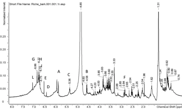
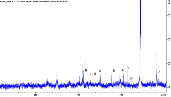
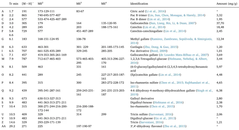
Ricinodendron heudelotii (Baill.) Heckel stem barks and seed extracts, a native food plant from Africa: Characterization by NMR and HPLC-DAD-ESI-MSn
Tam metin
Şekil

Benzer Belgeler
Results: The stem and root extracts showed no activity at the maximum concentration, while the leaf extract at 100 µg/mL showed remarkable cell growth inhibition against
Among variables for the analysis the following are chosen: domestic and foreign inflation rates, investment cost overrun factor, % change in the real price of glycerin,
Soil contains a living, complex ecosystem • Plants obtain most of their water and minerals.. from the upper layers
Seçtiğim türküler, Ruhi'nin radyo, televizyon programlarından almış olduğu eserlerdir. Gene bu türküler kulüplerde, dost evlerinde verdiği konserlerin
Frontal mukosellerin komşulukları nedeniyle orbita medial-üst duvarını tahrip ederek intraorbital alana uzanması sıklıkla bildirilmesine rağmen frontal kemiğin
Benzer şekilde dokuz randomize kontrollü çalışmanın (n=705, 2 hafta-22 ay süreli) incelendiği bir başka meta- analizde, düşük Gİ içerikli diyetlerin uygulanması ile HbA1c
Bu süreçte, egemen kılınan “küreselleşme” söylemi ile, artık tek kutuplu bir dünyada yaşandığı, liberal kapitalist siste- min (piyasa ekonomisinin)
gerekmektedir. Doktor kontrolü ve önerisi ışığında, tedavi amacı ile kullanıldığı zaman çekinilmemelidir. Bu ilaçların büyük çoğunluğu yeşil reçete adı
