Ankara Üniv Vet Fak Derg, 52, 2005 193 Ankara Üniv Vet Fak Derg, 52, 193-196, 2005
Short Communication / Kısa Bilimsel Çalışma
Bilateral hydronephrosis and hydroureter in a German shpherd dog
Mehmet ŞAHAL1, Rıfkı HAZIROĞLU2, Yunusemre ÖZKANLAR3, Latife BEYAZ4
1 Ankara Üniversitesi, Veteriner Fakültesi, İç Hastalıkları Anabilim Dalı, Ankara; 2Ankara Üniversitesi, Veteriner Fakültesi, Patoloji
Anabilim Dalı, Ankara; 3Atatürk Üniversitesi, Veteriner Fakültesi, İç Hastalıkları Anabilim Dalı, Erzurum; 4Erciyes Üniversitesi,
Veteriner Fakültesi, Patoloji Anabilim Dalı, Kayseri.
Summary: A six month-old German shepherd dog was referred to clinic with complaints of urinary incontinence, polidipsia,
polyuria, apathy, anorexia and continuous weakness. Abdominal pain, leucocytosis, slight anemia, mild elevation in serum urea and elevation in creatinin were detected. Extremely dilated renal pelvis and grossly thinned renal parenchyma were detected on ultrasonography and intravenous pyelography. Following the conformation of the diagnosis by the exploratory laparatomy, performed for diagnosis, the dog was euthanased with the owner's request because of advanced bilateral hydronephrosis and hydroureter. At necropsy, bilateral hydronephrosis and hydroureter with stenosis in both ureters were observed. On the histopathological examination, there were tubuler dilation and medullar fibrosis in both kidneys. In the stenotic areas of ureters; transitional cell hyperplasia, collagen bundles and haemorrhagic areas were observed. In this case, bilateral hydronephrosis and hydroureter were considered to be developed congenitally.
Key words: German shepherd dog, hydronephrosis, hydroureter.
Alman kurt köpeğinde bilateral hidronefroz ve hidroüreter olgusu
Özet: Bu olgunun materyalini idrar kaçırma, poliüri, polidipsi, apati, anoreksi ve ilerleyen zayıflık şikayetleri ile kliniğe
getirilen 6 aylık bir Alman kurt köpeği oluşturdu. Olguda abdominal ağrı, lökositozis, hafif anemi, serum üre ve kreatinin değerlerinde artış belirlendi. Ultrasonografide ve intravenöz pyelografide, aşırı derecede dilate olmuş pelvis renalis ve renal paranşimde incelme tespit edildi. Tanı amaçlı olarak yapılan deneysel laparatomide ileri derece bilateral hidronefroz ve hidroureter tanısı konuldu ve köpek sahibinin isteği üzerine ötenazi uygulandı. Nekropside, bilateral hidronefroz ve hidroüreter ile birlikte her iki üreterde stenoz gözlendi. Histopatolojide, her iki böbrekte tubuler dilatasyon ve medullar fibrozis vardı. Üreterlerin stenotik alanlarında, transitional hücre hiperplazisi, kollajen bundle’ler ve hemorajik alanlar gözlendi. Bu olguda, bilateral hidronefroz ve hidroüreterin doğmasal olarak geliştiği düşünüldü.
Anahtar sözcükler: Alman kurt köpeği, hidronefroz, hidroüreter.
Hydronephrosis refers to dilation of the renal pelvis and calyces resulting in progressive atrophy and cystic enlargement of renal parenchyma. When there is complete obstruction of the outflow of urine at some point below the convergence of the two ureteral streams, it may be unilateral or bilateral (2,6). Most causes are acquired and associated with lower urinary tract disease, generally urolithiasis. Furthermore, it is known mainly as a disease of adult dogs (2,7). However, congenital hydronephrosis and hydroureter are rarely described in puppies (10). It may be caused by ectopic ureter and structure of ureters and urethra (9). Congenital bilateral hydronephrosis has been reported in a German shepherd dog without association of obstructive lesion (10). The diagnosis of advanced stages of hydronephrosis and hydroureter is readily apparent with ultrasonography because of dilated pelvic diverticula and proximal ureter. If renal function is still adequate, an excretory urogram should always be compared with the ultrasound findings.
In this case, bilateral congenital hydronephrosis and hydroureters associated with obstructive congenital anomaly were reported in a German shepherd dog.
A six month-old, female German shepherd dog was referred to the Department of Internal Medicine, Faculty of Veterinary Medicine, University of Ankara with a history of apathy, anorexia, urinary incontinence, polidipsia, polyuria, and rarely vomiting and continuous weakness for 3 months of progressive illness. On the clinical examination, increased body temperature (39.7 °C), pale mucous membranes, incoordination, and weakness were observed. Superficial lymph nodes were within normal size. There were vesicular sounds in the auscultation of the lung and heart frequency. There was a pain on the abdominal palpation. Leucocytosis (36.300 µl), mild anemia (erythrocyte 4.180.000 µl), moderate increases of serum urea (60.8 mg/dL) and creatinin (2.3 mg/dL) levels were detected by the laboratory examination. Urinalysis was within physiological ranges.
Mehmet Şahal - Rıfkı Hazıroğlu - Yunusemre Özkanlar - Latife Beyaz 194
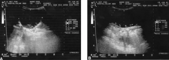
Şekil 1A-B. Ultrasnografide, sol ve sağ böbreğin görünümü. A. Sol böbrek B. Sağ böbrek
Figure 1A-B. Appearance of left and right kidneys in ultrasonogram. A. Left kidney B. Right kidney
There was a restriction to the passage of radio-opague medium through the bladder. By the ultrasonog-raphic examination with the 4.5 MHz convex transducer, kidneys were enlarged like a head of a child; pelvis renalis was greater than normal size (58 mm for left kidney, 50 mm for right kidney). Renal parenchyma could not be observed and urine was appeared as an anecogenic structure in both dilated kidneys (Figure 1A-B). At the intravenous pyelography; the contrast material (Diatrizoate meglumine, Diatrizoate sodium,
Urogra-finTM, Shering, USA) reached the kidneys but not to the
ureters at the first hour. However, renal parenchyma and calices renalis were not detected (Figure 2). The diagnosis was confirmed by the exploratory laparotomy which was severe bilateral hydronefrosis and hydrou-reter. Then, euthanasia was performed after discussing with the owner. At necropsy, both ureters and kidneys were enlarged and there were obstruction at ureters which were 1-2 cm in length in close proximity to the vesicoureteral orifice (Figure 3).
Şekil 2. İntravenöz piyelografide, birinci saatte, böbreklerin görünümü.
Figure 2. Appearance of the kidneys in intravenous pyelography at first hour
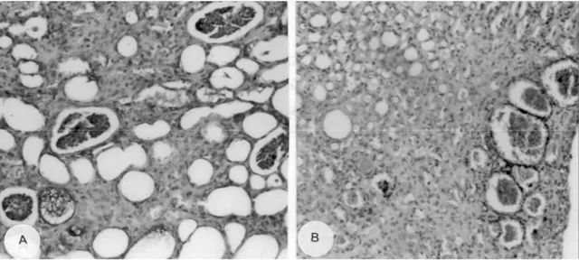
Macroscopically, right and left hydronephrotic kidneys were enlarged to 8.5 cm x 8 cm x 6 cm and 8 cm x 7.5 cm x 5 cm in diameter respectively. The cortex had lost its smoothness and there were some brownish structures protruding outwards. The cortex was thin and there were small cyst-like structures on the cut surfaces. There were rudimentary tissue debris arrested as white cordlike projection through out radially across the pelvic wall (Figure 4A and B). The renal pelvis was dilated and filled with light yellow in color content. Ureters were about 2.5 cm in scale and there was stenosis on the vesico-ureteral orifice profoundly. Histopathologically, glomeruli were atrophic and their basement membranes were also thickened and there were severe tubular degeneration. Tubular lumina were cysticly dilated and occasionally filled with calcifying cast in the some area. Fibrosis was obvious in the intersititium (Figure 5A and
Şekil 3. Her iki böbrekte, hidroüreter (A) ve hidronefrozun (B) makroskobik görünümü.
Figure 3. Macroscopic appearance of hydroureter (A) and hydronephrosis (B) in both kidneys.
Ankara Üniv Vet Fak Derg, 52, 2005 195
Şekil 4A-B. Böbreklerin kesit yüzeyi, pelvis renalis şiddetli derecede dilate. A. Sağ böbrek B. Sol böbrek Figure 4A-B. Cut surfaces of the kidneys, pelvis renum severely dilated. A. Right kidney B. Left kidney
Şekil 5A-B. Renal paranşimin histopatolojik görünümü. Figure 5A-B. Histopathologic appearance of renal parenchyma.
B). Uretral mucosa were contained some areas of haemorrhagiae in both ureters. The collagen bundles and transitional cell hyperplasia were observed in the sclerotic areas.
Hydronephrosis is known to be dilatation of the renal pelvis and calyces resulting in progressive atrophy and cystic enlargement of renal parenchyma. The cause is some form of urinary obstruction, existing at any level form the urethra to the renal pelvis. The obstruction may be caused by anomalous development of the lower urinary passage or it may be acquired. Most cases of hydronephrosis occur as a result of obstructive reason in lower part of urinary system. The position of the obstruction is responsible for dilation through upper sites of the urinary system. Depending on the site of obstruction, hydronephrosis may be unilateral or bilateral and there may be some degree of hydroureter and
dilation of the bladder (7). Hydronephrosis may be caused by ectopic ureter (5), mass in ureters (1) urolithiasis (2), tumors (8), and congenital abnormality (9,10). Congenital hydronephrosis and hydroureter have been rarely described in dogs and it is mostly seen in German Shepherd puppies (4,10). Congenital bilateral hydronephrosis unrelated with an obstructive lesion has been reported in a German Shepherd puppy with less severe findings in contrast to the present case, and in healing process of that dog, urinary acidifiers were successfully applied (10). Bilateral hydronephrosis and hydroureter were diagnosed in a nine-year old female German shepherd dog, but the reason was not found out in that dog (3). The case of young German shepherd dog presented in this case has been diagnosed to have bilateral congenital severe hydronephrosis and hydroureter associated with obstructive congenital
Mehmet Şahal - Rıfkı Hazıroğlu - Yunusemre Özkanlar - Latife Beyaz 196
abnormality in both ureters in close proximity to the ureterovesical junction, suggesting that the breed of German shepherd dog may be prone to congenital abnormality in both ureters and kidneys.
The diagnosis in advanced stages of hydronephrosis and hydroureter were readily apparent with ultrasonog-raphy because of the dilated pelvic diverticula. If kidneys are still functioning, an excretory urogram should always be accompanied with blood chemistry (serum urea and creatinin). Therefore, urogram results and ultrasound findings should be compared. In present case, certain diagnosis was confirmed by ultrasonographic, radiograp-hic, and serum biochemistry findings. Furthermore, an exploratory laparotomy was performed to confirm macroscopical appearance. Surgical correction had not been feasible because of the excessive dilated kidneys and ureters.
Congenital bilateral ureteral stenosis and hydronephrosis with the excessive collagen bundles and disorganized smooth muscle fibers in the stenotic areas has been reported in a ten day old Britanian spaniel/ Beagle puppy (9). In the present case, the dog also had collagen bundles and transitional cell hyperplasia in both ureters occurring in the sclerotic areas. Furthermore, bilateral hydronephrosis, and hydroureter and identical changes in both sides may support the idea that the disease was developed congenitally. Clinical signs observed in 3-6 months of age, although there was a pressure by urine on kidneys during prepartum or postpartum. It was surprising that the disease developed congenitally and showed the advanced level changes in the kidneys, although there was an increase in urea level with no complete obstruction. This condition might have given an opportunity to the dog to live up to 6 months of age with no severe clinical signs.
In conclusion, although changes in kidneys were in advanced level and kidneys were able to function normally for six months, the disease might have been developed congenitally in this young German shepherd dog.
References
1. Brovida C, Castagnaro M (1989): An unusual case of
hydronephrosis in a bitch. J Small Anim Pract, 30,
367-370.
2. Finco DR (1995): Obstructive uropathy and
hydronephrosis. 889-894. Canine and Feline Nephrology
and Urology. In: CA Osborne (Ed) Philadelphia, Williams. 3. Gopegui R, Espada Y, Majo N (1999): Bilateral
hydroureter and hydronephrosis in a nine-year-old female German Shepherd dog. J Small Anim Pract, 40, 224-226.
4. Gorisek J, Vukelic E (1959): Contribution to the clinical
diagnosis of hydronephrosis in the dog. Vet Archiv, 29,
233-236.
5. Holt PE, Moore AH (1995): Canine ureteral ectopia: an
analysis of 175 cases and comparison of surgical treatments. Vet Rec, 136, 345-349.
6. Jones TC, Hunt RD, King NW (1997): Veterinary
Pathology. 467-469. 6th Edition. Lippincott Wilkins.
Baltimore.
7. Maxie MG (1993): The urinary system. 447-525. In: KVF Jubb, PC Kenedy, N Palmer (Eds), Pathology of Domestic Animals, Vol.2 Academic Press Inc. San Diego.
8. Mischke R, Buhl K, Freund J, Fischer C, Trautwein M (2002): An unusual case of malignant histiocytosis in a
Bemese Mountain Dog with hydronephrosis caused by tumour cell infiltration of the ureter. Praktischer Tierarzt,
83, 20-126.
9. Pullium JK, Dillehay, DL, Webb S, Pinter MJ (2000):
Congenital bilateral ureteral stenosis and hydronephrosis in a neonatal puppy. Cont Top Lab Anim Sci, 39, 12-15.
10. Rawlings CA (1969): Bilateral hydronephrosis and
hydroureter in a young dog. J Am Vet Med Assoc, 155,
26-29.
Geliş tarihi : 07.02.2005 / Kabul tarihi: 21.02.2005
Yazışma adresi:
Prof. Dr. Mehmet Şahal
Ankara Üniversitesi, Veteriner Fakültesi, İç Hastalıkları Anabilim Dalı, 06110, Dışkapı, Ankara.