Porcine circovirus type 2 infection in Turkey
Taner KARAOĞLU1, T.Çiğdem OĞUZOĞLU1, V.Soydal ATASEVEN2, M.Tolga TAN3,
Feray ALKAN1
1Department of Virology, Faculty of Veterinary Medicine, Ankara University, 2Department of Virology, Faculty of Veterinary
Medicine, Mustafa Kemal University, Hatay, 3Department of Virology, Faculty of Veterinary Medicine, Adnan Menderes University,
Turkey.
Summary: Porcine circovirus is the smallest known DNA virus and is identified and characterized by two types: PCV type 1
(PCV 1) and PCV type 2 (PCV 2). Porcine circovirus type 1 (PCV 1) was first identified in 1974 and was recognized as a non-disease-causing agent that frequently occured in laboratory tissue cultures. As for PCV 2, it is defined as an antigenically and genomically different PCV which was commonly seen in swine populations and led to clinic disorders at the end of the 1990s. PCV2 infection in pigs can cause a wide variety of clinical signs and syndromes. This study aims to analyze the presence of PCV 2 related to different clinic cases and syndromes found on some pig farms. To this end, 86 nasal swab samples from two different pig farms and 12 lung tissue pieces from wild boars were collected to detect whether PCV 2 was present. In the study, 98 samples were used in total. The one step PCR technique was applied to the samples obtained. The samples were analyzed in terms of PCV presence; 38 of the samples were identified as PCV positive. Thirty-one of the 38 samples which were identified as PCV positive were identified as PCV 2 in the discriminant diagnosis, while 7 were evaluated as PCV 1.
Key words: Circovirus, PMWS, PCR, porcine, Turkey.
Türkiye’de domuzlarda circovirus 2 enfeksiyonu
Özet: Porcine circovirus bilinen en küçük DNA virusu olup PCV tip 1 (PCV 1) ve PCV tip 2 (PCV 2) olmak üzere 2 tipi
identifiye ve karakterize edilmiştir. Porcine circovirus tip 1 (PCV 1) ilk defa 1974 yılında identifiye edilmiştir ve laboratuvarda doku kültürlerinde sıklıkla karşılaşılan ve patojen olmayan bir ajan olarak tanımlanmıştır. PCV tip 2 ise 1990’lı yılların sonlarında domuz populasyonlarında yaygın olarak görülen ve klinik hastalık tablosuna neden olan, antijenik ve genomik olarak farklı bir PCV olarak tanımlanmıştır. PCV tip 2 enfeksiyonu domuzlarda çeşitli klinik belirtilere ve sendromlara sebep olabilmektedir. Bu çalışmada bazı domuz yetiştiriciliklerinde farklı klinik hastalık tabloları ve sendromlarla ilişkili bulunan PCV tip 2’nin varlığının araştırılması amaçlanmıştır. Bu amaçla 2 farklı domuz yetiştiriciliğinden toplam 86 adet nasal swap ve 12 adet yaban domuzuna ait akciğer doku parçaları örneklenmiştir. Araştırmada toplam 98 adet örnek kullanılmıştır. Elde edilen örneklere tek basamak PCR tekniği uygulanmıştır. Örnekler öncelikle PCV varlığı yönünden araştırılmış, örneklerden 38 adedi PCV yönünden pozitif olarak tespit edilmiştir. PCV pozitif olarak tespit edilen 38 örneğin 31 adedi ayırıcı tanıda PCV 2 olarak tespit edilmiş, 7 örnek ise PCV 1 olarak değerlendirilmiştir.
Anahtar sözcükler: Circovirus, domuz, PMWS, PZR, Türkiye.
Introduction
Porcine Circovirus (PCV) is a very common disease among pig populations (31). The agent is the smallest DNA virus, which has two different types identified and characterised as PCV 1 and PCV 2 (20). Nonpathogenic PCV 1 was identified in 1974 for the first time. The virus was originally identified as a contaminant in the porcine kidney cell line (33). PCV1 can readily infect pigs but has not been associated with any clinical disease.
PCV 2, a prototype of the genus Circovirus within the family Circoviridae causes wasting, growth retardation, anemia, diarrhea, pneumonia and enlarged lymph nodes among pigs (3, 22, 23, 24). PCV 2 infected pigs develop
different syndromes. Porcine post-weaning multisystemic wasting syndrome (PPMWS) (6, 11), porcine dermatitis nephropathy syndrome (PDNS) (27) and reproductive disorders (34) are all caused by PCV 2, although only PPMWS has a strong negative effect on pig breeding.
Porcine Postweaning Multisystemic Wasting Syndrome (PPMWS): PPMWS occurs in piglets after weaning. In most pig-breeding countries, PMWS virus is the major cause of wasting disease of pigs. In 1991, the disease was reported in Western Canada for the first time (7, 13) and it has since been identified in North America, Europe and Asia (1).
Progressive weight loss, respiratory symptoms and pallor (1, 2, 6, 12), poor body condition, respiratory
distress, skin lesions, generalized lymphadenopathy, muscle wasting, dyspnea, jaundice and icterus are the characteristic symptoms of the disease (13). Enlargement of the lymph nodes is the visible symptom of the early clinical phase (7, 26). On post mortem analysis big and obvious lesions are observed on lungs, kidneys, liver and lymph nodes (13). Generally, the disease is diagnosed by PCR, immunohistochemistry, in-situ hybridisation and antibody detection specific to the virus.
Porcine Dermatitis Nephropathy Syndrome (PDNS): PDNS was first defined in the UK in 1993 (28) and then identified in many pig-producing countries (8). Affected animals develop lesions on skin, kidneys, lungs, stomach, body cavities and lymph nodes (8, 28). Systemic necrotizing vasculitis and glomerulonephritis are the characteristic histopathological lesions. Although the aetiology of PDNS is still obscure, it is associated with PRRSV (29) and Pasteurella multocida (30) infections.
PCV2 is significantly different from PCV1 and can be easily distinguished by laboratories through testing blood samples or tissues. PCV 2 can be detected by immunohistochemistry (9, 26), in-situ hybridisation (21, 26), indirect IFA (1), PCR (17, 21), PCR combined with restriction fragment polymorphism (10) and virus isolation (32).
Different methods have been developed to detect PCV 2 in tissues. Immunostaining and in-situ hybridisation are the methods reported to detect the agent on tissues (9, 22, 26) from pigs with wasting disease and reproductive disease (19, 34). PCR is more sensitive than in-situ hybridisation in the detection of PCV 2 in tissue samples (4).
This study aims to analyze the presence of PCV 2 related to different clinical cases and syndromes found in some swine breeding.
Material and Method
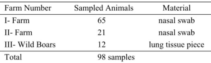
Sampled Animals: In this research 86 swab samples from 2 different pig-farms and 12 lung samples from wild boars were collected to detect PCV 2 (Table-1).
Table 1. Account of animals sampled Tablo 1. Örneklenen hayvan sayısı
Farm Number Sampled Animals Material
I- Farm 65 nasal swab
II- Farm 21 nasal swab
III- Wild Boars 12 lung tissue piece
Total 98 samples
History of herds: The two pig farms had different management conditions. The first farm had some management problems additional to breeding problems such as reproductive failure, low birth weight, stillbirths,
mummified pigs/foetal death and respiratory problems such as coughing, pneumonia/dyspnea rapid breathing, sneezing and blindness. Sixty-five swab samples were collected from this farm. The other farm was smaller and had fewer reproductive and respiratory problems. Twenty-one samples were collected from this farm. All the sampled animals were under 2 years old. There was no clinical history of the wild boars. The wild boars studied were thought to be older than 2 years.
Viral nucleic acid isolation: Swab samples and lung tissue pieces, brought to the laboratory, were examined with High Pure Viral Nucleic Acid Isolation kit (Roche-Cat. No.11 858 874 001) to isolate viral nucleic acid.
Detection of PCV by one-step PCR: Each of the nucleic acid extraction materials was amplified through PCR. To detect porcine Circovirus, one step-PCR (VeTek PCV Detection Kit, Cat No: D40062) method was applied. This kit is a generic kit for the detection of both PCV 1 and PCV 2 without differentiation. In this step all animals were examined for the presence of PCV.
Detection of PCV 2 by one-step PCR: Each of the PCV PCR positive samples was examined by one step PCR for PCV 2. For this purpose a VeTek PCV 2 detection Kit (Cat. No. D40110) was used.
Detection of amplified products: All of the one step PCR products were run on 1,5 % agarose gel electrophoresis, and the results were examined using an ultraviolet (UV) transilluminator.
Results
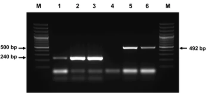
Results of One step PCR for PCV: Thirty-eight of 98 samples produced 240 bp PCR product like the positive control of the kit and were defined as positive (Table 2, Fig.1: lane 1,2,4,6). These 38 samples were all collected from the first farm, in which the animals had reproductive and respiratory problems. PCV could not be detected in the samples from the second farm or among the wild boars.
Fig 1. Electrophoresis of PCR product using PCV Detection Kit Lane M: 100 bp Molecular ladder
Lane 1,2,4,6: PCV positive sample Lane 3,5 : PCV negative sample Lane P: PCV positive control.
Şekil 1. Circovirus tespit kiti kullanılarak elde edilen PCR ürünlerinin elektroforez görüntüleri.
Table 2. PCR results of the sampled animals Tablo 2. Örneklenen hayvanların PCR sonuçları
Farm Number Sampled Animals PCV PCV 2
I- Farm 65 38 31
II- Farm 21 - -
III- Wild Boars 12 - -
Results of One-step PCR for PCV 2: The 38 samples which tested positive for PCV were analysed for PCV 2 using one step PCR. Thirty one of the 38 samples had an amplified product of 492 bp, the same as the positive control (Table 2, Fig.2: lane 5). Seven of the 38 samples tested negative for PCV 2 using PCR (Fig.2, lane 4).
Fig 2. Electrophoresis of PCR product using by PCV and PCV 2 Detection Kit
Lane M: 100 bp Molecular ladder Lane 1: PCV generic positive control
Lane 2: PCV positive sample (sample number is 26) Lane 3: PCV positive sample (sample number is 5) Lane 4: PCV-2 negative sample (sample number is 5) Lane 5: PCV-2 positive sample (sample number is 26) Lane 6: PCV-2 positive control
Şekil 2. PCV ve PCV 2 tespit kiti ile elde edilen PCR ürünlerinin elektroforez görüntüleri
Discussion and Conclusion
PCV 2 virus is a common cause of clinical signs such as wasting, growth retardation, anemia, pneumonia (3, 22, 23, 24), abortions and stillbirths (34) among pigs. The virus is associated with PPMWS (6, 11), PDNS (27) and reproductive disorders (34). PPMWS and PDNS are major health problems of late nursery and fattening stages, having a negative impact on pig-breeders (8).
In this research, we aim to detect PCV 2, recently agreed to be the primary agent of PPMWS (15), associated with late gestation abortions, stillbirths and respiratory problems (34). This is the first reported study in this field in Turkey.
For this purpose 98 pigs of a variety of ages were sampled. Sixty five of the sampled animals were herded in the first farm in which the animals had respiratory and reproductory problems. The second farm was less crowded and had fewer complaints than the first farm. Twenty-one animals were sampled in this farm. Twelve wild boars were also sampled for this research. PCV 2
was not detected at the second farm or among the wild boars.
The one-tube PCR detection of PCV kit is a generic kit that detects both PCV 1 and PCV 2 without distinction. PCV PCR negative samples were not examined to detect PCV 2 by PCR. Thirty-eight of 65 samples from the first farm, that experienced respiratory and reproductory problems, were identified as positive since they produced 240 bp PCR product like the positive control (Fig. 1, lane 1, 2, 4, 6). The positive samples were tested using one tube PCR for PCV 2. Thirty-one of the samples produced 492 bp PCR product and were determined to be positive (Fig. 2, lane 5, 6).
Seven samples which were positive for PCV (Fig. 2, lane 3) but negative for PCV 2 (Fig. 2, lane 4), implying that these samples were positive for PCV 1.
As a result of the PCR for the detection of PCV, 38 samples were found to be positive. Detection of PCV 2 in 31 out of 38 samples implied that the rest of the samples were positive for PCV 1. PPMWS is characterised by wasting, respiratory symptoms, diarrhea, paleness or icterus in pig populations after weaning (2, 6, 7, 9, 12). Simultaneous single or multiple coincidental bacterial infections increase the mortality among pigs during or after weaning (16, 18). PPMWS is usually combined with viral pathogens such as PRRS, swine influenza virus, porcine parvovirus (PPV) or bacterial pathogens such as Haemophilus parasuis, Actinobacillus pleuropneumonia, Streptococcus suis and Mycoplasma hyopneumonia (16, 25).
Huang et al. (14) collected lymph nodes, tonsils, lungs and spleen from 58 sick piglets. By multiplex PCR they detected PCV 2 in 30 samples (51.7 %), porcine pseudorhabdovirus in 1 sample (1.7%), PCV 1 and PCV 2 mix infection in 8 samples (13.8 %), PCV 2 and porcine parvovirus mix infection in 3 samples. None of these agents had been detected in the remaining 8 samples.
Cao et al. (5) researched on 137 piglets suffering PMWS. They used multiplex PCR method to detect PCV 2, PPV and PPRV. They detected PCV 2 in all the samples. Also, they defined 43 samples positive for PPV, all samples negative for PPRV, 11 samples negative for PPV but positive for PPRV, 35 samples positive for both PPV and PPRV. In the light of these results, PCV 2 is an important agent of PPMWS and agents such as PPRV and PPV are also associated with PPMWS.
In a herd, the detection of PCV 2 and the symptoms of respiratory and reproductive systems strengthen the likelihood of PPMWS. The first farm was experiencing not only management problems, but also reproductive failures such as, low birth weight, stillbirths, mummified pigs/foetal death and respiratory problems such as coughing, pneumonia / rapid breathing, sneezing and blindness. PPRS was also detected on this farm, along
with PCV 2 (unpublished data). Detection of PCV 2, respiratory and reproductive problems is indicative of PPMWS.
Stress is accepted as an important contributing factor to PPMWS. Disease stress, feed stress, environmental stress, crowded herds, earlier weaning and insufficient health care can also be factors for PPMWS separately or altogether.
The stress level should be minimized and the management quality for animal welfare and breeding should be maximized to reduce control PPMWS.
With further studies, this infectio agent’s role and its interaction with other agents will be well understood.
References
1. Allan GM, and Ellis JA (2000): Porcine circoviruses: a
review. J Vet Diagn Invest, 12, 3-14.
2. Allan GM, Kennedy S, McNeilly F, Foster JC, Ellis JA, Krakowka SJ, Meehan BM, Adair BM (1999)
Experimental reproduction of severe wasting disease by co-infection of pigs with porcine circovirus and porcine parvovirus. J Comp Pathol, 121, 1-11.
3. Allan GM, McNeilly F, Kennedy S, Daft B, Clarke EG, Ellis JA, Haines DM, Meehan BM, Adair BM (1998):
Isolation of porcine circovirus-like viruses from pigs with a wasting disease in the United states of America and Europe. J Vet Diagn Invest, 10, 3-10.
4. Calsamiglia M, Segales J, Quintana J, Rosell C, and Domingo M (2002): Detection of porcine circovirus types
1 and 2 in serum and tissue samples of pigs with and without postweaning multisystemic wasting syndrome. J
Clin Microbiol, 1848-1850.
5. Cao S, Chen H, Zhao J, Lü J, iao S, Jin M, Guo A, Wu B, and He Q (2005): Detection of porcine circovirus
type-2, porcine parvovirus and porcine pseudorabies virus from pigs with postweaning multisystemic wasting sydrome by multiple PCR. Vet Res Comm, 29, 263-269.
6. Clark EG (1996): Pathology of the post-weaning
multisystemic wasting syndrome of pigs. Proc West Can
Assoc Swine Pract, 22-25.
7. Clark EG (1997): Postweaning multisystemic wasting
sydrome. Proc Annu Meet Am Assoc Swine Pract,
499-501.
8. Drolet R, Thibault S, D’Allaire S, Thomson JR, Done SH (1999): Porcine dermatitis and nephropathy syndrome
(PDNS): An overview of the disease. J Swine Health Prod,
7, 283-285.
9. Ellis J, Hassard L, Clark E, Harding J, Allan G, Wilson P, Strokappe J, martin K, McNeilly F, Meehan B, Todd D, Haines D (1998): Isolation of circovirus from lesions of
pigs with postweaning multisystemic wasting syndrome.
Can Vet J, 39, 44-51.
10. Fenaux M, Halbur, PG, Gill M, Toth TE, Meng XJ (2000): Genetic characterization of type 2 porcine
circovirus (PCV 2) from pigs with postweaning multisystemic wasting sydrome in different geographic regions of North america and development of a differential PCR-restriction fragment length polymorphism assay to detect and differentiate between infections with PCV 1 and PCV 2. J Clin Microbiol, 38, 2494-2503.
11. Harding JC (1996): Post-weaning multisystemic wasting
syndrome (PMWS):Preliminary epidemiology and clinical presentation. Proc West Can Assoc Swine Pract, 21.
12. Harding J (1997): Post-weaning multisystemic wasting
syndrome (PMWS): Preliminary epidemiology and clinical presentation. Proc West Can Assoc Swine Pract, 28, 503.
13. Harding JCS, Clark EG (1997): Recognizing and
diagnosing post-weaning multisystemic wasting sydrome (PMWS). Swine Health Prod, 5, 201-203.
14. Huang C, Hung JJ, Wu CY, Chien MS (2004):
Multiplex PCR for rapid detection of pseudorabies virus, porcine parvovirus and porcine circoviruses. Vet
Microbiol, 101, 209-214.
15. Kennedy S, Moffett D, McNeilly E, Meehan, Ellis J, Krakowka S, Allan GM (2000): Reproduction of lesions
of postweaning multisystemic wasting syndrome by experimental infection of conventional pigs with porcine circovirus type 2 alone or in combination with porcine parvovirus. J Comp Pathol, 122, 9-24.
16. Kim J, Chung H.-K, Jung T, Cho W.-S, Choi C, Chae C (2002): Postweaning multisystemic wasting syndrome of
pigs in Korea: prevalance, microscopic lesions and coexisting microorganisms. J Vet Med Sci, 64, 57-62.
17. Larochelle R, Antaya M, Morin M, Magar R (1999):
Typing of porcine circovirus in clinical specimens by multiplex PCR. J Virol Methods, 80, 69-75.
18. Madec F, Eveno E, Morvan P, Hamon L, Blanchard P, Cariolet R, Amenna N, Morvan H, Truong C, Mahe D, Albina E, Jestin A (2000): Post-weaning multisystemic
wasting syndrome (PMWS) in pigs in France: clinical observations from follow-up studies on affected farms.
Livestock Prod Sci, 63, 223-233.
19. Mc Neilly F, Kennedy S, Moffet D, Meehan BM, Foster JC, Clarke EG, Ellis JA, Haines DM, Adair BM, Allan GM (1999): A Comparison of in situ hybridization and
immunohistochemistry for the detection of a new porcine circovirus in formalin-fixed tissues from pigs with post-weaning multisystemic wasting syndrome (PMWS). J Virol
Methods 80, 123-128.
20. Mc Nulty M, Dale J, Lukert P, Mankertz A, Randles J, Todd D (2000) Circoviridae. In Virus Taxonomy: Classification and nomenclature of viruses the seventh report of the International committee on taxonomy of viruses. MHV van Regenmortel, CM Fauquet, DHL Bishop, CH Calisher, EB Carstens, MH Estes, SM Lemon, J Maniloff, MA Mayo, DJ McGeoch, CR Pringle, RB Wickner, eds. San Diego: Academic Press, pp. 299-303. 21. Meehan BM, McNeilly F, Todd D, Kennedy S,
Jewhurst VA, Ellis JA, Hassard LE, Clark EG, Hanes DM, and Allan GM (1998): Characterization of novel
circovirus DNAs associated with wasting sydromes in pigs.
J Gen Virol, 79, 2171-2179.
22. Morozov I, Sirinarumiter T, Sorden SD, Halbur PG, Morgan MK, Yoon KJ, Paul PS (1998): Detection of a
novel strain of porcine circovirus in pigs with postweaning multisystemic wasting syndrome. J Clin Microbiol, 36,
2535-2541.
23. Onuki A, Abe K, Togashi K, Kawashima K, Tanehi A, Tsunemitsu H (1999): Detection of porcine circovirus
from lesions of a pig with wasting disease in Japan. J Vet
24. Ouardani M, Wilson L, Jette R, Montpetit C, Dea S (1999): Multiplex PCR for detection and typing of porcine
circoviruses. J Clin Microbiol, 37, 3917-3924.
25. Pallares FJ, Halbur PG, Oprıessnıg T, Sorden SD, Villar D, Janke BH, Yaeger MJ, Larson DJ, Schwartz KJ, Yoon KJ, Hoffman LJ (2002): Porcine circovirus
type 2 (PCV 2) coinfections in US field cases of postweaning multisystemic wasting syndrome (PMWS). J
Vet Diagn Invest, 14, 515-519.
26. Rosell C, Segales J, Plana-Duran J, Balasch M, Rodriguez-Arrioja GM, Kennedy S, Allan GM, MNeilly F, Latimer KS, Domingo M (1999):
Pathological, immunohistochemical, and in-situ hybridization studies of natural cases of postweaning multisystemic wasting syndrome (PMWS) in pigs. J Comp Pathol, 120, 59-78.
27. Rosell C, Segales J, Ramos-Vara JA, Folch JM, Rodriguez-Arrioja GM, Duran CO, Balasch M, Plana-Duran j, Domingo M (2000): Identification of porcine
circovirus in tissue of pigs with porcine dermatitis and nephropathy syndrome. Vet Rec, 146, 40-43.
28. Smith WJ, Thomson JR, Done S (1993): Dermatitis/
nephropathy syndrome of pigs. Vet Rec, 132, 47.
29. Thibault S, Drolet R, Germain MC, D’Allaire S, Larochelle R, Magar R (1998): Cutaneous and systemic
necrotizing vasculitis is swine. Vet Pathol, 35, 108-116.
30. Thomson JR, Lainson FA, Thomson N, Donachie W (1998): A study of Pasteurella multocida as a possible
aetiological agent in porcine immune complex glomerulonephritis and dermatitis syndrome. In: Dore S,
Thomson J, and Varley M (eds), Proceedings of the 15th IPVS Congress, vol. 3, p.396. Nottingham University press, Nottingham.
31. Tischer I, Bode l, Peters D, Pociuli S, Germann B (1995): Distribution of antibodies to porcine circovirus in
swine populations of different breeding farms. Arch Virol,
140, 737-743.
32. Tischer I, Gelderblom H, Vettermann W, and Koch MA (1982): A very small porcine circovirus with circular
single-stranded DNA. Nature, 295, 64-66.
33. Tischer I, Rasch R, Tochtermann G (1974):
Characterization of papovavirus- and picornavirus-like particles in permanent pig kidney cell lines. Zentralbe
Bakteriol Org A, 226, 153-167.
34. West KH, bystrom JM, Wojnarowicz C, Shantz N, Jacobson M, Allan GM, Haines DM, Clark EG, Krakowka S, McNeilly F, Konoby C, Martin K, Ellis JA (1999): Myocarditis and abortion associated with
intrauterine infection of sows with porcine circovirus 2. J
Vet Diagn Invest, 11, 530-532.
Geliş tarihi: 26.04.2010 / Kabul tarihi: 06.08.2010
Address for correspondence:
Doç.Dr. Taner Karaoğlu
Ankara Üniversitesi, Veteriner Fakültesi Viroloji Anabilim Dalı, Dışkapı, Ankara. Tel: +90 312.3170315/366