Versatility of high resolution ultrasonography in the assessment of granulomas and radicular cysts: a comparative in vivo study
Tam metin
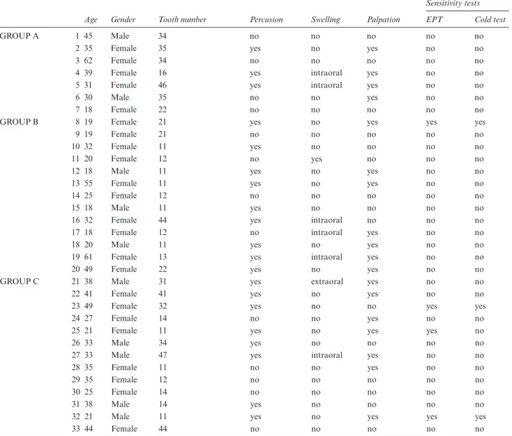
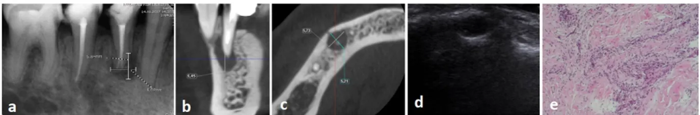
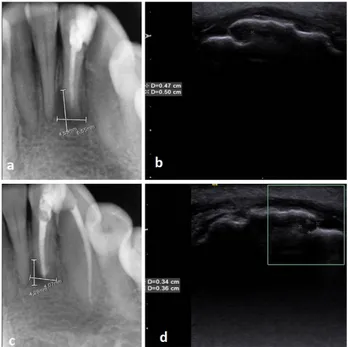
Şekil
Benzer Belgeler
(1987) made, "An Analytical Study of Traditional Muslim System of Education and its Relevance in the Modern Indian Context."3oi. Objectives: The objectives of the
而且需耗費您寶貴的時間清潔與保養,萬一在此時有感染或發炎的情形,更是得不償失。 每三十分鐘閱讀休息五分鐘:
[r]
Charpy, commandant le corps d ’occupation français, J l'amiral Duménil, commandant l ’escadre française du Levant, avec M“ Duménil, le général Filloneau, les
ARDL modeli sonuçlarına göre seçilen dönem için Türkiye’de, enflasyon ve ekonomik büyüme arasında hem kısa dönemde hem de uzun dönemde negatif yönlü ilişki olduğu
From the same figure, it is possible to notice the change in the density of solid waste for the two restaurants at the University of Tikrit, which is the subject of the study, as
The anthropometric pelvic measurements which are intercrestal diameter (IC), interspinous diameter (IS), intertrochanteric diameter (IT), intertuberous diameter (ITb),
In this study, we aimed to compare large hepatic hy- datid cysts (diameter ≥10 cm) and multiple cysts (>4) in giant hydatid cysts in terms of diagnosis, treatment,
