Abstract
I
ntRoductIonPilonidal sinus disease (PSD) occurs in the sacrococcygeal region. Hair shaft penetration through the skin leads to inflammation and infection and causes the disease. It is more often seen in young adult males. Factors such as the deep intergluteal fold and curved anatomic shape of the sacrococcygeal region may predispose pathogenesis of PSD.[1] Chronic PSD is accompanied with recurrent infection and prolonged inflammation which result in scarring and gradual spread of the disease toward neighboring soft tissue. These reactions cause the formation of multiple sinus tracts and result in persistent and continuous drainage that leads to substantial morbidity.[2] Many treatments have been advocated for PSD, but no consensus exists on this topic. Removal of the diseased tissue together with some healthy tissue is adopted an effective method for the complete eradication of the disease since recurrent disease constitutes a greater therapeutic challenge.[3,4] Therefore, primary closure may not be feasible after excision in some circumstances, and various flaps including Limberg flap, V-Y advancement flap, and Z-plasty have been described for closure of defects after excision.[5-7]
The aim of the current study is to describe the inferior gluteal artery perforator flap (IGAPF) as a new alternative method for closure of large PSD excision defects and to present surgical outcomes.
P
atIents andM
ethodsFifteen male patients treated surgically with IGAPF for PSD in our plastic, and reconstructive surgery departments between March 2014 and May 2017 were presented. None of the patients had any comorbidities. Descriptive data (age and gender), duration of follow-up, complications, and recurrence were noted together with an assessment of the size of the surgical defect. Our study follows the principles of the Declaration of Helsinki.
Favorable inferior gluteal perforating arteries were chosen as described by Allen et al.[8].Accordingly, a vertical line is
Introduction: The aim of the current study was to introduce the use of the inferior gluteal artery perforator flap (IGAPF) as a new alternative
surgical technique for closure of sacral defects after pilonidal sinus surgery. Patients and Methods: Inferior gluteal artery perforators were
used on 15 male patients operated in the plastic and reconstructive surgery department of our tertiary care centers between March 2014 and May 2017. Age, size of the defect, duration of follow-up, complications, and recurrence rate were assessed. Results: The average age was
30.2 (range: 17–54) years, and the mean duration of follow-up was 8.2 (range: 7–16) months. No recurrence was detected within the follow-up period, and the only remarkable complication reported was total flap necrosis attributed to venous congestion in one patient. The mean size of the defects after excision was 21.6 cm2. Conclusion: Our preliminary results imply that IGAPF can be a safe and effective alternative for
closure of large defects after pilonidal sinus surgery. Further controlled trials on larger series are warranted to establish the advantages and disadvantages of this alternative technique.
Keywords: Inferior gluteal artery perforator flap, perforator flap, pilonidal sinus disease, sacral defect
Address for correspondence: Dr. Yavuz Keçeci,
119/4 Sokak 1/1 C Blok Daire 8, Evka 3, Bornova, Izmir, Turkey. E‑mail: [email protected]
Access this article online Quick Response Code:
Website:
http://www.turkjplastsurg.org
DOI:
10.4103/tjps.tjps_36_18
This is an open access journal, and articles are distributed under the terms of the Creative Commons Attribution‑NonCommercial‑ShareAlike 4.0 License, which allows others to remix, tweak, and build upon the work non‑commercially, as long as appropriate credit is given and the new creations are licensed under the identical terms.
For reprints contact: [email protected]
How to cite this article: Bali ZU, Ahmedov A, Özkan B, Mazican M,
Keçeci Y. Inferior gluteal artery perforator flap for closure of sacral defects after pilonidal sinus surgery. Turk J Plast Surg 2019;27:9-13.
Inferior Gluteal Artery Perforator Flap for Closure of Sacral
Defects after Pilonidal Sinus Surgery
Zulfikar Ulas Bali, Anvar Ahmedov1, Burak Özkan2, Mustafa Mazican3, Yavuz Keçeci
Department of Plastic and Reconstructive Surgery, Manisa Celal Bayar University, Manisa, 1Department of Plastic and Reconstructive Surgery, Evliya Çelebi Hospital,
drawn over the buttock, between the ischial tuberosity and the posterior superior iliac spine on the volume-rendered image. A horizontal line that bisects the vertical line delineates four quadrants. Inferior gluteal artery perforator vessels originate in the inferolateral quadrant [Figure 1].
Preoperative Color Duplex Doppler ultrasonography examination (Philips Ultrasound, Andover, MA, USA) with a 12-3 MHz linear transducer was routinely performed for identification of the exact localization of the perforator vessels.
Surgical procedure
Patients were operated in prone position. After shaving of sacrococcygeal area, surface was cleaned with povidone-iodine, and methylene blue was injected to identify the sinus tracts. The excision margins were determined in a vertical ellipse as to include at least 1 cm of healthy tissue margin. Excision was carried out by means of a scalpel and wide local excision was aimed for removal of all tissues overlying the presacral fascia. Hemostasis was accomplished using electrocautery.
Preparation and harvesting of IGAPF were made as described in the literature.[8,9] A skin island was planned according to the size of the defect. A sterile pad was used as a template to confirm the accurate size and shape of the recipient site. The skin paddle of the flap was located on the perforator mark and designed with an extra 0.5 cm width around the border of the template to make certain that the flap had adequate skin to cover the defect without tension. Rotation arc and movement of the flap were simulated with sponges. Flap elevation started with an incision at the lateral side of the flap. The fat can be beveled around the flap to take the maximum amount of soft tissue in the flap if it is necessary. The incision was deepened down to the gluteus maximus muscle, and the dissection proceeded medially. The flap was elevated as a fasciocutaneous flap. Extreme care is necessary, during elevation, not to injure the musculocutaneous perforators. Loupe magnification was used for dissection of the perforators. Marginal perforators supposed to restrict the transposition or the advancement of the flap were ligated and cut. After finding them, the preoperatively selected perforating vessels were skeletonized by blunt dissection of the surrounding muscle fibers. They were followed up to the direction of its origin from the inferior gluteal artery until the flap had a freely enough movement to reach and cover the defect area. Then, the flap was tunneled beneath a skin bridge between the defect and the flap donor site. Donor site was closed primarily. Steps of surgical procedure, including the excision of the PSD and preparation and transfer of IGAPF, are shown in Figures 1-3. No drains were used since no dead spaces were created by the procedure. Patients remained prone or on their contralateral side by changing their position every 2 h in the postoperative period for approximately 2 weeks until the flap was healed. Subsequent to removal of the sutures, patients were instructed for oil massage for flaps, silicone gel application for scars, and depilation or shaving in the natal cleft. Figure 4 demonstrates the appearance of sacrococcygeal region at 6 months postoperatively.
R
esultsThe average age of patients was 30.2 (range: 17–54 years). Physical examination demonstrated draining sinuses with scar
Figure 1: Preoperative plan for excision of pilonidal sinus disease and
design of the inferior gluteal artery perforator flap
Figure 2: Preparation of the inferior gluteal artery perforator flap before
transfer to the recipient site after excision of pilonidal sinus
Figure 3: View after transfer of inferior gluteal artery perforator flap and
formation and skin discoloration. The mean follow-up period of the patients was 8.2 months (range: 4–16). The mean size of the defect after excision of PSD was 21.6 cm2 (range: 15–36). No recurrence was detected in the follow-up period. The only complication was total flap necrosis because of venous congestion seen in one patient. An overview of descriptive, clinical, operative, and histopathological data derived from our series is presented in Table 1.
d
IscussIonThe treatment for PSD is mostly surgical; however, no consensus exists on the selection criteria for the most appropriate method.[10] Predisposing factors for PSD include obesity, occupations requiring prolonged sitting, young males with positive family history, deep natal cleft, poor local hygiene, and excessive body hair.[1,2]
Pilonidal sinus does not exist on convex surfaces, and the main reason for an unsuccessful surgery is the deepness of the gluteal fold, which produces warm, damp, bacteria-friendly environment.[4] Therefore, the goal of surgical treatment is to excise the diseased tissue until the level of presacral fascia and to reduce the deepness of the gluteal fold that houses the problem to prevent recurrence. Nevertheless, the modality of management for the surgical defect is still under debate.[10] Surgical methods mainly consist of primary closure, leaving the wound open, and flap closure. Healing by the secondary intention causes prolonged duration of hospitalization with requiring daily wound dressing, loss of productivity, increased postoperative morbidity, and poor cosmetic outcome.[5] On the other hand, primary closure is a simple technique, but it is not feasible in large defects, and it has high rates of recurrence. Furthermore, suture line is at the midline, and wound dehiscence is not uncommon because of wound tension.[3] Common flap methods used for PSD reconstruction are V-Y advancement flap, Limberg flap, Z-plasty, and W-plasty.[5-7] In the Z-plasty and W-plasty procedures, a part of the suture line locates in the midline of the wound, and this may cause recurrence. A transposition flap technique, Limberg flap, is
appropriate only for the repair of rhomboid defects. Moreover, the Limberg flap needs excessive mobilization and necessitates experience with more excision of healthy skin. The V-Y advancement flap can be used in repairing large defects. It can be elevated safely without dissecting of the pedicle, and it has a shorter learning curve.[7] Nevertheless, especially in bilateral flaps, suture line is at or near the midline. These flaps can produce increased tension at wound margins. In all these flap procedures, adjacent tissue is used.
Musculo-fascio-cutaneous flaps have been described particularly for the management of larger defects and recalcitrant disease. In spite of the high cure rates, increased blood loss, sacrificing a part of functional muscle, and longer duration of hospitalization are the disadvantages of musculo-fascio-cutaneous flaps.[11] Due to the lacking of these disadvantages, perforator flaps raised on perforator arteries have been preferred to musculo-fascia-cutaneous flaps. Use of superior gluteal artery perforator flap has been defined for the repair of PSD excision defects.[12] However, the neighboring flap, IGAPF, has not been reported yet for the reconstruction of PSD surgical defects.
On the other hand, an healthy tissue can be left between flap donor site and defect area in perforator-based flaps. Furthermore, they have the advantage of transferring a completely healthy and bulky tissue to the defect area.[13] The subcutaneous fat provides sufficient bulk tissue without muscle sacrifice in the repair of deep defects. It allows a tension-free closure without leaving any dead space and improves wound healing and increases patient comfort. Besides removing existing pilonidal sinus, it also eliminates some of the predisposing factors for the development of another sinuses. This is accomplished by flattening the gluteal fold and locating the scars away from the midline. Furthermore, donor sites are closed primarily in most cases. In our series, donor sites were closed primarily in all patients.
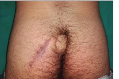
Figure 4: View at 6 months after surgery
Table 1: Overview of descriptive, histopathological, operative, and clinical data of our series
Patient
number Age Gender Follow‑up (months) Complication defect (cmSize of 2)
1 22 Male 12 - 36 (6×6)
2 19 Male 12 - 28 (7×4)
3 20 Male 13 - 24 (6×4)
4 41 Male 11 - 36 (6×6)
5 39 Male 7 - 24 (6×4)
6 22 Male 16 Total necrosis 28 (7×4)
7 44 Male 8 - 18 (4.5×4) 8 21 Male 16 - 15 (5×3) 9 23 Male 4 - 24 (6×4) 10 28 Male 5 - 21 (6×3.5) 11 45 Male 7 - 18 (4.5×4) 12 17 Male 8 - 28 (7×4) 13 54 Male 4 - 18 (4×4.5) 14 31 Male 5 - 22 (5×4.5) 15 27 Male 5 - 20 (5×4)
Besides these advantages, perforator flap elevation requires scrupulous technique and skill. Long-learning curve and partial or total necrosis risk are other disadvantages of these flaps. Perforator flap surgery should be avoided when the perforator arteries of the region are damaged due to the previous operation. Contralateral site perforator flaps or other conventional local flaps can be preferred in such cases. Superior gluteal artery perforators were described previously for reconstruction of pilonidal sinus defects.[12] There are some differences between superior and inferior gluteal artery perforators. Ahmadzadeh et al. revealed that the mean area nourished by the inferior and superior gluteal vessels is 177 ± 38 cm2 and 69 ± 56 cm2, respectively.[14] Georgantopoulou et al. found that IGAPF has a longer pedicle length than superior gluteal artery perforator flap.[15] Song et al. also reported that, in about two-thirds of the cases, inferior gluteal artery has a dominant pattern.[16] With this knowledge, we considered that perforator flaps of the inferior gluteal artery can be preferred to those of superior gluteal artery in local usage. Thus, this flap contributes surgeons as another perforator flap option for PSD surgery. Furthermore, we could find close perforators on the IGA route in these patients as seen in Figure 1.
Perforator vessels are marked in radiology department by color Duplex Doppler ultrasound. Lethaus et al. reported that color Doppler ultrasound is more precise and reliable than hand-held Doppler in detecting the anatomical position of perforating vessels.[17] Furthermore, hand-held Doppler has been found to be unreliable in detecting perforators in the gluteal region.[18] Therefore, color Doppler ultrasonography was chosen as the imaging modality for preoperative vascular mapping in the presented series.
We have come across with flap necrosis in one case due to venous congestion in the second day of the operation. We waited the congestion to recover, but we discovered a small volume of hematoma around the pedicle when we took the patient for the second operation for the exploration. Because the flap had completely filled the dead space, we had not used suction drains in the first operation. It seemed that a small volume of hematoma led to flap loss. This considered us that meticulous hemostasis and Penrose drain placement for 1 or 2 days would be precautionary against this complication since this flap is nourished only from the perforator vessels. Furthermore, when venous congestion develops on the perforator flap, the existence of hematoma, even a small volume, should be ruled out.
In our series, no recurrence was observed, and this can be explained by several factors. First, scar placement away from the midline might avoid forming a penetration area for hairs in the midline. Moreover, flattening gluteal fold owing to IGAPF setting may decrease moisture, local warmth, and hair accumulation. The lesser number of hair follicles at the flap site in comparison to the reconstructed area, which is seen in Figure 4, may be another factor that contributes to the avoidance of recurrence.
We suggest that the use of IGAPF as a new alternative method for the management of large defects after PSD excision. Furthermore, IGAPF may reduce recurrence rates since a moist and concave area is not formed at the operation site after the transposition of IGAPF. Classically, the perforator branch of the inferior gluteal artery nourishing the gluteal sulcus is used for reconstruction purposes.[8,19] Nevertheless, this donor site undergoes compression at sitting position and causes discomfort for the patients. In this presented series, IGAPF was harvested cranial to this site, so this type of flap design might be more comfortable to the patients since it is free from exposure to such a pressure.
Results of the current study demonstrate that our technique offers a safe and effective alternative for PSD defect repair. It allows a tension-free closure. This bulky flap does not leave a dead space and increases patient comfort and improves wound healing. Documentation of cost-effectivity and safety compared to other treatment modalities require controlled trials. Besides removing existing pilonidal sinus, it eliminates some of the predisposing factors for the development of another sinuses. This is accomplished by locating the scars away from the midline and reducing the depth of the gluteal fold. Even though this presented series consists of only primary cases, this procedure may be convenient for recurrent cases after an unsuccessful operation. This topic as well as the long-term cosmetic and functional outcomes need to be investigated in further studies.
The main limitations of the present study comprise a short duration of follow-up, a small sample size, and absence of a control group. However, the main objective of this report is not to compare the treatment methods but to present a new closure method for the PSD defects.
c
onclusIonIGAPF seems to be a promising alternative for the repair of large surgical defects of PSD. Further multicentric, prospective, controlled trials with a longer duration of follow-up are mandatory for making more accurate conclusions and establishing criteria for the selection of patients.
Financial support and sponsorship
Nil.
Conflicts of interest
There are no conflicts of interest.
R
efeRences1. da Silva JH. Pilonidal cyst: Cause and treatment. Dis Colon Rectum 2000;43:1146-56.
2. Akinci OF, Bozer M, Uzunköy A, Düzgün SA, Coşkun A. Incidence and aetiological factors in pilonidal sinus among Turkish soldiers. Eur J Surg 1999;165:339-42.
3. Iesalnieks I, Fürst A, Rentsch M, Jauch KW. Primary midline closure after excision of a pilonidal sinus is associated with a high recurrence rate. Chirurg 2003;74:461-8.
operation leading to cures. Arch Surg 2002;137:1146-50.
5. Fazeli MS, Adel MG, Lebaschi AH. Comparison of outcomes in Z-plasty and delayed healing by secondary intention of the wound after excision of the sacral pilonidal sinus: Results of a randomized, clinical trial. Dis Colon Rectum 2006;49:1831-6.
6. Mentes BB, Leventoglu S, Cihan A, Tatlicioglu E, Akin M, Oguz M,
et al. Modified limberg transposition flap for sacrococcygeal pilonidal
sinus. Surg Today 2004;34:419-23.
7. Sahasrabudhe P, Panse N, Waghmare C, Waykole P. V-y advancement flap technique in resurfacing postexcisional defect in cases with pilonidal sinus disease-study of 25 cases. Indian J Surg 2012;74:364-70. 8. Allen RJ, Levine JL, Granzow JW. The in-the-crease inferior gluteal
artery perforator flap for breast reconstruction. Plast Reconstr Surg 2006;118:333-9.
9. Unal C, Ozdemir J, Yirmibesoglu O, Yucel E, Agir H. Use of inferior gluteal artery and posterior thigh perforators in management of ischial pressure sores with limited donor sites for flap coverage. Ann Plast Surg 2012;69:67-72.
10. Ersoy OF, Karaca S, Kayaoglu HA, Ozkan N, Celik A, Ozum T, et al. Comparison of different surgical options in the treatment of pilonidal disease: Retrospective analysis of 175 patients. Kaohsiung J Med Sci 2007;23:67-70.
11. Rosen W, Davidson JS. Gluteus maximus musculocutaneous flap for the treatment of recalcitrant pilonidal disease. Ann Plast Surg 1996;37:293-7.
12. Acartürk TO, Parsak CK, Sakman G, Demircan O. Superior gluteal
artery perforator flap in the reconstruction of pilonidal sinus. J Plast Reconstr Aesthet Surg 2010;63:133-9.
13. Leow M, Lim J, Lim TC. The superior gluteal artery perforator flap for the closure of sacral sores. Singapore Med J 2004;45:37-9.
14. Ahmadzadeh R, Bergeron L, Tang M, Morris SF. The superior and inferior gluteal artery perforator flaps. Plast Reconstr Surg 2007;120:1551-6.
15. Georgantopoulou A, Papadodima S, Vlachodimitropoulos D, Goutas N, Spiliopoulou C, Papadopoulos O, et al. The microvascular anatomy of superior and inferior gluteal artery perforator (SGAP and IGAP) flaps: A fresh cadaveric study and clinical implications. Aesthetic Plast Surg 2014;38:1156-63.
16. Song WC, Bae SM, Han SH, Koh KS. Anatomical and radiological study of the superior and inferior gluteal arteries in the gluteus maximus muscle for musculocutaneous flap in Koreans. J Plast Reconstr Aesthet Surg 2006;59:935-41.
17. Lethaus B, Loberg C, Kloss-Brandstätter A, Bartella AK, Steiner T, Modabber A, et al. Color duplex ultrasonography versus handheld Doppler to plan anterior lateral thigh flaps. Microsurgery 2017;37:388-93.
18. Coşkunfirat OK, Ozgentaş HE. Gluteal perforator flaps for coverage of pressure sores at various locations. Plast Reconstr Surg 2004;113:2012-7.
19. Levine JL, Miller Q, Vasile J, Khoobehi K, Craigie J, Wise MW, et al. Simultaneous bilateral breast reconstruction with in-the-crease inferior gluteal artery perforator flaps. Ann Plast Surg 2009;63:249-54.