NOVEL BIOLOGICAL MATERIALS FOR FOOD AND
ENVIRONMENTAL APPLICATIONS
A THESIS
SUBMITTED TO THE MATERIALS SCIENCE AND NANOTECHNOLOGY
PROGRAM OF GRADUATE SCHOOL OF ENGINEERING AND SCIENCE
BILKENT UNIVERSITY
IN PARTIAL FULFILLMENT OF THE REQUIREMENTS
FOR THE DEGREE OF
MASTER OF SCIENCE
By
Özgün Candan Onarman Umu
I certify that I have read this thesis and that in my opinion it is fully adequate, in scope and in quality, as a thesis of the degree of Master of Science.
………. Assist. Prof. Dr. Turgay Tekinay
I certify that I have read this thesis and that in my opinion it is fully adequate, in scope and in quality, as a thesis of the degree of Master of Science.
………. Prof. Dr. Mahinur S. Akkaya
I certify that I have read this thesis and that in my opinion it is fully adequate, in scope and in quality, as a thesis of the degree of Master of Science.
………. Assist. Prof. Dr. Turgay Çakmak
Approved for the Graduate School of Engineering and Science: ……….
Prof. Dr. Levent Onural
iii
ABSTRACT
NOVEL BIOLOGICAL MATERIALS FOR FOOD AND
ENVIRONMENTAL APPLICATIONS
Özgün Candan Onarman Umu
M.S. in Materials Science and Nanotechnology
Supervisor: Assist. Prof. Dr. Turgay Tekinay
July, 2012
Probiotics are microorganisms that have many health benefits to their host, such as promoting normal intestinal microflora, inhibiting the growth of pathogenic microorganisms, improving digestion and stimulation of gastrointestinal immunity. Probiotic microorganisms include bacteria, fungi and yeast, and they are highly desirable to be used as animal feed supplements. For this application, Bacillus species are preferred since they are resistant to extreme environmental conditions due to their spore-forming capacity in addition to having other important probiotic characteristics. In the first chapter of this study, 84 independent bacterial colonies were obtained from different bovine chyme samples and among them 29 were determined as belonging to genus Bacillus. These isolates were principally screened for their antimicrobial activity against a group of selected bacteria including pathogenic organisms such as Salmonella enterica, Klebsiella pneumoniae, Pseudomonas aeruginosa and Staphylococcus aureus. 7 strains (STF4, STF8, STF9, STF10, STF15, STF25 and STF26) with
iv
highest antimicrobial activity were further tested for other probiotic properties. They were resistant to the gastrointestinal conditions and most of the antibiotics tested. In addition, they were checked for the presence of plasmids and performed cytotoxicity tests. We propose novel Bacillus strains that have potential to be used as probiotic organisms.
TNT is a hazardous nitroaromatic compound that can be found in soil, sediment and water due to extensive contamination from military munitions after the World War II. It has many negative health effects on almost all of the living organisms (e.g. bacteria, fungi, algae, animal and human). So far, bacteria, fungi and plants are commonly used for biodegradation process but only a little is known about effect of algae on this issue. However, algae can be used as a good alternative for bioremediation and biosensor purposes as they do not require advance technology and are effective in terms of cost. 5 different microalgae strains (STA1, STA2, STA3, STA4 and STA5) were tested in terms of survival in different TNT concentration and biodegradation capability of TNT. These strains were isolated from water contaminated with TNT obtained from the Brass Factory affiliated with Mechanical and Chemical Industry Corporation (MKE) located in Kırıkkale, Turkey. Even though these strains did not use TNT as carbon source for growth; they utilize it at different degrees for other metabolic activities. Moreover, the growth of STA2 strain was not inhibited by high TNT concentrations (up to 50 mg/L TNT).
v
ÖZET
GIDA VE ÇEVRE UYGULAMALARI İÇİN YENİ BİYOLOJİK
MALZEMELER
Özgün Candan Onarman Umu
Malzeme Bilimi ve Nanoteknoloji Programı, Yüksek Lisans
Tez yöneticisi: Assist. Prof. Dr. Turgay Tekinay
Temmuz, 2012
Probiyotikler, üzerinde yaşadıkları canlılara normal bağırsak mikroflorasına katkı sağlama, patojen mikroorganizmaların büyümelerini engelleme, sindirimi geliştirme ve sindirim sistemi bağışıklığını uyarma gibi bir çok yarar sağlayan canlılardır. Probiyotik canlılar; bakteri, mantar ve mayayı içerir ve bu canlılar hayvan yem katkı maddesi olarak büyük ölçüde talep görür. Bu uygulama için, önemli olan probiyotik özellikleri bulundurmaları yanında spor oluşturma kapasiteleri sayesinde Bacillus türleri tercih edilir. Bu çalışmanın ilk bölümünde farklı büyükbaş hayvan kimüslerinden 84 bağımsız koloni elde edildi ve bunların arasından 29 tanesinin Bacillus cinsine ait olduğu belirlendi. Bu izolatların öncelikle, Salmonella enterica, Klebsiella pneumoniae, Pseudomonas aeruginosa ve Staphylococcus aureus gibi patojenik bakterileri de içeren seçilmiş bir grup bakteriye karşı gösterdikleri antimikrobiyel aktiviteleri test edildi. En yüksek antimikrobiyel aktiviteyi gösteren 7 suş (STF4, STF8, STF9, STF10, STF15 ve STF26) diğer probiyotik özellikler için test edildi. Bu
vi
suşlar sindirim sistemi şartlarına ve test edilen çoğu antibiyotiğe karşı dirençli çıktılar. Ayrıca plasmid varlığı test edildi ve sitotoksisite test edildi. Probiyotik canlılar olarak kullanılabilecek potansiyeli olan yeni Bacillus suşları ileri sürdük.
TNT zararlı nitroaromatik bir bileşiktir, toprak, sediment ve su özellikle II. Dünya Savaşı’ndan sonra askeri cephaneler yüzünden geniş çapta bu bileşikle kirlenmiştir. TNT neredeyse bütün canlılarda (bakteri, mantar, alg, hayavan ve insan) bir çok sağlık problemine yol açar. Bugüne kadar bu bileşiğin temizlenmesinde genel olarak bakteri, mantar ve bitkiler kullanılmıştır ve bu konuda algler hakkında çok fazla bilgi bulunmamaktadır. Ancak, algler yüksek teknoloji gerektirmediği ve maliyet olarak uygun olduklarından dolayı biyolojik temizleme ve biyoalgılayıcı için iyi alternetiflerdir. 5 farklı mikroalg suşu (STA1, STA2, STA3, STA4 ve STA5,) farklı TNT konsantrasyonlarında yaşayabilirlikleri ve TNT’yi biyolojik olarak parçalayabilme kapasiteleri açısından test edilmiştir. Bu suşlar Makina ve Kimya Endüstrisi Kurumu’na bağlı Kırıkkale, Türkiye’de bulunan Pirinç Fabrikası’ndan elde edilen TNT’li sudan izole edilmişlerdir. Bu algler, TNT’yi büyüme için karbon kaynağı olarak olmasa bile başka metabolik faaliyetler için farklı derecelerde kullanmışlardır. Üstelik, STA2 alg suşunun büyümesi yüksek TNT konsantrasyonlarında (50 mg/L TNT’ye kadar) inhibe edilmemiştir.
Anahtar Kelimeler: probiyotik bakteri, Bacillus, yem katkı maddesi, TNT, mikroalg.
vii
ACKNOWLEDGEMENT
I would like to express the deepest appreciation to my supervisor Assist. Prof. Dr. Turgay Tekinay for his guidance, encouragement, support and warm personality during the course of this research.
I am very grateful to my dear friends Diren Han and Pınar Angün for their partnership and support in the first part of this study and unselfish and unfailing friendship from the day I knew them.
I also want to express my gratitude to Burcu Gümüşcü for her support and help for the second part of this study, Selma Bulut for her guidance and help for SEM photos, Zeynep Erdoğan for the help on HPLC studies and Zeynep Ergül Ülger for guidance and support in laboratory.
I want to thank to my other group members; Turgay Çakmak, Ömer Faruk Sarıoğlu, Alper Devrim Özkan, Ebuzer Kalyoncu, Berna Şentürk, Ahmet Emin Topal, Pelin Tören, Tolga Tarkan Ölmez and Ayşe Özdemir.
I would like to acknowledge to State Planning Organization (DPT) of Turkey for its support to UNAM Institute of Materials Science and Nanotechnology, MKEK and Ministry of Science, Industry and Technology of Turkey for their financial support by a SANTEZ project (No: 480 STZ 2009-2).
Finally, I would like to express my sincere gratitude to my dear husband Sinan Uğur Umu, he was always with me and nothing is enough to say for him to explain his meaning to me. I also would like to express my sincere gratitude to my parents, brother and sister for their presence, endless understanding and support. I am so glad you exist and accompany me.
viii
ix
TABLE OF CONTENTS
ABSTRACT ... iii ÖZET ... v ACKNOWLEDGEMENT ... vii TABLE OF CONTENTS ... ixLIST OF FIGURES ... xii
LIST OF TABLES ... xiv
CHAPTER 1 ... 1
Screening and Selection of Novel Bacillus Probiotic Strains Isolated from Bovine Chyme ... 1
1.1. INTRODUCTION ... 1
1.1.1. The History and the Definition of Probiotics ... 1
1.1.2. Probiotics in Animal Feed-As an Alternative to Antibiotics ... 4
1.1.3. The Effects of Probiotics on Health ... 4
1.1.4. Animal Probiotics ... 5
1.1.5. Mechanism of Probiotics ... 7
x
1.1.7. Safety of Probiotics ... 9
1.1.8. Development of Novel Probiotics ... 10
1.2. MATERIALS AND METHODS ... 12
1.2.1. Bacillus Isolation and Growth Conditions ... 12
1.2.2. Probiotic Properties of Isolates ... 13
1.2.3. Effect of Isolates on Intestinal Cells ... 18
1.3. RESULTS AND DISCUSSION ... 21
1.3.1. Bacillus Isolation and Growth Conditions ... 22
1.3.2. Probiotic Properties of Isolates ... 22
1.3.3. Effect of Isolates on Intestinal Cells ... 36
1.4. CONCLUSION ... 39
1.5. FUTURE PROSPECTS ... 40
CHAPTER 2 ... 41
2,4,6-trinitrotoluene (TNT) biodegradation and survival capability of microalgea isolated from TNT-contaminated water ... 41
2.1. INTRODUCTION ... 41
2.1.1. Microalgae ... 41
xi
2.1.3. Objective ... 46
2.2. MATERIAL AND METHODS ... 47
2.2.1. Algae and culture conditions ... 47
2.2.2. Absorbance Measurement ... 49
2.2.3. HPLC Analysis ... 49
2.2.4. Acetic Acid Concentration Determination ... 50
2.2.5. Growth with TNT ... 50
2.2.6. Growth in TAP medium with TNT ... 50
2.3. RESULTS AND DISCUSSION ... 51
2.3.1. Isolation of Microalgae ... 51
2.3.2. Acetic Acid Concentration Determination ... 52
2.3.3. Growth with TNT ... 55
2.4. CONCLUSION ... 69
2.5. FUTURE PROSPECTS ... 70
xii
LIST OF FIGURES
Figure 1. Cross-section of a Bacillus spore (79) ... 6
Figure 2. A,B,C,D,E,F represent antimicrobial inhibition degrees, ‘-, ±, +, +±, ++ and +++’ respectively. All antimicrobial results were discussed accordingly. ... 24
Figure 3. Phylogenetic tree of 7 isolates (STF4, STF8, STF9, STF10, STF15, STF25 and STF26) ... 25
Figure 4. SEM images of isolates. (a), (b), (c), (d), (e), (f) and (g) represent STF4 (Paenibacillus xylanexedens), STF8 (Bacillus subtilis), STF9 (Bacillus subtilis), STF10 (Bacillus licheniformis), STF15 (Bacillus pumilus), STF25 (Bacillus licheniformis) and STF26 (Bacillus pumilus) respectively. ... 27
Figure 5. Agarose gel electrophoresis image of plasmid profile of isolates. A, B, C, D, E, F and G represent STF4 (Paenibacillus xylanexedens), STF8 (Bacillus subtilis), STF9 (Bacillus subtilis), STF10 (Bacillus licheniformis), STF15 (Bacillus pumilus), STF25 (Bacillus licheniformis) and STF26 (Bacillus pumilus) respectively. ... 35
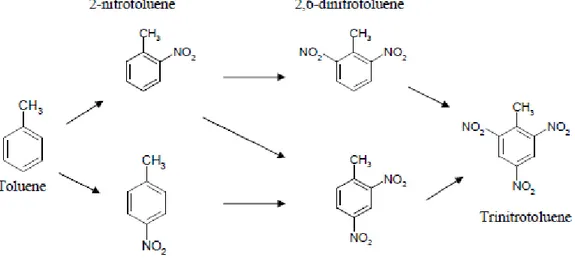
Figure 6. Formation of TNT from toluene (69). ... 42
Figure 7. Image of Pink Water ... 43
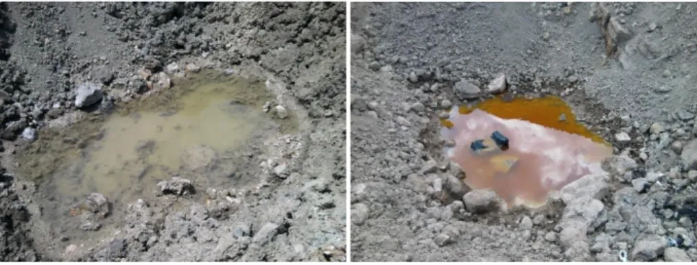
Figure 8. Images of water contaminated with TNT obtained from the Brass Factory affiliated with Mechanical and Chemical Industry Corporation (MKE) located in Kırıkkale, Turkey ... 51
xiii
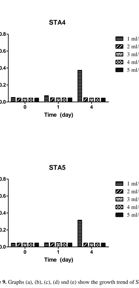
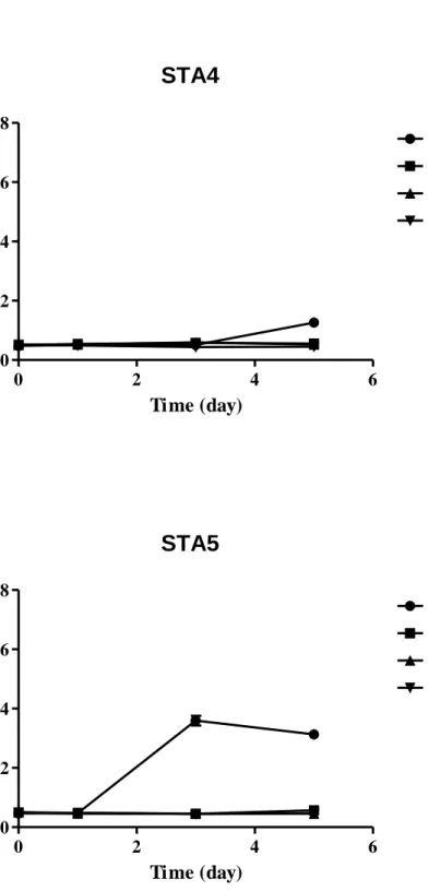
Figure 9. Graphs (a), (b), (c), (d) snd (e) show the growth trend of STA1, STA2, STA3, STA4 and STA5 respectively in TAP medium with different acetic acid concentrations ... 54
Figure 10. Graphs of (a), (b), (c), (d) and (e) represent the absorbance curves of STA1, STA2, STA3, STA4 and STA5 for nitrogen free medium, TAP medium and 25 mg/L, 50 mg/L and 100 mg/L TNT concentrations respectively ... 57
Figure 11. The bars of (a), (b), (c), (d) and (e) represent the medium TNT concentrations for STA1, STA2, STA3, STA4 and STA5 respectively ... 60
Figure 12. Graphs of (a), (b), (c), (d) and (e) represent the absorbance curves of STA1, STA2, STA3, STA4 and STA5 for TAP medium and 3 mg/L, 5 mg/L and 10 mg/L TNT concentrations respectively. ... 63
Figure 13. The bars of (a), (b), (c), (d) and (e) represent the medium TNT concentrations for STA1, STA2, STA3, STA4 and STA5 respectively. ... 66
Figure 14. Graphs (a) and (b) represent the absorbance curves of STA2 and STA3 respectively for nitrogen free medium, TAP medium and TAP medium with 3 mg/L, 5 mg/L, 10 mg/L and 50mg/L TNT concentrations. ... 68
xiv
LIST OF TABLES
Table 1. Microorganisms used in this study ... 14
Table 2. Inhibitory activity of isolates* ... 23
Table 3. Identification of selected isolates ... 26
Table 4. Identification and characterization of selected isolates ... 30
Table 5. The Microbiological Breakpoints Used by SCAN Categorizing Bacterial Species as Resistant ... 32
Table 6. Minimum inhibitory concentrations (MIC) for isolates ... 33
Table 7. Adhesion, Invasion and Cytotoxic Abilities* of Bacillus Isolates for HT-29 ... 38
1
CHAPTER 1
Screening and Selection of Novel Bacillus Probiotic Strains
Isolated from Bovine Chyme
Part of this study was submitted to be published as “Screening and Selection of Novel Bacillus
Probiotic Strains with High Antimicrobial Activity Isolated from Bovine Chyme” Ozgun C. O. Umu,Diren Han, Pinar Angun, Alper D. Ozkan and Turgay Tekinay.
1.1. INTRODUCTION
1.1.1. The History and the Definition of Probiotics
Probiotic as a word comes from the Greek ‘pro bios’ meaning ‘for life’ (33, 96). The history of probiotics began with the history of man; cheese and fermented milk were consumed by Greeks and Romans. The consumption of fermented products were preferred especially for children and convalescents (33).
The definition of the probiotic has been changed and enhanced in terms of content through the years. Firstly, Lilly and Stillwell (56) described probiotics as “substances secreted by one microorganism which stimulates the growth of another”. Later, probiotics have been generally defined as living organisms including bacteria and fungi providing benefits to host beyond inherent basic nutrition (23, 59, 65, 95).
Some of the published definitions are listed below (91).
Substances produced by microorganisms that promote the growth of other microorganisms (56).
2
Organisms and substances that contribute to intestinal microbial balance (72).
A live microbial feed supplement that beneficially affects the host animal by improving its intestinal microbial balance (32).
A viable mono- or mixed-culture of microorganisms that, applied to animal or man, beneficially affects the host by improving the properties of the indigenous microflora (42).
Living microorganisms that, upon ingestion in certain numbers, exert health benefits beyond inherent basic nutrition (38).
A microbial dietary adjuvant that beneficially affects the host physiology by modulating mucosal and systemic immunity, as well as improving nutritional and microbial balance in the intestinal tract (90).
A preparation of or a product containing viable, defined microorganisms in sufficient numbers, that alter the microflora (by implantation or colonization) in a compartment of the host and by that exert beneficial health effects in this host (96).
Live microorganisms that, when administered in adequate amounts, confer a health benefit on the host (29).
Currently, the lactic acid bacteria are the best-known probiotics, especially Lactobacillus sp. and Bifidobacterium sp.. However, Escherichia coli, Streptococcus sp., Enterococcus sp., Bacteroides sp., Bacillus sp., Propionibacterium sp. and various fungi are the other organisms that are used as probiotic (75). Moreover, mixtures of more than one bacterial strain can also be
3
used in some probiotic preparations (83). Probiotic-containing products are available for human nutrition, as animal feed supplements and also for aquaculture (83, 86, 104).
There are many criteria that influence the efficacy of a potentially useful probiotic. These are viability and reproducibility of probiotic on a large scale and during use and storage, ability of probiotic to survive in the intestinal ecosystem; and beneficial effect of probiotic on the host (33).
The potential benefits being claimed include improved nutrition and growth and prevention of various gastrointestinal disorders (23). Organisms considered as potential probiotic must have some characteristics. These are mentioned below (91).
Probiotics must be alive. The dead cells may also contribute physiologic benefits but they should not be called as probiotic.
Probiotics are administered. The native beneficial bacteria are not probiotics in their native state.
Probiotics must provide health benefit confirmed by tests on host.
Pharmaceuticals or topical agents are also admitted as probiotic like food supplements or oral delivery.
The mechanism of action should not be restricted by the definition of probiotics. The delivery of substances may also be probiotic activity even though the microorganism cannot survive in intestinal tract.
The composition of preparation of probiotic must be known. Deposit of a probiotic strain into an internationally recognized culture collection is
4
recommended. Moreover, the strain level should be defined for both consumers and scientists.
1.1.2. Probiotics in Animal Feed-As an Alternative to Antibiotics
The gastrointestinal tract infections and their consequences are still serious and difficult to treat clinical problems in both humans and livestock, despite of numerous therapeutic improvements, especially in the field of antibiotics (4, 5, 13, 83). Moreover, intense use of antibiotics has led to a dramatic increase in the number of antibiotic-resistant bacteria.
The antibiotics are not permitted to be used as feed supplement for livestock and these factors arose the concern of alternative products by researchers and feed companies (13, 93). European Parliament and the Council of the European Union encourage the development of alternative products to replace antibiotics as feed supplements for growth promotion (81, 93). An effective and safe alternative to antibiotic implementation is deliberately feeding probiotics which protect the animal from pathogens by improving microbial balance in the gastrointestinal tract to exclude potentially harmful bacteria (13, 68, 75, 93). Moreover, probiotics eliminate the negative effect of antibiotics which is the further delay of recolonization by normal colonic flora (83).
1.1.3. The Effects of Probiotics on Health
Probiotics influence health of host organisms by improving normal intestinal microbiota, preventing growth of pathogenic microorganisms, controlling infections (prevention of many diseases such as inflammatory bowel disease,
5
mastitis and miscarriage), promoting digestion and intake of feed, inducing the immune system decreasing lactose intolerance, lowering serum cholesterol, reducing diarrheal incidence, acting as antibiotics, suppressing tumors and protecting against colon/bladder cancer (50, 59, 83, 95). Therefore, the use of probiotics on livestock enhances the growth of animals, improves efficiency of feed conversion and decreases the rate of mortality (50, 59).
1.1.4. Animal Probiotics
All animals have micro flora in their intestines to be protected from pathogens and to regulate digestive system. Although protective flora which establishes itself in gut is very stable, it can be influenced by some dietary and environmental factors. The most important factors are excessive hygiene, antibiotic therapy, pathogens and stressful conditions (59). Therefore, protection and restoring is required for gut micro flora, and it is provided by supplementation of probiotic bacteria with feed.
Moreover, in growing animals, the stabilization of the intestinal microflora leads to a high performance level and economic benefits by increasing daily weight gain and the reduction of both fattening period and feed expenditure. Also, medication and the corresponding costs may be reduced by the use of probiotics (79).
Less well known than the Lactobacilli and Bifidobacteria, there are certain species of the spore-forming Bacillus genus that are being used as probiotics, notably Bacillus clausii, Bacillus subtilis, Bacillus pumilus, Bacillus coagulans
6
(often mislabeled as ‘Lactobacillus sporogenes’) and B. cereus (45, 91). The use of B. subtilis is approved for use as a food supplement in at least one European country (Italy), but for other species, this is not the case with the exception of B. clausii that is licensed as a prophylactic.
Bacillus sp. is sporulated living microorganisms. Spore form is a natural stable form allowing them to survive in harsh environment, protected from extreme heat, cold and mechanical strain without any loss in their vital potential. In spore form, various cell walls (Figure 1) protect the nucleus from external stresses.
Figure 1. Cross-section of a Bacillus spore (79)
This natural protection enables the Bacillus products to withstand massive strains during feed production and storage, such as high temperatures, pressure, shear forces or oxidation impacts (61, 64, 79, 100). Therefore, for all types of feed Bacillus spores are suitable and Bacillus sp. is advantageous. Moreover, their viability is not affected negatively in low pH values in stomachs of animals. Fermentation conditions during production of Bacillus products affect the quality and stability of spores (79).
External spore wall Internal spore wall Cell wall
Spore cortex
Membrane of the cytoplasma Cytoplasma with nucleus structures
7
1.1.5. Mechanism of Probiotics
Despite the fact that probiotics provide health benefits to their hosts, little is known about the molecular mechanisms of the benefits (3) and also much work remains to classify the mechanisms of action of particular probiotics against particular pathogens. Moreover, probiotics may use different mechanisms to inhibit different pathogens as well as combinations of different mechanisms which make the system very difficult and complex (43, 83).
The mechanisms of probiotics can be categorized based on specific effects of the bacteria on the microbial milieu, intestinal epithelium, immune response, allergic diseases, distant mucosal sites, and cancer (67).
Antimicrobial effects of probiotics (33, 43, 83, 95);
Modification of microflora to suppress pathogens Production of antimicrobial substances
Competition with pathogens to prevent their adhesion to the intestine (20, 35, 52)
Inhibition of epithelial invasion of pathogens
Competition for nutrients necessary for pathogen survival Antitoxin effect (11, 12, 77)
Effect of probiotics on the intestinal epithelium (33, 83);
Promotion of tight contact between epithelial cells forming a functional barrier
8
Reduction of the secretory and inflammatory consequences of bacterial infection
Enhancement of the production of defensive molecules such as mucins Increase in brush border enzyme production
Immune effects of probiotics (31, 33, 43, 48, 57, 63, 76, 78, 83, 88, 95);
Probiotics as vehicles to deliver anti-inflammatory molecules to the intestine
Enhancement of signaling in host cells to reduce inflammatory response Switching in immune response to reduce allergy
Inducing antibody response to reduce infection Reduction the production of inflammatory substances.
1.1.6. Characteristics of Probiotics
A potential probiotic strain is expected to have some desired properties in order to provide health benefits to its host. The most important properties of probiotics are listed below (67) although a potential probiotic strains does not need to fulfill all such selection criteria (71).
Human origin is desired for safety for human use (37). This criterion is important for species depending on health effects.
Acid and bile tolerance is important to predict good survival during gastrointestinal transit for oral consumption and also to maintain adhesiveness and metabolic activity through this tract (37).
9
Adhesion to mucosal surface is important to improve immune system, compete with pathogens, maintain metabolic activity, and prevent pathogens to adhere and colonize. The microorganisms of probiotics are tested for their ability to colonize on intestinal epithelia to permanently establish in the host over time without the need for periodic reintroduction of the bacteria (8, 15, 22). Identification and characterization of strains accurately and documentation of safety is important for food, feed and clinical use, there must be no invasion and no degradation of intestinal mucus.
Good technological properties are desired. Probiotics should be stable and active during processing and storage; if viable organisms are required, phage resistance, strain stability, cultivable in large scales and oxygen resistance are important and they should have no negative effects on product flavor.
Activity against pathogenic bacteria is an important criteria, this can be by several different mechanisms; directly by producing bacteriocins or antibiotics, by a competitive mechanism of adhesion, by competitive nutrition or indirectly by modulating the local immune system. (25, 50, 75, 98).
1.1.7. Safety of Probiotics
Safety is an important requirement for probiotics. Antibiotic resistance and pathogenicity are the main safety concerns of probiotics. The passage of antibiotic resistance genes to other pathogenic microorganisms most probably by plasmids (5, 45, 73, 97) and pathogenicity (89) may result with serious health problems to host .
10
1.1.7.1. Antibiotic gene resistance of probiotics
Antibiotic resistance may exist among probiotic microorganisms as with any bacteria (90). Chromosomal, transposon or plasmid located genes may be responsible from the resistance. When dealing with selection of probiotic strains, it is recommended that probiotic bacteria should not harbour transmissible resistance genes to other intestinal and/or food borne microorganisms. The transmissible antibiotic resistance gene carrier bacteria should not be used in foods and animal feeds (105).
1.1.7.2. Pathogenicity of probiotics
The microorganisms can be grouped into three in terms of safety. These are nonpathogenic microorganisms, opportunistic pathogens, and pathogens. Every microorganism that can live and grow in a host can cause an infection under certain conditions such as the case of severely immunocompromized hosts. Most intestinal microorganisms are not pathogenic to host who is healthy. The normal immune of host influence the growth and metabolism of potential pathogens. Current probiotics are generally considered as safe. Moreover, non-viable or inactivated microbial preparations are rarely give harm to host and they are least likely to cause safety concerns(89).
1.1.8. Development of Novel Probiotics
High meat quality, high product yield, improvement of growth and health of animals can be provided using probiotic supplementary in diet of animals. Probiotics are also effective on prevention of viral illnesses, pathogen growth,
11
digestive tract problems, and immune deficiency problems. Therefore, these organisms have significance in food industry, farming and for pets.
Recently, by development of biotechnology, importance of probiotics has taken more attention. As a result, number of scientific studies about isolation of new probiotic species has increased. Inspired by this, in this part of the thesis, isolation and characterization of new probiotic bacteria strains of Bacillus sp. from chyme samples of animals is aimed. For animals, probiotics provide many health benefits and also economical gains such that they increase daily weight gain and reduce of feed expenditure. Moreover, Bacillus sp. has many advantages over other probiotic bacteria strains because of its spore formation ability. It can easily survive in extreme physical and chemical conditions such as the one of gastrointestinal tract and the probiotic production processes such as storage.
12
1.2. MATERIALS AND METHODS
1.2.1. Bacillus Isolation and Growth Conditions
Microorganisms were isolated from chyme samples of male cattle between 2 and 4 years of age. The small intestine was removed as a whole and chyme samples were collected by squeezing the intestinal contents into sterile tubes. Isolation of the microorganisms was performed as described by Lalloo et al. (53) with slight modifications. 1 g of chyme fluid from each sample was suspended into 3 ml of 0.9% NaCl solution and inoculated into 9 ml of nutrient broth (NB). Samples were incubated at 37ºC for 24 h, followed by incubation at 45ºC for 10 min to initiate spore formation. 50% (v/v) ethanol was added to a volume of 20 ml and the suspension was incubated at 20ºC for 1 h. Centrifugation was performed at 12000 g for 30 sec, the supernatant was decanted and the pellets were incubated at 105ºC for 5 min. Dry pellets were resuspended into 20 ml of 0.9% NaCl solution and serially diluted in ten-fold increments.
For Bacillus isolation, MYP (Mannitol Egg Yolk Polymyxin) agar which is composed of 225 ml melted base (10 g/L peptone from casein, 1 g/L meat extract, 10 g/L D-mannitol, 10 g/L NaCl, 0.025 g/L phenol red and 12 g/L agar), 2.5 ml of 0.1% Polymyxin B Solution and 12.5 ml of 50% Egg-yolk Emulsion was used. Polymyxin in this agar inhibits the growth of gram-negative bacteria while Bacillus species are differentiated according to their ability to ferment mannitol or degrade lecithin. B. subtilis ferment mannitol result in acid production and produce yellow colonies while B. cereus is typically mannitol negative and result in pink colonies. 150 µl of each dilution was spread onto
13
MYP agar plates. Plates were incubated at 37ºC for 24 h. Single colonies having distinct morphologies were observed on MYP agar plates, collected and transferred onto Luria-Bertani (LB) agar plates. Cultures were incubated at 37ºC, 125 rpm and stored at 4ºC to maintain viability. For long-term storage, cultures were maintained at -80ºC in 30% glycerol.
1.2.2. Probiotic Properties of Isolates
1.2.2.1. Antimicrobial Activity
Antimicrobial activity assay was adapted from the method described by Saravanakumari et al. (94). Overnight incubated cultures of the isolates were horizontally streaked onto LB agar and incubated at 37°C for 48 h. After the incubation period, the plates were exposed to chloroform vapor for 90 min for the inactivation of active cells. The plates were aerated for 20 min to completely remove the residual chloroform by vaporization and the plate covers were changed (5). After that, representative bacteria (Table 1) were streaked vertically on the horizontally streaked cells and the plates were incubated at 37°C for 24 h. The inhibition on the growth line of bacteria (Table 1) was assessed for antimicrobial activity of isolates.
14 Table 1. Microorganisms used in this study
Bacterial species Strains Origina
Bacillus subtilis RSHM 03013 (ATCC 6633) RSHM
Staphylococcus aureus RSHM 96090/07035 (ATCC 25923) RSHM Escherichia coli RSHM 888 RSHM Pseudomonas aeruginosa RSHM 03015 (ATCC 29212) RSHM Klebsiella pneumoniae RSHM 06017 (ATCC 10031) RSHM Salmonella enterica RSHM 4059 (ATCC 13311, NCTC 74) RSHM
a
RSHM, National Type Culture Collection Laboratory, Ankara, Turkey.
1.2.2.2. Sporulation Efficiency
Sporulation efficiencies of the isolates were measured as described by Barbosa et al. (5). The isolates were inoculated (0.1% v/v) in Difco Sporulation Medium (DSM) and incubated at 125 rpm and 37°C for 24 h. After 24 h, serial dilutions were prepared in ten-fold increments and spread onto LB agar before and after heat exposure at 80°C for 20 min. The plates were incubated at 37°C overnight and colony forming unit (cfu) numbers were determined. Growth before and after heat was compared via the cfu numbers obtained to assess the sporulation capacities of the isolates.
15 Spore Formation
For the purpose of testing the tolerance of spores to simulated gastric fluids and bile salts, spore formation procedure was performed. Cultures were grown in 50-ml DSM medium at 37°C, 250 rpm for 1 day and subsequently centrifuged at 1500 g for 5 min to refresh the medium of spores. Harvested spores were then inoculated into DSM medium and incubated at 37°C, 250 rpm for 2 days. After the incubation period, spores were centrifuged at 5000 g for 30 min. The spore pellets were washed with 20 ml sterile distilled water and suspended again into 5-ml sterile water. Spores of each isolate were stored at 4°C for further use.
1.2.2.3. Resistance to Simulated Gastric Fluids
Vegetative cells of the isolates were tested for survival capacity in simulated gastric fluids. The method was performed as described by Barbosa et al., Hong et al. and Patel et al. (5, 46, 73) with slight modifications. Briefly, overnight grown bacteria were inoculated in LB broth (pH=2.0, adjusted with 37% HCl) and the cultures were incubated at 37°C, 125 rpm for 30 min. Samples were then serially diluted, spread onto Luria-Bertani (LB) agar and incubated at 37°C for 24 h. Colonies were counted and compared with those of control cultures.
The tolerance of spores to simulated gastric fluids was also tested as described in Barbosa et al., Duc et al., Hong et al. and Patel et al. (5, 23, 46, 73). ~ 108 to 109 purified spores per ml (48-h spores) were resuspended into 0.85% NaCl solution (pH = 2 adjusted with HCl), incubated and spread onto LB agar plates as described for vegetative cells. Controls were spores that were resuspended into 0.85% NaCl solution only. Cfu counts were carried out and compared with
16
those of the controls’ to assess the tolerance of spores to simulated gastric fluid conditions.
1.2.2.4. Bile Salt Tolerance
Bile salt tolerances of vegetative cells were tested as described by Barbosa et al. (5). Overnight cultures of isolated bacteria were inoculated into fresh LB supplemented with 0.2% bile salts (50% cholic acid sodium salt, 50% deoxycholic acid sodium salt; Sigma-Aldrich, USA) and incubated at 37°C, 125 rpm. Controls, which are bacteria inoculated into fresh LB, were incubated in parallel. Aliquots were taken after 3 h of incubation. In order to determine the number of cfu, samples were serially diluted and plated onto LB agar plates. Cfu counts were done after incubation at 37°C for 24 h.
Bile salt tolerances of spores were also assayed as described by Barbosa et al. (5). Spores were purified as described by Henriques et al. (44) and ~ 108 to 109 purified spores per ml (48-h spores) were resuspended into an isotonic buffer (Bott and Wilson salts: 1.24% K2HPO4, 0.76% H2PO4, 0.1% trisodium citrate, 0.6% [NH4]2SO4, pH=6.7) containing 0.2% bile salts (50% cholic acid sodium salt, 50% deoxycholic acid sodium salt; Sigma-Aldrich Co.). The spores were incubated at 37°C, 125 rpm. Controls, which are spores resuspended into Bott and Wilson salts only, were incubated in parallel. Aliquots were taken at 0, 1, 3h and cfu count was done.
17
1.2.2.5. Biofilm Formation
Biofilm formation abilities of isolates were tested as described by Barbosa et al. (5) with slight modifications. Instead of Sterlini-Mandelstam medium, a biofilm growth medium consisting of Luria-Bertani (LB) medium supplemented with 0.15 M ammonium sulfate, 100 mM potassium phosphate, pH=7, 34 mM sodium citrate, 1 mM MgSO4 and 0.1% glucose (39) was used in this study. Isolates were grown in this biofilm growth medium to exponential phase and transferred into 3 ml of the same medium. Cultures were incubated at 37ºC without agitation to an optical density of ~0.01 at 600 nm. An uninoculated culture tube and a culture tube inoculated with exponential phase cells were prepared 10 min before washing to be used as controls. Tubes were rinsed with water and stained with a 1% crystal violet solution for 15 min. Excess stain was then removed by washing. A ring of staining on the medium suggests that bacteria are capable of forming biofilm.
1.2.2.6. Antibiotic Susceptibility
Antibiotic resistances of the isolates were tested as described by Chaiyawan et al. and Santini et al. (13, 93) with modifications. Ampicillin, kanamycin, chloramphenicol, erythromycin, vancomycin, neomycin, ciprofloxacin, streptomycin and gentamicin were the antibiotics tested. Serial dilutions of antibiotics were prepared in concentrations ranging between 1 µg/ml and 256 µg/ml. 160 µl of fresh LB broth, 20 µl of appropriate dilutions of antibiotics, and 20 µl of overnight grown isolates were added in each well of 96-well microtiter plates. Before adding bacteria, overnight cultures were diluted to 106
18
cfu/ml in fresh LB broth by means of OD620 measurements. The microtiter plates were incubated at 37ºC for 24 hours and the minimum inhibitory concentrations of each antibiotic were determined by measuring the growth of isolates at OD620 and comparing them to control wells which are only bacteria and LB broth, as positive and negative controls respectively.
Plasmid DNA Extraction and Purification
Isolates were grown overnight at 37°C and 250 rpm in LB broth. 16-18 h grown bacteria were centrifuged at 12000 g for 3 min to obtain cell pellets. Plasmid DNA was extracted from the pellet of each isolate by using the QIAprep Spin Miniprep Kit (QIAGEN). Gel electrophoresis was performed to detect whether the isolates carry plasmid DNA or not.
1.2.3. Effect of Isolates on Intestinal Cells
1.2.3.1. Adhesion and Invasion
Adhesion and invasion assays of Bacillus isolates were performed as described by Rowan et al. (84, 85) with slight differences. HT-29 cell monolayers were grown in DMEM supplemented with 10% FBS and 1% penicillin-streptomycin at 37ºC in 5% CO2 environment. Cells were seeded in 24-well plates containing approximately 3x10⁵ cells per well and incubated overnight at 37ºC in 5% CO2. Before adhesion and invasion assays, HT-29 cell monolayers were washed three times with DMEM and inoculated with 1 ml of bacterial cultures that were previously washed with DMEM containing 10% FBS. 24-well plates were incubated at 37 ºC in 5% CO2
19
were washed with DMEM to remove nonadherent bacteria. For adhesion assay, cells with adhered bacteria in 1 ml of DMEM supplemented with 10% FBS were incubated for another 2 h at 37ºC in 5% CO2. For the invasion assay, infected cells were incubated for 2 h in 1 ml of DMEM containing 10% FBS and 100 µg of gentamicin per ml at 37ºC in 5% CO2. Cells were then washed three times with DMEM and lysed with 1 ml of 1% (v/v) Triton X-100 for 5 min at 37ºC. Samples from lysed cells were serially diluted and spread onto BHI agar plates for cfu count.
1.2.3.2. Cell Cytotoxicity
Cytotoxic effects of isolates were tested as described by Rowan et al. and Hong et al. (46, 84, 85) with modifications. An MTT based in vitro toxicology assay kit (MTT; Sigma) was used to measure total cellular metabolic activity, the presence of which directly corresponds to the lack of cytotoxic effects. HT-29 cell monolayers were grown in DMEM supplemented with 10% FBS and 1% penicillin-streptomycin at 37ºC in 5% CO2 environment. The cells were then transferred to 96 well-plates with approximately 5x10⁴ cells in each well and incubated overnight at 37ºC in 5% CO2 atmosphere. Filter sterilized (by 0.2 µm pore size membranes) supernatants of overnight grown bacteria (BHI, 37ºC, and 125 rpm) were added to test plates before and after heat treatment (95ºC, 10 min). 4 replicates were done for supernatant of each isolate. 1% Triton X-100 (Sigma) and PBS were used as positive and negative controls, respectively. Cells infected with bacterial supernatant were incubated overnight at 37ºC in 5% CO2. After the incubation period, media in each well were removed and 100 µl
20
of DMEM supplemented with PBS containing 0.5 % MTT was added to wells. Plates were incubated at 37°C for 4 h. 100 µl of MTT solubilization solution (Sigma) was also added to wells after 4 h. The absorbance of the wells was measured at 570 nm by using microplate reader (SpectraMax M5, Spectra Lab.). Background absorbance of well plates was also measured (at 690 nm) and subtracted from 570 nm measurements. Toxicities of the supernatants on HT-29 cells were determined by using the equation (85):
(
21
1.3. RESULTS AND DISCUSSION
Use of Bacillus sp. as probiotics is increasing since these species are spore-formers which make them more effective in surviving in harsh conditions. Bacillus species have been used in some commercial probiotic products (21). Bactisubtil (B. cereus strain IP5832), Enterogermina (B. clausii), Biosubtyl Nha Trang (B. pumilus), Bispan (B. polyfermenticus) and Toyocerin (Bacillus cereus var. toyoi) are some examples of commercially available Bacillus probiotics (21, 23). Moreover, the preparation of natto, a fermented soybean product originating in Japan, involves the use of B. subtilis to facilitate the fermentation process, indicating that Bacillus species have been safely used in foodstuff for a considerable period of time (46).
It was reported previously that Bacillus species that have different probiotic characteristics were isolated from the GIT of broilers and fish (5). In this study, Bacillus strains were isolated from cattle chyme and screened them for probiotic potential, including antimicrobial activity. Isolation of novel strains with high antimicrobial activity against pathogenic bacteria such as S. aureus, P. aeruginosa, S. enterica, K. pneumoniae and other probiotic characteristics from bovine chyme is important for the development of probiotic bacteria as feed supplements. Moreover, the commercial product Toyocerin (Bacillus cereus var. toyoi) which is the first probiotic to receive authorisation in the European Union to be used as an additive in animal nutrition and is one of the most studied at scientific level was tested in terms of antimicrobial activity and resistance to antibiotics and compared with isolates.
22
1.3.1. Bacillus Isolation and Growth Conditions
For potential Bacillus probiotic isolation, chyme samples of male cattle between 2 and 4 years of age were used as sources. Samples were collected from the chyme of 8 different cattle and 84 independent isolates were obtained. Isolates were grown on MYP agar, 29 of them were found to be the members of the genus Bacillus. When heat treated and grown in DSM broth, all 29 isolates were observed as spore-formers.
1.3.2. Probiotic Properties of Isolates
1.3.2.1. Antimicrobial Activity
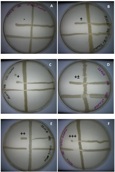
Antimicrobial effects of the selected Bacillus isolates and the commercial probiotic product Toyocerin (Bacillus cereus var. toyoi) were tested against different indicator strains, including both gram-negative and gram-positive bacteria (Table 1). Although all isolates showed some antimicrobial activity, 7 (STF4, STF8, STF9, STF10, STF15, STF25 and STF26) showed strong inhibitory activity among the 29 strains and these 7 strains also showed higher antimicrobial activity than Toyocerin against some of the indicator strains (Table 2). Representative photos of inhibition zones were shown in Figure 2.
23 Table 2. Inhibitory activity of isolates*
Isolates Indicator Strains
S. aureus P. aeruginosa E. coli B. subtilis K. pneumoniae S. enterica
STF1 - - - + - - STF2 - - - - STF3 - - - - STF4 ++ ++ ++ - ++ - STF5 + ± - ± ± - STF6 ± ± - + ± ± STF8 ++ ± + +++ - ± STF9 + - - ++ - - STF10 +± ± ± ++ - - STF11 - - - - ± - STF12 ± + - ± - ± STF13 + - - ± +± ± STF14 + - - - + ± STF15 +++ ± ± ± - - STF16 ± - - ± - ± STF18 ± - - - - ± STF19 ± - - ± ± ± STF20 - - + ± ± ± STF21 ± + - - - - STF22 ± +± - - ± ± STF23 - + - + +± - STF24 - + + - - - STF25 ± ± + +++ - - STF26 +++ ± +++ +± ++± ++ STF27 + - - + - - STF28 - ± - ± + - STF29 ± ± - ± - ± Toyocerin + + + +± + +
*The signs reflect the degrees of inhibitory effect on growth from “– ”
(no inhibition) to “+++” (increased inhibitory activity by increased inhibition zone). All results were performed in triplicate.
24
Figure 2. A,B,C,D,E,F represent antimicrobial inhibition degrees, ‘-, ±, +, +±, ++ and +++’ respectively. All antimicrobial results were discussed accordingly.
A
±
+++
+±
+
++
B D C F E--
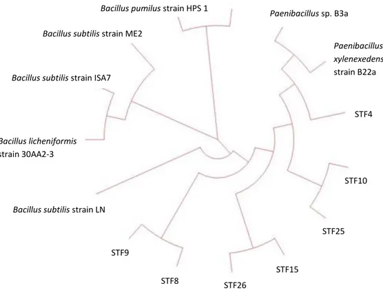
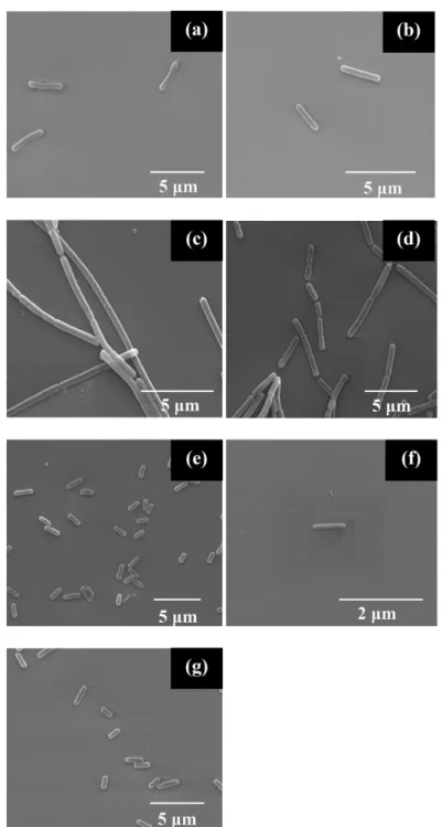
25
These 7 isolates were therefore chosen for further study. These isolates were identified by 16S rRNA sequencing (Figure 3, Table 3) and imaged by SEM to analyze their cell morphologies (Figure 4). STF4, STF8, STF15, STF26 were all effective in preventing the growth of S. aureus; however, only STF4 showed inhibitory effect on P. aeruginosa. In addition, STF8, STF9, STF10 and STF25 displayed a clear inhibition zone on the growth line of B. subtilis, but only STF26 could effectively inhibit the growth of S. enterica. In addition to STF26, STF4 also showed strong antimicrobial activity on E. coli and K. pneumoniae compared to other isolates.
Figure 3. Phylogenetic tree of 7 isolates (STF4, STF8, STF9, STF10, STF15, STF25 and STF26)
Bacillus pumilus strain HPS 1
Bacillus subtilis strain ISA7
Bacillus pumilus strain I5
Paenibacillus xylenexedens strain B22a Paenibacillus sp. B3a Bacillus licheniformis strain 30AA2-3
Bacillus subtilis strain LN
Bacillus subtilis strain ME2
STF9 STF4 STF10 STF25 STF15 STF26 STF8
26 Table 3. Identification of selected isolates
Isolate 16S rRNA sequence analysis
Closest known species Maximum
Identity (%) STF4 Paenibacillus xylanexedens 96 STF8 Bacillus subtilis 96 STF9 Bacillus subtilis 97 STF10 Bacillus licheniformis 99 STF15 Bacillus pumilus 97 STF25 Bacillus licheniformis 99 STF26 Bacillus pumilus 95
27
Figure 4. SEM images of isolates. (a), (b), (c), (d), (e), (f) and (g) represent STF4 (Paenibacillus xylanexedens), STF8 (Bacillus subtilis), STF9 (Bacillus subtilis), STF10 (Bacillus licheniformis), STF15 (Bacillus pumilus), STF25 (Bacillus licheniformis) and STF26 (Bacillus pumilus) respectively.
28
1.3.2.2. Sporulation Efficiency
Sporulation efficiencies of the isolates STF4, STF8, STF9, STF10, STF15, STF25 and STF26 were tested by inducing spore formation in a sporulation medium and counting colony numbers before and after heat exposure (80°C). Colony numbers were compared and survival percentages were subsequently calculated. The results were designated as percentages in Table 4. All isolates were spore-formers although their sporulation efficiencies were different.
1.3.2.3. Resistance to Simulated Gastric Fluids
Vegetative cells of the isolates STF4, STF8, STF9, STF10, STF15, STF25 and STF26 were tested for survival in simulated gastric fluids. All of the tested isolates were resistant to simulated gastric fluid conditions although their survival rates were different (Table 4).
Tolerance of purified spores of STF4, STF8, STF9, STF10, STF15, STF25 and STF26 to low pH condition of gastric fluids was tested after 30 min and 1 h of incubation. After 30 min, there was no reduction in the number of spores compared to controls. After 1 h incubation, there were 2-log reduction in STF15 and STF25 and no difference in STF4, STF8, STF9, STF10 and STF26 spores compared to controls.
1.3.2.4. Bile Salt Tolerance
Isolates were analyzed for their resistance to intestinal conditions by testing survival of both vegetative cells and spores in bile salts medium. When vegetative cells were exposed to bile salts, all isolates survived (Table 4).
29
When spores of the isolates were exposed to bile salts, at 0 and 1 h, no reduction was observed in the number of spores. After 3 h incubation in LB supplemented with bile salts, 2-log reduction was observed in STF25; 1-log in STF4, STF8, STF9 and STF10; slight reduction in STF26, and no reduction was noted in STF15.
1.3.2.5. Biofilm Formation
Biofilm production is desired for prospective probiotics, since it allows the probiotic to remain longer in the GIT (46). Biofilm formation provides protection for probiotic bacteria against the harsh conditions of the GIT, supports spore formation and prevents pathogenic bacteria to hold on to the GIT surface (5).
When grown in LB broth, some isolates namely STF9, STF10, STF15 and STF26 formed highly viscous structures on the bottom of the tubes. After the biofilm assay these isolates formed a notable ring of crystal violet and these rings suggest that the isolates have capacity to form biofilms (Table 4).
30
Table 4. Identification and characterization of selected isolates
Isolate Sporulation efficiency %a Survival in bile saltb Survival in simulated gastric fluidb Biofilm Formationc Vegetative form Vegetative form Paenibacillus xylanexedens STF4 79.9 + + - Bacillus subtilis STF8 127.02 +++ ++++ - Bacillus subtilis STF9 22.75 +++ +++ + Bacillus licheniformis STF10 173.91 +++ ++ + Bacillus pumilus STF15 15.28 + +++++ + Bacillus licheniformis STF25 35.2 + ++++ - Bacillus pumilus STF26 36.7 ++ ++++ + a
Sporulation efficiency is given as the percentage of the survivors. bPlus signs reflect the degrees of survival in simulated gastric fluid, “+” denoting the lowest survival and “+++++” the highest. c“+” reflect presence of biofilm formation while “-” reflect absence of biofilm formation. All tests were performed in triplicate.
31
1.3.2.6. Antibiotic Susceptibility and Plasmid Presence
Antibiotic susceptibility is considered as one of the most important characteristics of probiotic bacteria since the resistance genes could be transferred to pathogenic bacteria in the intestinal tract and also passed onto humans through the food chain (5, 45, 73, 97). This transfer mostly occurs by plasmid-borne resistance genes (97).
Antibiotic selection and determination of resistance or sensitivity of isolates against chosen antibiotics were performed according to the microbiological breakpoints used by SCAN (Scientific Committee on Animal Nutrition) (Table 5) (105). Minimum inhibitory concentration (MIC) values were displayed in Table 6. Of the isolates tested, all except STF4 were resistant to ampicillin, vancomycin and gentamicin; 4 isolates (STF8, STF9, STF15, and STF25) were resistant to kanamycin while another 4 (STF9, STF15, STF25, and STF26) were resistant to chloramphenicol. STF4 and STF8 were sensitive to erythromycin whereas other isolates displayed resistance to this antibiotic. MIC values of STF4 and STF26 against neomycin were lower than the breakpoints mentioned in SCAN report; therefore, they were considered as sensitive to neomycin. 3 cultures (STF15, STF25, and STF26) showed resistance to ciprofloxacin and only STF26 exhibited low MIC values to streptomycin. Moreover, Toyocerin also showed resistance to most of the antibiotics (ampicillin, chloramphenicol, erythromycin, vancomycin, ciprofloxacin and streptomycin).
32
Table 5. The Microbiological Breakpoints Used by SCAN Categorizing Bacterial Species as Resistant
Antibiotic Enterococcus
faecium
Pediococcus Lactobacillus Bacillus
Ampicillin 8 3 2* 2* Streptomycin 1024 32 16 64 Kanamycin/neomycin 1024 32 32 64 Entamycin 500 4 1 8 Chloramphenicol 16 16 16 16 Tetracycline 16 16 16 16 Erythromycin 4 4 4 4 Quinupristin/dalfopristin 4 4 4 4 Vancomycin 4 R 4* 4 Trimethoprim 8 16 32 8 Cipro/enrofloxacin 4 16 4* 1 Linezolid 4 4 4 4 Rifampin 4 8 32 324 R = resistant
33
Table 6. Minimum inhibitory concentrations (MIC) for isolates
Isolates Minimum Inhibitory Concentration (MIC) µg/ml
A K CH E V N CIP S G STF4 <1 <1 <1 <1 <1 4 <1 64 <1 STF8 16 64 8 2 4 64 <1 >128 16 STF9 64 64 64 8 4 64 <1 128 8 STF10 128 <1 8 4 8 64 <1 128 16 STF15 16 64 64 16 4 64 8 >128 8 STF25 128 64 64 128 128 128 16 128 16 STF26 32 16 32 32 32 32 4 32 16 Toyocerin 2 32 128 16 16 8 8 64 4
A, K, CH, E, V, N, CIP, S and G refer to Ampicillin, Kanamycin, Choloramphenicol, Erytromycin, Vancomycin, Neomycin, Ciprofloxacin, Streptomycin and Gentamycin respectively. All experiments were performed triplicate.
34
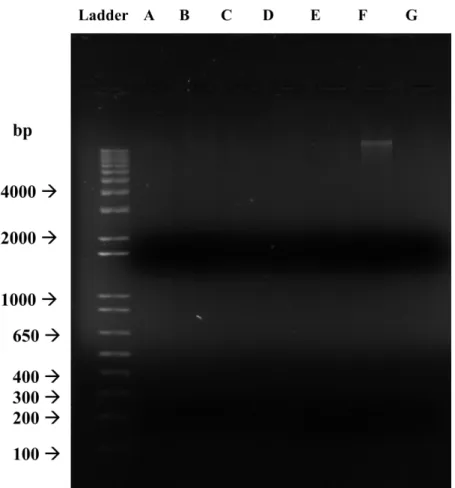
After the observation of that most of the isolates display resistance to antibiotics, plasmid isolation and visualization was performed to determine whether this resistance is due to plasmid-borne resistance genes. If such genes are not integrated into the genomic DNA and are instead carried on plasmids, they may be transferred to other bacteria in the environment and confer them resistances to the same antibiotic. This constitutes a potential health hazard if the recipient bacteria are pathogenic, and the absence of plasmid DNA should therefore be confirmed for an antibiotic-resistant strain to be recommended as a prospective probiotic. For this purpose, plasmid extraction and gel electrophoresis were performed. No plasmid presence was detected in the isolates STF4, STF8, STF9, STF10, STF15 and STF26; only the sample from STF25 displayed a band after gel electrophoresis (Figure 5). Our results suggest that all the samples tested, with the possible exception of STF25, carry potential antibiotic resistance genes in their genomic DNA and bear no risk of transferring them to other pathogenic or non-pathogenic bacteria that may be present in the environment (46).
35
Figure 5. Agarose gel electrophoresis image of plasmid profile of isolates. A, B, C, D, E, F and G represent STF4 (Paenibacillus xylanexedens), STF8 (Bacillus subtilis), STF9 (Bacillus subtilis), STF10 (Bacillus licheniformis), STF15 (Bacillus pumilus), STF25 (Bacillus licheniformis) and STF26 (Bacillus pumilus) respectively.
36
1.3.3. Effect of Isolates on Intestinal Cells
1.3.3.1. Adhesion and Invasion
The ability to adhere to and colonize in the GIT is an important requirement for potential probiotics, as the capacity to adhere increases the mean time of bacteria in the GIT and allows greater periods of probiotic activity after oral introduction of the probiotic strain (47, 87). Therefore, probiotic strains displaying stronger adhesion to the GIT are considered to better modify the normal intestinal microbiota, prevent the growth of pathogenic bacteria by competitive exclusion and induce immune system compared to the non-adherent ones (73, 87). HT-29 cell line is used as a model for the testing of adhesion of probiotic bacteria, as these cells differentiate into enterocytes (73, 87). Thus, we used HT-29 colon carcinoma cell line for the assessment of adhesion.
Recent research suggests that members of Bacillus species might induce important systemic diseases such as septicemia, peritonitis, endocarditis, liver failure and meningitis (85). Therefore, invasion studies were conducted for our candidate probiotics.
Abilities of Bacillus isolates to adhere to HT-29 cells were tested. We observed that all isolates adhered to the HT-29 cells (Table 7). STF9 could not grow in BHI broth; therefore, we could not perform the test for this isolate. STF4 showed the strongest adherence with a percentage of 2.5% while STF15 showed the lowest with 0.002%. Invasion properties of Bacillus isolates were also assayed as mentioned above. Assay results demonstrate that none of the bacteria
37
were capable of invading HT-29 cells, with the possible exception of STF9, which failed to grow in the assay medium (Table 7).
1.3.3.2. Cell Cytotoxicity
MTT assay was used for detection of toxic materials in the supernatants of bacteria. This assay was chosen as it has been determined to be more reliable and sensitive than others (28). In contrast to previous methods which were based on visual evaluation of toxin induced cell damage, the use of MTT has the advantage of removing the subjective visual assessment from the assay. MTT is a yellow, water soluble tetrazolium salt which is cleaved in the mitochondria of metabolically active cells. In the presence of living cells, MTT solution displays a color change as the insoluble purple formazan is formed due to intramitochondrial metabolization of MTT (6, 28, 30, 85). MTT-based assay is highly sensitive and suitable for assessing cell viability and cytotoxicity, since only living cells produce formazan reaction products (6, 30, 85).
The cell cytotoxicities of the supernatants of 18 h grown isolates in BHI were assayed by using the MTT test. STF9 could not grow in BHI broth; therefore, we could not perform the test for this isolate. STF8 and STF15 had heat-stable toxic materials in their cell free supernatants; however, the amounts of these materials were different. Also, STF4, STF25 and STF26 contained low amounts of heat-stable toxic materials in their cell-free supernatants. However, heat treatment of the culture supernatants (STF4, STF25 and STF26) eliminated toxicity in HT-29, which suggests that toxic materials are protein-based.
38
Moreover, STF10 supernatant showed no toxicity to HT-29 cell line, but when it was heated, the test result showed the presence of low toxic material (Table 7).
Table 7. Adhesion, Invasion and Cytotoxic Abilities* of Bacillus Isolates for HT-29
Isolate Bacterial Species
Adhesion (%)
Invasion (%)
Cytotoxicity (%)
Normal Heat treated
STF4 Paenibacillus xylanexedens 2,500 0 2,9 0 STF8 Bacillus subtilis 0,010 0 95,2 68,6 STF9** Bacillus subtilis - - - - STF10 Bacillus licheniformis 0,010 0 0 3,8 STF15 Bacillus pumilus 0,002 0 11,6 7,5 STF25 Bacillus licheniformis 0,100 0 7,8 0 STF26 Bacillus pumilus 0,008 0 8,5 0
* All results are given as the percentage of the survivors. ** "-" sign indicates that no growth on BHI was observed. All tests were performed triplicate.
39
1.4. CONCLUSION
The aim of this chapter of study was to screen many Bacillus sp. from the GIT of bovine in terms of their probiotic properties and select the most effective Bacillus probiotic to be used as animal feed supplement. For this purpose, different strains were isolated from chyme samples of bovines and probiotic characterization tests were applied. As a result, among the isolates, STF4, STF10, STF15 and STF26 appeared to have the highest potential as probiotic candidates compared to others by considering their antimicrobial effects, resistance to simulated gastrointestinal conditions, antibiotic susceptibility, colonization capacity and lack of toxicity. Moreover, regarding antimicrobial activity, the selected strains are more effective against some of the indicator strain than the commercial product Toyocerin.
40
1.5. FUTURE PROSPECTS
The growth conditions of the selected strains can be optimized to increase biomass because these strains are characterized for use as probiotic product. Therefore, maximum biomass production is necessary to decrease the cost. In fact, the growth conditions (temperature, pH, carbon source, nitrogen source and some salts percentages) of one of the strains which is Bacillus pumilus STF26 was optimized in another study.
Moreover, the selected potential probiotic bacteria were tested for most of the properties of a probiotic. However, for further use of these bacteria as probiotic products some additional research is necessary. These future studies that needs to be done include in vivo animal experiments and investigation of technical properties such as strain stability, bacteriophage resistance and shelf life.
Finally, animal digestive system conditions are similar to human; therefore, the selected potential probiotic Bacillus sp. may also be used as human probiotics. For human use purpose, further studies like clinical tests are required.
41
CHAPTER 2
2,4,6-trinitrotoluene
(TNT)
biodegradation
and
survival
capability of microalgea isolated from TNT-contaminated water
2.1. INTRODUCTION
2.1.1. Microalgae
Microalgae are a diverse group of prokaryotic and eukaryotic photosynthetic microorganisms that grow rapidly due to their simple structure (55). They can be found in most of the terrestrial and aquatic habitat such as oceans, fresh water, even on bare rock and soil.
Algal biomass can be used for food, animal feed, biofertilizer, soil conditioner, as feed in aquaculture, biological purification of waste water, production of a variety of compounds such as polysaccharides, lipids, proteins, carotenoids, pigments, vitamins, sterols, enzymes, antibiotics, pharmaceuticals, and other chemicals such as hydrogen, hydrocarbon, biofuel (7).
Microalgae are the principal primary producers of aquatic ecosystems (60); therefore, the tolerance of them to contaminated environments depends. The tolerance of microalgae to stressed environments is important because of their ecological value. However, knowledge about the mechanisms allowing algal adaptation to such extreme conditions is little and more research is necessary on this issue for future (60). Algae can be an appealing alternative technology as
42
phytosensors and phytoremediation systems for controlling TNT pollution. Plants and algae may be photosensors producing a phenotyping response to specific environmental stimuli although in phytoremediation generally plants are used for in situ treatment of contaminated sites polluted by a variety of hazardous chemicals (9, 18, 74). The plants and algae that are genetically engineered may be more appropriate for biosensor applications than hand--held biosensors or microbes in many conditions. Moreover, they do not involve advance technology and are cost effective (74).
2.1.2. 2,4,6-trinitrotoluene (TNT)
2,4,6-Trinitrotoluene (TNT) is a highly explosive, single-ring nitroaromatic compound that is a crystalline solid at room temperature (19). It is formed by nitration of toluene with three sequential reactions (Figure 6). The products of these reactions are mono-, di- and lastly trinitrotoluene respectively.
43
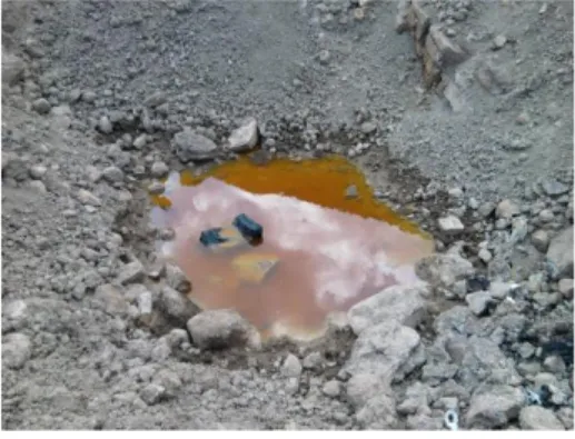
TNT is spread into soil and water systems mainly due to its use of high amounts during the two World Wars (17); furthermore, it has been used as dyes, pesticides and explosives historically. As a result, the soil, sediment and water were contaminated extensively with this toxic explosive by production, use and disposal of it (10, 14, 27). Figure 7 shows the image of pink water which is the waste stream generated during the process of loading, assembling, packaging, and demilitarization of munitions by army ammunition plants (AAPs) typically contains 2,4,6-trinitrotoluene (TNT) with photochemical activity that imparts the pink color (49, 66, 92).
Figure 7. Image of Pink Water
In surface water, 2,4,6-trinitrotoluene can be rapidly broken down into other chemical compounds by sunlight with photochemical activity (103). However, it accumulates in groundwater and sediments, and breaking down of TNT is more slowly by microorganisms in these sites.
2,4,6-trinitrotouluene (TNT) is the most dangerous one of the nitroaromatic compounds having mutagenic properties, persistency and contaminated sites