Human Mutation. 2020;41:e7–e45. wileyonlinelibrary.com/journal/humu © 2020 Wiley Periodicals LLC
|
e7D A T A A R T I C L E
Revisiting the complex architecture of ALS in Turkey:
Expanding genotypes, shared phenotypes, molecular
networks, and a public variant database
Ceren Tunca
1,2| Tuncay
Şeker
3| Fulya Akçimen
2| Cemre Co
şkun
2|
Elif Bayraktar
1| Robin Palvadeau
1| Seyit Zor
3| Cemile Koço
ğlu
2|
Ece Kartal
2| Nesli Ece
Şen
2| Hamid Hamzeiy
2| Asl
ıhan Özoğuz Erimiş
2|
Utku Norman
4| O
ğuzhan Karakahya
4| Gülden Olgun
4| Tahsin Akgün
5|
Hacer Durmu
ş
6| Erdi
Şahin
6| Arman Çakar
6| Esra Ba
şar Gürsoy
7|
Gülsen Babacan Y
ıldız
7| Bar
ış İşak
8| Kay
ıhan Uluç
8| Ha
şmet Hanağası
6|
Ba
şar Bilgiç
6| Nilda Turgut
9| Fikret Aysal
10| Mustafa Erta
ş
6| Cavit Boz
11|
Dilcan Kotan
12| Halil
İdrisoğlu
6| Aysun Soysal
13| Nurten Uzun Adatepe
14|
Mehmet Ali Akal
ın
14| Filiz Koç
15| Ersin Tan
16| Piraye Oflazer
6| Feza Deymeer
6|
Öznur Ta
ştan
17| A. Ercüment Çiçek
4,18| Er
şen Kavak
3| Ye
şim Parman
6|
A. Nazl
ı Başak
1,21
Suna andİnan Kıraç Foundation, Neurodegeneration Research Laboratory (NDAL), Research Center for Translational Medicine (KUTTAM), Koç University School of Medicine, Istanbul, Turkey
2
Suna andİnan Kıraç Foundation, Neurodegeneration Research Laboratory (NDAL), Department of Molecular Biology and Genetics, Boğaziçi University, Istanbul, Turkey
3Genomize Inc., Boğaziçi University Technology Development Region, Istanbul, Turkey 4
Department of Computer Engineering, Bilkent University, Ankara, Turkey
5
Department of Anesthesiology and Reanimation, American Hospital, Istanbul, Turkey
6
Department of Neurology, Istanbul Medical School, Istanbul University, Istanbul, Turkey
7
Department of Neurology, Faculty of Medicine, Bezmialem Vakıf University, Istanbul, Turkey
8
Department of Neurology, Marmara University School of Medicine, Istanbul, Turkey
9
Department of Neurology, Namık Kemal University School of Medicine, Tekirdağ, Turkey
10
Department of Neurology, Medipol University School of Medicine, Istanbul, Turkey
11
Department of Neurology, Karadeniz Technical University School of Medicine, Trabzon, Turkey
12
Department of Neurology, Faculty of Medicine, Sakarya University, Sakarya, Turkey
13
Department of Neurology, Bakırköy Research and Training Hospital for Neurologic and Psychiatric Diseases, Istanbul, Turkey
14
Department of Neurology, Cerrahpaşa Medical School, Istanbul University‐Cerrahpaşa, Istanbul, Turkey
15
Department of Neurology, Çukurova University Medical School, Adana, Turkey
16
Department of Neurology, Hacettepe University Medical School, Ankara, Turkey
17Department of Computer Science and Engineering, Sabancı University, Istanbul, Turkey 18
Department of Computational Biology, Carnegie Mellon University, Pittsburgh, Pennsylvania
A. Nazlı Başak, Suna and İnan Kıraç Foundation, Neurodegeneration Research Laboratory (NDAL), KUTTAM, Koç University School of Medicine, 34450 Istanbul, Turkey. Email:[email protected]
Present address
Fulya Akçimen, Department of Human Genetics, McGill University, Montréal QC, Canada
Cemre Coşkun, Faculty of Biology, Ludwig‐Maximilians‐University of Munich, Munich, Germany
Cemile Koçoğlu, Neurodegenerative Brain Diseases Group, Center for Molecular Neurology, VIB, Antwerp, Belgium Ece Kartal, Structural and Computational Biology Unit, European Molecular Biology Laboratory, Heidelberg, Germany Nesli EceŞen, Experimental Neurology Department, University Hospital Frankfurt, Frankfurt am Main, Germany
Hamid Hamzeiy, Computational Systems Biochemistry, Max‐Planck Institute of Biochemistry, Martinsried, Germany Piraye Oflazer, Department of Neurology, Koç University School of Medicine, Istanbul, Turkey Feza Deymeer, Department of Neurology, MemorialŞişli Hospital, Istanbul, Turkey.
Funding information
TÜBITAK, Grant/Award Number: 109S075; Bogaziçi University Research Funds, Grant/Award Number: 15B01P1; Suna and İnan Kıraç Foundation, Grant/Award Number: 2005–2020
Abstract
The last decade has proven that amyotrophic lateral sclerosis (ALS) is clinically
and genetically heterogeneous, and that the genetic component in sporadic
cases might be stronger than expected. This study investigates 1,200 patients
to revisit ALS in the ethnically heterogeneous yet inbred Turkish population.
Familial ALS (fALS) accounts for 20% of our cases. The rates of consanguinity
are 30% in fALS and 23% in sporadic ALS (sALS). Major ALS genes explained the
disease cause in only 35% of fALS, as compared with ~70% in Europe and North
America. Whole exome sequencing resulted in a discovery rate of 42% (53/127).
Whole genome analyses in 623 sALS cases and 142 population controls,
se-quenced within Project MinE, revealed well
‐established fALS gene variants,
solidifying the concept of incomplete penetrance in ALS. Genome
‐wide
association studies (GWAS) with whole genome sequencing data did not
in-dicate a new risk locus. Coupling GWAS with a coexpression network of
disease
‐associated candidates, points to a significant enrichment for cell
cycle
‐ and division‐related genes. Within this network, literature text‐mining
highlights DECR1, ATL1, HDAC2, GEMIN4, and HNRNPA3 as important genes.
Finally, information on ALS
‐related gene variants in the Turkish cohort
sequenced within Project MinE was compiled in the GeNDAL variant browser
(
www.gendal.org
).
K E Y W O R D S
ALS, ALS variant database, genetics, clinical exome sequencing, coexpression network analysis, genome-wide association study, motor neuron disease, next generation sequencing,
Turkish peninsula
1 | I N T R O D U C T I O N
Amyotrophic lateral sclerosis (ALS) is a progressive neurode-generative disease of upper and lower motor neurons, leading to muscle wasting. The average age of onset (AO) is 55–60 years, however juvenile cases exist (Ghasemi & Brown, 2018). Two‐ thirds of patients present with spinal‐onset, the rest shows bulbar‐onset with dysarthria and/or swallowing problems. Cog-nitive and behavioral changes are seen in almost 50% of ALS cases (van Es et al.,2017). The disease results in death within 2–5 years due to respiratory failure, which can only be slightly ex-tended with exceptional medical care (Brown & Al‐Chalabi,2017; Kiernan et al.,2011).
ALS genetics is complex; the familial form (fALS) is rare (5–10%), sporadic cases or isolated (singlet) patients (sALS) constitute 90% of cases. Familial and sporadic ALS are clinically indistinguishable and well‐established fALS genes are implicated in sporadic disease, pointing to “apparently” sporadic cases with incomplete penetrance (Brown & Al‐Chalabi, 2017).
In populations in which consanguinity is common, juvenile aty-pical disease accompanies classical, resulting in a more hetero-geneous genetic background that makes the differential diagnosis in the clinic challenging.
The Human Genome map, advances in next generation se-quencing (NGS), genome‐wide association studies (GWAS), and applications of whole exome sequencing (WES) changed para-digms in identifying ALS‐associated alleles even with low power, both in family‐based studies and in large admixed populations (Brenner et al., 2018; Cirulli et al., 2015; Smith et al., 2017). Moreover, GWAS performed using single nucleotide polymorph-isms (SNPs) from whole genome sequencing (WGS) data, enabled association of rare variants with rare diseases, such as ALS (Nicolas et al., 2018; van Rheenen et al., 2016). Project MinE Sequencing Consortium, a large multinational ALS colla-boration, is established based on this purpose, to define new disease‐causing genes and risk loci associated with true sporadic disease to target novel therapeutics (van Rheenen et al.,2018).
2 | D A T A S P E C I F I C A T I O N S
Data type Tables and figures Data acquisition
method
Sanger sequencing, NGS
Data format Filtered and analyzed Experimental factors None
Experimental features Pathogenic genomic variation analysis in a large Turkish ALS cohort using conventional and next generation sequencing methods. Variant interpretation through in silico tools and clinician‐researcher
collaboration. Genotype‐phenotype correlations in ALS and ALS‐like disease. Data collection and sharing in public databases for ALS. Data source location Suna andİnan Kıraç Foundation
Neurodegeneration Research Laboratory, Koç University Hospital, Davutpaşa Street No. 4, 34010, Istanbul, Turkey
Data accessibility Data in this paper is published within the paper and deposited to ClinVar public database (https://www.ncbi.nlm.nih. gov/clinvar/?term=SUB7287039).
3 | I M P A C T O F D A T A
The last decade has seen an unprecedented and exponential progress in data output, based on advances in genetics/genomics and on large international collaborations. Consequently, our knowledge of the genetic factors behind ALS has improved in an unparalleled fashion and the scientific scenario of ALS has dramatically changed. Today, the disease is accepted to be part of a continuum with other neurological diseases and a crossroads between genetic, neurometabolic and environmental factors.
This manuscript, five years apart from our previous publica-tion on ALS in Turkey (Özoğuz et al.,2015), is not only an update with a triple increase in patient numbers, but it supersedes our earlier work by reflecting a whole new picture, being much more comprehensive in its scope, upgraded in cutting edge techniques, applying genome‐wide and bioinformatic approaches to extract candidate disease genes and pathways, followed by a population‐ specific database.
Our understanding of the etiopathology of neurological diseases stem from the identification of disease genes and pathways. With its well‐selected and large cohort, this study not only represents a dis-tinct resource for ALS in Turkey, it also reveals the genetic variation in a highly inbred and admixed population, is thus expected to con-tribute to human disease at large.
4 | E X P E R I M E N T A L D E S I G N , M A T E R I A L S
A N D M E T H O D S
This study includes 1,200 Turkish patients recruited from hospitals across Turkey between 2002 and 2019; 246 cases with a family history of ALS, plus 80 affected family members and 954 isolated ALS cases. Sample collection was approved by Boğaziçi University Ethics Committee. Genetic counseling was given to patients at the local institutions during blood collection and signed informed consent was obtained from all subjects. DNA samples from healthy relatives were obtained for research purposes only with their written approval. Genomic DNA was isolated from whole blood using the MagNa Pure Compact System (Roche, Switzerland).
4.1 | Screening for common ALS genes
Conventional screening for the hexanucleotide repeat expansion in the C9orf72 gene was performed in all patients with or without fa-mily history of ALS, whereas SOD1, TARDBP, and FUS were screened only in familial cases. The C9orf72 repeat expansion was tested using repeat‐primed polymerase chain reaction (PCR) and flanking PCR was performed to identify the zygosity and the size of the repeats within the normal range. All five exons of SOD1 were analyzed in fALS. Additionally, exon 4 of the SOD1 gene was screened in all cases with consanguineous parents, independent of family history. Geno-mic variant analyses in TARDBP and FUS genes were restricted to their hotspots, exon 5 for TARDBP and exons 14 and 15 for FUS. Genotyping experiments were performed with GoTaq® Flexi DNA Polymerase (Promega), MyTaq™ DNA Polymerase (Bioline), FastStart Universal Master Mix (Roche, Switzerland) and One Taq® 2× Master Mix (New England Biolabs). The sequences of primers are available upon request. Sanger sequencing was outsourced (Macrogen Inc., Korea) and CLC Main software (Qiagen, Germany) was used for analysis.
4.1.1 | Bisulfite sequencing in the promoter region
of
C9orf72
The 5mC levels of the C9orf72 promoter regions harboring 26 CpG sites were detected using direct bisulfite sequencing assay (BST‐ PCR). EZ DNA Methylation‐Gold Kit (Zymo Research) was used for bisulfite conversion of genomic DNA according to the manufacturer's protocol. The converted genomic DNA was amplified using nested PCR with primers targeting the converted sequence in the promoter region. ZymoTaq Premix was used for these consecutive amplifica-tions. Methylation levels were detected by direct evaluation of Sanger sequencing results. Commercially available human methy-lated (100%) and nonmethymethy-lated (0%) standards were used as con-trols, 50% control was prepared by mixing equal amounts of commercial standards (Zymo Research). The number of methylated
CpG sites was calculated for each individual and two‐tailed Fisher's exact test was used to assess the association between promoter hypermethylation for the expansion carriers. The maximum number of methylated CpG sites among controls (2/26) was considered as the threshold for hypermethylation.
4.2 | WES
WES was applied in 250 individuals; 127 probands, 32 affected, and 91 healthy family members (Macrogen Inc., Korea). Selection criteria of the patients subjected to WES were (a) close consanguinity in the parents of the affected individual, (b) atypical clinical features, and (c) early/juvenile disease‐onset. In addition, WES was also applied to cases with a positive family history of disease in the upper genera-tions, if screening in four common dominant genes did not reveal any disease‐associated variants. Suspected inheritance pattern was au-tosomal dominant for 25, auau-tosomal recessive for 79 families, and in 23 cases the inheritance pattern could not be certified. Clinical in-formation of the cases subjected to WES, their suspected inheritance patterns and initial clinical diagnoses are listed in Appendix (TableA1).
Bioinformatic analyses of the samples were initially performed using in‐house Burrows‐Wheeler Aligner (BWA) (H. Li & Durbin,
2009) and Genome Analysis Toolkit (GATK; McKenna et al., 2010) pipeline. More recently, the online SEQ Platform, a cloud‐based genomics software, was used and all samples have been retro-spectively analyzed with this platform (Genomize Inc., Turkey). The SEQ Platform enables calculation of real‐time minor allele frequency (MAF) for variants using NDAL‐ and SEQ‐specific cohorts.
For the in‐house pipeline, paired‐end sequencing reads obtained from sequencing platforms were aligned to the human reference genome GRCh37 plus the decoy using BWA‐MEM algorithm. Quality control and variant calling from binary sequence alignment/map format files were performed with HaplotypeCaller tool of GATK. The ANNOVAR software was used for structural and functional annota-tion of variants (Wang, Li, & Hakonarson,2010). MAFs were re-cruited from 1000 Genomes Project (1000 G) and National Heart, Lung and Blood Institute Exome Sequencing Project (NHLBI‐ ESP6500; Auton et al.,2015; EVS,2014). Functional consequences of variants were predicted via several sources (e.g., SIFT, PolyPhen2, and GERP++) and DANN scores were assigned to each variant.
For variant prioritization, the association of candidate genes with known human phenotypes was obtained from the OMIM database. Annotated variants were filtered using the VarSifter software (ver-sion 1.7) or SEQ Platform according to the inheritance mode and MAF (>0.01; Teer, Green, Mullikin, & Biesecker,2012). Functional predictions were used for evaluation, but not for filtration. Center (NDAL)‐specific MAF lower than 0.01 was used as a parameter during variant prioritization in the WES data of 600 Turkish patients and healthy family members. American College of Medical Genetics (ACMG) guideline verdict was determined for each candidate for further evaluation. Segregation analysis for candidate variants was
performed by Sanger sequencing in the index case and in all available family members.
4.3 | WGS
Whole genome sequencing of 632 Turkish sALS cases and 151 neurologically healthy controls was performed within the scope of Project MinE. Samples were selected on the basis of definitive, late‐ onset ALS diagnosis, without a family history. The mean AO for the patients included in Project MinE was 51 years, in agreement with 52‐year‐old mean AO of the total sALS cohort; control subjects had a mean age of 55 years. C9orf72 hexanucleotide repeat expansion was excluded in all patients before WGS. Project MinE guidelines were followed for sample selection and preparation (van Rheenen et al., 2018). PCR‐based library free paired‐end sequencing was performed on the Illumina HiSeq 2000 platform with an average of 40× coverage per sample (Illumina FastTrack Services, San Diego). Alignment to the hg19 reference genome and variant calling were performed using the Isaac pipeline and provided by Illumina as aligned reads in BAM files and individual‐based gVCF files containing the single nucleotide variations (SNVs), short indels and structural variations (Raczy et al.,2013).
Protein coding variants in all ALS‐causing and ‐associated genes reported (Ghasemi & Brown, 2018) were screened in annotated variant files. Candidate variants identified in sALS patients were further analyzed for pathogenicity using prediction tools, VarSome software (Kopanos et al.,2019) and our in‐house exome database.
4.3.1 | WGS sample processing and quality control
All variants across the individuals were merged with AGG tool (Illumina). Individual/variant‐level quality control was performed using PLINK (version: 1.9; Purcell et al., 2007) and VCF‐tools (Danecek et al.,2011) (version:0.1.16). Samples with a deviated inbreeding coefficient (>3 SD) from the mean of the distribution, as well as related/duplicate samples and those with missingness rate higher than 10% were not included for further analyses (623 cases and 142 healthy controls remaining). A pruned set of high‐quality SNPs were prepared using missingness rate (<10%), MAF (>5%) and Hardy–Weinberg equilibrium (p < 1 × 10⁻⁶ for controls and p < 1 × 10⁻¹² for cases) thresholds. SNPs within the MHC or LCT loci or, the inversions on chromosome 8/17 were excluded. Prin-cipal components (PCs) for each individual were calculated using PLINK.4.3.2 | Genome‐wide association study
Variants with MAF > 5% in the whole cohort were tested for asso-ciation using a binary logistic regression in PLINK. First 10 PCs and gender were used as covariates.
4.3.3 | Gene
‐based burden testing
All variants were annotated using Ensembl Variant Effect Predictor version 92 (McLaren et al.,2016) and classified into two functional groups for gene‐based association testing: (a) disruptive variants (stop‐gained, stop‐loss, start‐loss, splice sites, and frameshift indels with high confident according to loftee prediction), and (b) missense variants predicted to be damaging using REVEL and MetaLR algo-rithms, on the basis of a combining approach (Dong et al., 2015; Ioannidis et al., 2016). ClinVar‐benign variants were excluded. ClinVar‐pathogenic variants were kept regardless of other elimina-tion criteria (Landrum et al.,2014). In the remaining list, variants with MAF≥ 1% in any public population databases (ExAC, gnomAD, 1000 G, and ESP6500; Auton et al., 2015; EVS,2014; Exome Ag-gregate Consortium,2016; Karczewski et al.,2019) and variants with MAF≥ 5% in our cohort were excluded.
Gene‐based burden testing was performed using the R‐package of SKAT‐O by aggregating disruptive and possibly damaging variants (missense variants) on genic regions and pathways (Ionita‐Laza, Lee, Makarov, Buxbaum, & Lin,2013). Pathway and associated lists were downloaded from the Broad Institute GSEA site (http://software. broadinstitute.org/gsea) using all canonical pathway dump (version 6.2). Tests were adjusted for gender and first 10 PCs.
4.3.4 | Gene coexpression network analysis
The ST‐Steiner algorithm (Norman & Cicek,2019) was used, which searches for a connected component (a tree) on a gene coexpression network or a cascade of gene‐coexpression networks. The algorithm
aims at maximizing the prizes of the selected genes and minimizes the cost of edges that are used to connect these genes.
To construct the gene coexpression network, we utilized the full BrainSpan microarray data set of the Allen Brain Atlas (Sunkin et al.,2013). This data set contains gene expression measurements of 524 brain samples from various brain regions obtained from 42 in-dividuals that represent various time points in neurodevelopment, starting from embryonic period up to adulthood. We used correlation threshold of 0.7 (Pearson correlation, r2). That is, an edge was added to a graph if two genes’ correlation exceeded this threshold. This is a common threshold choice in the literature (Çiçek, 2017; Liu et al.,2014; Liu, Lei, & Roeder,2015). After pruning for the genes that do not exist in the data, the final network contains 547,056 edges and 8,499 nodes.
As our edge cost 1−r2was used. For the node (gene) prizes, we
used the negative log10 transformed p‐values derived from gene‐
based SKAT‐O analysis. The ST‐Steiner algorithm also inputs a list of terminal nodes, which have to be included in the tree (due to a very large artificial prize) and joined by other nodes. These are the genes with high level of risk confidence for ALS: SOD1, TARDBP, SQSTM1, HNRNPA1, FUS, VCP, OPTN, PFN1, ATXN2, NEFH, SETX, ALS2, DCTN1, ANG, ELP3, FIG4, TAF15, SPG11, NEK1, PON1, PON3, TBK1, DAO, CHRNA3, CHRNB4, CREST (SS18L1), CHRNA4, NTE (PNPLA6) (Ghasemi & Brown,2018).
There are four hyperparameters to set in the ST‐Steiner Algo-rithm. The first parameter isω, which is the number of trees in an estimated forest. We set this parameter to 0 to obtain a single connected component with the assumption of a single functional cluster of genes as done in Norman and Cicek (2019).λ and α were set to zero, since the algorithm was run on a single network and T A B L E 1 Clinical characteristics of the
Turkish ALS cohort under study Total ALS fALS sALS
Number
Probands 1,200 246 (20%) 954 (80%) Affected family members 80 80 –
Male:female ratio 1.5 1.2 1.6 Consanguinity 301 (25%) 75 (30%) 226 (24%) Dementia 30 (2.5%) 13 (5%) 17 (2%) Age of onset Juvenile (<25 years) 101 (8%) 33 (13%) 68 (7%) Middle (25–45 years) 292 (24%) 61 (25%) 231 (24%) Late (>45 years) 718 (60%) 123 (50%) 595 (62%) Not available 89 29 60
Mean age of onset (total ±SD) 50 ± 15.4 47 ± 16.9 51 ± 15.1 Site of onset
Limb 773 (64%) 163 (66%) 610 (64%) Bulbar 212 (18%) 36 (15%) 176 (18%) Limb + bulbar 84 (7%) 12 (5%) 72 (8%)
Not available 131 35 96
Abbreviations: ALS, amyotrophic lateral sclerosis; fALS, familial ALS; sALS, sporadic ALS; SD, standard deviation.
these parameters are used when a cascade of networks is employed. The third parameterβ was used to put the node prizes and the edge costs on the same scale and adjust the size of the predicted sub-network. After a line search to obtain a network, which includes approximately three predictions for every ground truth (terminal) gene, it was set to 0.17. Edge thickness denotes the correlation threshold (thicker = higher correlation).
Functional annotation clustering of the candidate genes pre-dicted in the coexpression network was performed using DAVID Functional Annotation Tool (version 6.8; Huang et al., 2007). All 8,499 nodes (genes) used to create the coexpression network were given to the algorithm as background. Literature‐mining for asso-ciation between the predicted genes in the network and ALS was evaluated by screening GeneRif and DisGeNet databases (all species considered; Jimeno‐Yepes, Sticco, Mork, & Aronson, 2013; Piñero et al.,2017). We determined the number of occurrences of genes in association with ALS based on specific search matching restricted words:“ALS,” “fALS”, “sALS,” and “amyotrophic lateral sclerosis.” The output counts were used as a score to denote the strength of the evidence for each gene with ALS in GeneRIF and DisGeNet databases (TableA2).
5 | D A T A
5.1 | 45% of fALS and 10% of sALS are explained by
known ALS genes in the Turkish cohort, indicating
genetic heterogeneity in fALS and incomplete
penetrance among sALS patients
A total of 1,200 Turkish patients diagnosed with ALS or ALS‐like motor neuron disease were analyzed within the scope of this study, adopting a combination of conventional and NGS approaches. The clinical summary of the study cohort is compiled in Table1. Sixty percent of the Turkish ALS cohort under study had an age of disease
onset beyond 45 years and the mean ages of onset were 47 for fALS and 51 for sALS patients. In 64% of our cases, spinal symptoms were detected as the initial clinical features, 18% reported to suffer from bulbar symptoms and 7% showed mixed site of onset. The male to female ratio in the present cohort was 1.5.
Four common ALS genes (C9orf72, SOD1, TARDBP, and FUS) contribute to 35% of fALS and 6.1% of sALS in Turkey and analysis of pathogenic exonic variants obtained from WES and WGS data in-creases these numbers to 45% in fALS and 10% in sALS (Figure1). This 10% of sALS cases explained by genomic variants in well‐ established and highly penetrant ALS genes, like C9orf72, SOD1, TARDBP, FUS, OPTN and VCP, are“apparently” sporadic, who are either (a) the only affected child of consanguineous couples, or (b) cases with low penetrance of the variant in the upper generations, or (c) carriers of de novo variants.
The GGGGCC hexanucleotide repeat expansion in the C9orf72 gene was detected in 42 families (plus 8 affected family members) and in 38 sporadic cases in 1,200 ALS patients (TableA3). Mean age of disease onset among the expansion carriers was 54.5, representing classical ALS. A higher frequency of bulbar‐onset ALS was observed among C9orf72 cases (23%), compared with 18% in all cases. In-trafamilial phenotypic variability was present among family members manifesting either ALS, ALS accompanied by frontotemporal de-mentia (ALS‐FTD) or solely FTD symptoms. Dementia was reported in nine expansion carriers and two affected family members (13%).
In ALS cases with or without the expansion and in controls, two, five, and eight repeats were found to be the predominant allelic variants in the Turkish population for the nonexpanded allele of the C9orf72 gene. In three cases the intermediate repeat sized GGGGCC(30‐35)was detected, which did not segregate with the
dis-ease in the two families tested. Bisulfite sequencing assay of the 26 CpG sites, located in the promoter region of C9orf72, revealed a significant increase in promoter hypermethylation for the expansion carriers (n = 52 expansion carriers and 31 age‐ and sex‐matched controls, Student's t test p < .0023); no significant correlation was
F I G U R E 1 Frequency of ALS gene variants in the Turkish cohort. The four major ALS genes account for 35% of fALS, NGS increases this number to 45% (left pie). The same four ALS genes solve 6.1% of sALS, NGS increases it to almost 9% (right pie). The dark blue areas in the pies, represent unsolved cases and also samples not yet analyzed by NGS. ALS, amyotrophic lateral sclerosis; fALS, familial ALS; NGS, next generation sequencing; sALS, sporadic ALS
observed between number of CpGs methylated and the AO of pa-tients (Hamzeiy et al.,2018).
Eighteen distinct pathogenic genomic variants in the SOD1 gene were identified in 57 patients (32 fALS index cases plus 13 affected family members and 12 sporadic cases; Figure2; Table2). The human reference transcript NM_000454.4 was used for the nucleotide numbering of SOD1; as an exception, the old nomenclature (excluding the initiation codon) was used for the amino acid changes. The SOD1‐ p.(Leu144Phe; c.435G>T) Balkan variant (Battistini, Benigni, Ricci, & Rossi,2013) was observed in 10 probands and seven affected family members, being the predominant SOD1 variant in the study cohort. The highly characterized SOD1‐p.(Asp90Ala; c.272A>C) genomic variant with a dual inheritance pattern, very common among Scan-dinavian populations in recessive form (Andersen et al.,1995), ex-plained the disease in nine consanguineous Turkish cases. The dual inheritance pattern known for the SOD1‐p.(Asp90Ala), was also true for three additional rare changes in SOD1; p.(Asn86Ser) (c.205T>C), p.(Leu117Val; c.352G>C), and p.Glu133Lys (c.400G>A), which were detected in 10 different pedigrees with or without family history of ALS (Table2). Apart from the highly penetrant and frequent patho-genic SOD1 variations, others identified in our cohort are present in relatively small families with few affected children in the same gen-eration (Table2). Examples of SOD1 variants with evidence of re-duced penetrance are the p.(Glu22Leu) (c.68A>T), p.(Glu40Gly) (c.122A>G), p.(His71Tyr) (c.214C>T), p.(Val87Met) (c.262G.A) and p.(Thr137Ala) (c.412A>G) variations with asymptomatic carriers in the families.
Pathogenic TARDBP (NM_007375.3) and FUS (NM_004960.3) genomic variants explained the disease in 20 probands and four af-fected family members (Figures1and2; Table2). The heterozygous FUS‐p.(Pro525Leu) (c.1574C>T) and FUS‐p.(Tyr526Cys) (c.1577A>G) variations were detected in four isolated juvenile cases without a family history, in whom de novo occurrence of the variants was
shown via variation‐negative parents. Additionally, the intermediate CAG repeat expansions in the ATXN2 gene, associated with an in-creased ALS risk, were reanalyzed in an extended cohort of 519 sALS cases as compared with 236 fALS and sALS patients in a previous study from our laboratory (Elden et al.,2010; Lahut et al.,2012). Analysis, using the control cohort of Lahut et al. (2012) (n = 420), confirmed increased ALS risk in carriers with (27–33) CAGs (21/519; Fisher's exact test p = .0086).
Analysis of exonic variants in the WES (n = 127) and WGS (n = 623) data revealed the presence of pathogenic gene variants in 74 cases, out of which, 19 were previously published by our group (Tables2and 3; Akçimen et al.,2019; Özoğuz et al.,2015; Tunca et al.,2018). Variant information and clinical features of the cases solved are compiled in Table3and in Appendix (TableA4). Homo-zygous OPTN variants were observed in a total of eight cases in the current Turkish cohort. In five families out of eight, the homozygous AA deletion in the OPTN gene leading to a premature stop codon (p.(Lys360Valfs*18), c.1078_1079delAA, NM_021980.4) was identi-fied. OPTN‐based disease in our cohort, presents as classical ALS with an earlier onset at 38 years on average (Table3). SPG11 and ALS2 genomic variants are the second most frequent causes of autosomal recessive ALS in the cohort, with average ages of onset of 14 and 1, respectively.
Rare autosomal dominant ALS genes predominating in our co-hort include VCP, ANG, and TBK1, identified in both familial and “apparently sporadic” cases without any reported disease history in the family (Table3). The novel heterozygous ERBB4 (p.Arg1096Cys, c.3286C>T, NM_001042599.1) and KIF5A (p.Asp1002Gly, c.3005A>G, NM_004984.2) pathogenic genomic variations were de-tected in two distinct families, in which the causative variants seg-regated with the disease in at least three affected family members. Among the ALS gene variants with unknown pathogenicity identified through WGS, two variations in the PON1 and PON3 genes, were
F I G U R E 2 Amino acid changes identified in SOD1, TDP‐43, and FUS proteins. Variant‐specific pie charts represent the variant's proportion in fALS (red) or sALS (blue) cases, smallest circle corresponding to one case and the largest to 11 cases. fALS, familial ALS; NES, nuclear export signal; NLS, nuclear localization signal; RRM, RNA recognition motif; sALS, sporadic ALS; ZFD, Zinc finger domain
T A B L E 2 Clinical data of patients with SOD1, TARDBP, FUS genomic variants Gene ALS ID Nucleotide change Protein changec Gender Age of onset Site of
onset Gene dosage Family
history Phenotype SOD1(NM_000454.4) 1398 c.13G>A p.(Ala4Thr) F 25 B het Yes Juvenile ALS
221 c.13G>T p.(Ala4Ser) M 20 L het Yes Juvenile ALS
308 F 44 L ALS
907 F NA NA ALS
1167a c.43G>A p.(Val14Met) M 42 L het No ALS
960 c.68A>T p.(Gln22Leu) F 30 L het Yes ALS 1327 c.95T>C p.(Val31Ala) M 45 B het Yes ALS
1547a F 64 L No ALS
1453 c.112G>C p.(Gly37Arg) M 41 L het Yes ALS 802 c.122A>G p.(Glu40Gly) F 39 L het Yes ALS
816 F 32 L Yes ALS
1450 c.205T>C p.(Ser68Pro)n M 54 L het Yes ALS
226 c.214C>T p.(His71Tyr)n M 19 L het Yes Juvenile ALS
707 F 57 L ALS
191 c.260A>G p.(Asn86Ser) M 28 L hom No ALS
623a F 42 L het No ALS
1207 M 48 L Yes ALS
102a c.262G>A p.(Val87Met) F 29 L het No ALS
147 c.272A>C p.(Asp90Ala) M 49 L hom Yes Lower limb dominant stereotyped Scandinavian phenotype 310 M 55 L Yes 429 F 45 L Yes 741 F 32 L Yes 810 F 29 L Yes 1256b M 44 L Yes 1359 M 35 L+B Yes 1545 F 64 L No 1579 M 51 L No
561 c.352C>G p.(Leu117Val) F 62 L het Yes ALS
1527 F 38 L ALS 1396 F 62 L Yes ALS 1412 F 40 L ALS 1472a F 36 L No ALS 1888 M 50 L Yes ALS 1882 F 58 L ALS
1439 F 24 L hom Yes juvenile ALS
355a c.376G>A p.(Asp125Asn) M 50 L het No ALS
1716b c.400G>A p.(Glu133Lys) F 37 L hom No ALS
1064a M 34 L het No ALS
1655b c.412A>G p.(Thr137Ala) F 49 L het No ALS
61 c.435G>T p.(Leu144Phe) F 52 L het Yes ALS
281a M 57 L No ALS 607 F 45 L Yes ALS 713 F 53 L ALS 724 M 52 L ALS 727 F NA L ALS 1773 M 60 L ALS 635 F 54 L Yes ALS 772 M 49 L Yes ALS 1059 F 51 L ALS 1063 F 56 L ALS 1235 F 59 L ALS
detected in Turkish sALS cases as compared to none of the controls (PON1, rs755475189, 2 patients/0 controls; PON3, rs147006695, 5 patients/0 controls; TableA5).
Apart from classical ALS genes, WES revealed variants in rare genes associated with diverse motor neuron phenotypes either with upper or lower motor neuron predominance (e.g., ZFYVE26, DNAJB2, PLEKHG5, TRPV4, FBXO38, and VRK1; Table 3 and Table A4). The C19orf12 genomic variant, implicated in neurodegeneration with brain iron accumulation, mimicking ALS, was detected in three cases with an autosomal recessive inheritance (AO: 9–24), who were initially diag-nosed with a probable juvenile ALS (Deschauer et al.,2012).
From the clinical exome sequencing perspective, WES‐only suc-cess rate of NDAL is calculated as 42% for patients diagnosed with ALS and ALS‐like disease in the Turkish cohort (Tables 2 and 3, TableA4). This rate increases to almost 50% in familial cases and in cases with consanguineous parents, regardless of family history.
5.2 | GeNDAL, a web‐based variant browser for
ALS
‐related genes
Fully anonymized information regarding ALS‐related variants with known or unknown pathogenicity identified in WGS analysis are T A B L E 2 (Continued) Gene ALS ID Nucleotide change Protein changec Gender Age of onset Site of
onset Gene dosage Family history Phenotype 935 M 37 L Yes ALS 1036 F 60 L Yes ALS 1633 M 64 L Yes ALS 1691 M 60 L Yes ALS 1715 F 34 L Yes ALS
97b c.446T>G p.(Val148Gly) F 46 L het Yes ALS
TARDBP (NM_007375.3) 1082 c.893G>T p.(Gly298Val)n F 66 L het Yes ALS
356b c.943G>A p.(Ala315Thr) M 58 L het Yes ALS
357 F 59 L Yes ALS (man in barrel)
600 F 57 L Yes ALS
1448 M 62 L Yes ALS
408a M 48 L No ALS
910 c.1042G>T p.(Gly348Cys) M 37 L het Yes ALS
919 M 42 L ALS
911 M NA L ALS
660 c.1060C>G p.(Gln354Glu)n F 42 L het No ALS
976 c.1144G>A p.(Ala382Thr) M 39 L het Yes ALS 277b c.1147A>G p.(Ile383Val) M 67 B het Yes ALS
311 F 42 NA ALS
FUS (NM_004960.3) 264b c.430_447del
p.(Glu144_-Tyr149del)
M 16 L het No Juvenile ALS
1034 (NC_000016.10) c.1394–1G>Tn
p.(=) F 47 L het Yes ALS
1208 c.1555C>T p.(Gln519*) F 42 L Yes ALS 485a c.1562G>T p.(Arg521Leu) M 39 L het No ALS
227b c.1571G>T p.(Arg524Met) F 32 L het Yes ALS
581 M 53 L Yes ALS
1647 F 29 L Yes ALS
549 c.1574C>T p.(Pro525Leu) M 14 L het (de novo) No Juvenile ALS with fast
progression
1610b M 17 L het (de novo) No
377a F 16 L het (de novo) No
1423b c.1577A>G p.(Tyr526Cys) M 12 L het (de novo) No Note: Bold indicates index cases.
Abbreviations: B, bulbar; het, heterozygous; hom, homozygous; L, limb; n, novel; NA, not available; WES, whole exome sequencing; WGS, whole genome sequencing.
aIdentified in the framework of Project MinE (WGS). b
Identified with WES.
presented in the Genome Browser of NDAL, GeNDAL (http://www. gendal.org). GeNDAL is a platform which allows the users to query variants by dbSNP ID, amino acid change, gene symbol, Human Genome Variant Server ID, transcript or sequence ontology defined by the Sequence Ontology Consortium (http://www.sequenceontology.org/). Detailed variant annotations and graphical representations of variant‐ related information from public databases (ClinVar, gnomAD, etc.) can be visualized (Figure3). In addition, the phenotypes can be distinguished as ALS or unaffected control. The GeNDAL database currently con-structed for ALS‐related gene variants will be complemented in future for other phenotypes in NDAL's cohort.
5.3 | Whole genome sequencing analysis of 623
Turkish sALS cases and 142 neurologically healthy
controls did not reveal significant risk loci
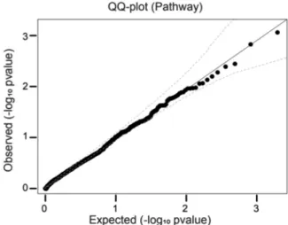
A joint cohort of ALS patients and control samples worldwide were analyzed by Project MinE Sequencing Consortium, which included 224 Turkish samples (van der Spek et al., 2019; van Rheenen et al.,2016). Analysis of WGS data of an expanded Turkish cohort consisting of 623 Turkish sALS patients and 142 neurologically healthy controls, revealed 47,971,649 novel variants which are not represented in gnomAD. Out of all variants detected, 23,410,513 had a MAF smaller than 0.1% in the cohort (TableA6). GWAS analysis and gene‐based burden testing (SKAT‐O) on this cohort did not re-veal a significant risk/protective variant or gene (Figure4). The top 25 SNPs detected in GWAS and the top ten genes from SKAT‐O are listed in TablesA7andA8in the Appendix. Even though there is a lack of association with new variants or genes from the GWAS, we scanned the literature for the candidate genes and none of them was associated with ALS or similar phenotypes.
On the basis of the hypothesis that ALS genes are working as a functional cluster, we conducted a gene coexpression network analysis (a) to search for other candidate genes which might also confer ALS risk, and (b) to investigate the function of the predicted cluster and its re-lation to ALS. The“guilt‐by‐association principle”‐based gene discovery approach has been applied on many complex neurologic/psychiatric disorders to discover more risk genes as in the autism spectrum dis-order (De Rubeis et al.,2014; Sanders et al.,2015) and schizophrenia (Torkamani, Dean, Schork, & Thomas,2010). For this purpose, the ST‐ Steiner algorithm (Norman & Cicek,2019) was used to create a network around established ALS genes using the SKAT‐O p‐values as the prize for the network analysis. The resulting subnetwork contains 98 newly predicted genes around the 28 ALS‐associated terminal genes (Figure5). Ninety‐five of 98 predicted genes had higher variation rates in patients and were significantly enriched for cell cycle (DAVID enrichment score: 15,03) and cell division genes (DAVID enrichment score: 6,8; TableA9; Huang et al.,2007). Coexpression network ana-lysis, coupled with literature text‐mining using GeneRIF and DisGeNet databases, pointed to DECR1 (case count: 3 and control count: 0), ATL1 (case count: 1 and control count: 0), HDAC2 (case count: 1 and control count: 0), GEMIN4 (case count: 1 and control count: 0) and HNRNPA3
(case count: 1, control count: 0) genes which are marked in Figure5. Finally, accumulation of all variants in WGS to canonical disease path-ways obtained from Broad Institute also did not suggest a significant association (FigureA1); top ranking pathways are listed in TableA10. Even with a Turkish sample size four times larger than the two initial Project MinE studies, neither GWAS nor network analysis point to any known or new significant association with ALS, indicating the crucial requirement for larger sample sizes.
6 | D I S C U S S I O N
6.1 | Clinical presentation of ALS in Turkey
For almost two decades, well‐established patient registers operating in European countries gather organized patient data to understand the epidemiology of ALS (Hardiman et al., 2017). These registers work countrywide and are unbiased in terms of origin, socioeconomic status and the disease stage of the patient in contrast to a local clinic. The incidence reported for ALS worldwide is argued to be misleading in the absence of long‐running patient registers which are more efficient in recognizing family history and hidden symptoms like FTD (Hardiman et al.,2017). Turkey's ALS patient registry operated by the Turkish ALS‐ MND Association (www.als.org.tr) since 2001 with headquarters in İs-tanbul andİzmir, is relatively recent and may not represent the whole country. Hence, information regarding the incidence and prevalence of ALS in Turkey and survival rates are still restricted and scattered. NDAL, being the only reference center for the molecular analysis of ALS in Turkey, has been recruiting patients from across the country for over 20 years and gathers available patient data to investigate the clinical and molecular basis of this complex neurodegenerative disease in an admixed population inhabiting the Turkish peninsula in the crossroads of many civilizations since several centuries.
This study, with 1,200 probands, offers an update on the phe-notypic and genetic landscape of ALS in Turkey. The fALS percentage of 20% (246/1,200), exceeding North American and European po-pulations (5–10%; Ghasemi & Brown, 2018), is explained by population‐specific and social factors, such as extensive kindreds consisting of many generations and offspring. A unique aspect in Turkey, common to countries in the Near and Middle East, is the high proportion of close consanguineous marriages, approaching 50% in the eastern parts of the country. Consanguinity in the ALS cohort under study is calculated as 30% in fALS and 23% in sALS. This suggests even a higher percentage for Mendelian inheritance in yet unexplained cases that are classified originally as sALS due to singlet patients in the family. Thus, familial ALS, harboring a simplex genetic component, seems to be above 20% among Turkish cases.
6.2 | Impact of common genes on ALS in Turkey
The C9orf72 hexanucleotide repeat expansion and pathogenic mis-sense variants in SOD1 together explain 30% of familial cases in ourTABL E 3 Variants identified via WES and WGS in rare genes Gene Transcript ID Variation Gene dosage AO Family history Consanguinity Phenotype DNA change Protein change ERBB4 NM_001042599.1 c.3286C>T p.(Arg1096Cys) Het 48 Yes No ALS KIF5A NM_004984.2 c.3005A>G p.(Asp1002Gly) n Het 50 Yes No ALS TBK1 NM_013254.4 c.922C>T p.(Arg308*) n a Het 46 No No ALS c.1436_1437delTG p.(Val479Glufs*4) Het 20 No No Juvenile ALS VCP NM_007126.5 c.463C>T p.(Arg155Cys) a Het 62 No No ALS c.475C>T p.(Arg159Cys) a Het 52 No No ALS c.572G>C p.(Arg191Pro) Het 60 Yes No ALS ‐FTD UBQLN2 NM_013444.3 c.1516C>T p.(Pro506Ser) Hemi 26 Yes No ALS c.1573C>T p.(Pro525Ser) Hemi 22 Yes Yes ALS TFG NM_001007565.2 c.854C>T p.(Pro285Leu) Het 47 Yes No ALS with sensory neuropathy ANG NM_001145.4 c.208A>G p.(Ile70Val) a Het 52 No No ALS c.208A>G p.(Ile70Val) a Het 40 No No ALS c.208A>G p.(Ile70Val) Het 28 No No ALS CHCHD10 NM_213720.2 c.176C>T p.(Ser59Leu) a Het 51 No No ALS FBXO38 NM_001271723.1 c.1577G>A p.(Arg526Gln) n Het congenital No Yes Juvenile MND TRPV4 NM_147204.2 c.943C>T p.(Arg315Trp) Het infancy Yes Yes Juvenile MND TRPM7 NM_017672.6 c.4445C>T p.(Thr1482Ile) Het teenage Yes No Juvenile ALS SETX NM_015046.7 c.5839G>A p.(Ala1947Thr) n Het 11 Yes No Juvenile ALS ERLIN1 NM_006459.4 c.281T>C p.(Val94Ala) n Hom 15 Yes Yes Juvenile ALS SPG11 NM_025137.4 c.1432C>T p.(Gln478*) n Hom 20 No Yes Juvenile ALS c.1966_1967delAA p.(Lys656Valfs*11) Hom 16 Yes Yes Juvenile ALS c.2250delT p.(Phe750Leufs*3) n Hom 16 Yes Yes Juvenile ALS c.7155T>G p.(Tyr2385*) n Hom 23 Yes Yes Juvenile ALS OPTN NM_021980.4 c.76delC p.(His26Thrfs*19) a Hom 35 No Same village ALS c.875dupC p.(Glu293Glyfs*19) n Hom 33 Yes Yes ALS c.1078_1079delAA p.(Lys360Valfs*18) Hom 32 Yes Yes ALS c.1078_1079delAA p.(Lys360Valfs*18) Hom 43 Yes Yes ALS c.1078_1079delAA p.(Lys360Valfs*18) Hom 31 Yes Yes ALS c.1078_1079delAA p.(Lys360Valfs*18) a Hom 42 No No ALS c.1078_1079delAA p.(Lys360Valfs*18) a Hom 42 Yes b No ALS c.1217delC p.(Thr406Lysfs*5) n a Hom 42 No No ALS (Continues)
TABLE 3 (Continued) Gene Transcript ID Variation Gene dosage AO Family history Consanguinity Phenotype DNA change Protein change ALS2 NM_020919.4 c.1718C>A p.(Ala573Glu) Hom 2,5 No Yes Juvenile ALS c.2761C>T p.(Arg921*) Hom 1 N o Yes juvenile ALS c.4381C>T p.(Arg1461*) n Hom 1 Yes Yes Juvenile ALS c.4808C>T p.(Pro1603Leu) Hom 1 N o Yes Juvenile ALS C19orf12 NM_001031726.3 c.32C>T p.(Thr11Met) Hom 24 No Yes Juvenile ALS c.194G>T p.(Gly65Val) Hom 10 Yes Yes Juvenile ALS c.194G>T p.(Gly65Val) Hom 9 N o Yes Juvenile ALS SYNE1 NM_182961.3 c.22930C>T p.(Gln7644*) n Hom 21 Yes Yes Juvenile ALS c.23524C>T p.(Arg7842*) Hom 17 Yes Yes Juvenile ALS ZFYVE26 NM_015346.3 c.2074delC p.(Leu692Serfs*52) n Hom 17 No Yes Juvenile MND c.2615 – 2617delGCTinsTGAA p.(Arg872Hisfs*17) n Hom 22 No Yes Juvenile MND DNAJB2 NM_006736.5 c.14A>G p.(Tyr5Cys) Hom 31 No Yes Juvenile ALS c.757G>A p.(Glu253Lys) Hom 22 No Yes Juvenile MND PLEKHG5 NM_198681.3 c.1648C>T p.(Gln550*) Hom 20 Yes Yes Juvenile ALS c.2120C>A p.(Pro707His) Hom 14 No Yes Juvenile ALS SIGMAR1 NM_147157.2 c.355G>A p.(Glu119Lys) Hom 2 N o Yes Juvenile ALS c.358A>G p.(Thr120Ala) n Hom 17 No Yes Juvenile ALS VRK1 NM_003384.3 c.961C>T p.(Arg321Cys) Hom 22 Yes Yes Juvenile ALS c.1135_1136delCA p.(Gln379Aspfs*23) n Hom 17 No Yes Juvenile MND DJ1 NM_007262.5 c.133C>T p.(Gln45*) Hom 24 Yes Yes ALSPDC IGHMBP2 NM_002180.2 c.638A>G p.(His213Arg) Hom 9 Yes Yes Juvenile MND SLC52A3 NM_033409.4 c.802C>T p.(Arg268Trp) Hom 1,5 Inconclusive Yes Madras MND Abbreviations: ALS, amyotrophic lateral sclerosis; ALSPDC, ALS ‐Parkinsonism ‐Dementia Complex; AO, age of onset; fALS, familial ALS; Het, heterozygous; Hemi, hemizygous; Hom, homozygous; MND, motor neuron disease; sALS, sporadic ALS. aCases solved in the framework of Project MinE (WGS). bPatient status changed from sALS to fALS upon diagnosis of a younger sister with ALS, nnovel variant.
cohort, with TARDBP and FUS solving another 5%. NGS data of “ap-parently sporadic”/isolated cases, unraveled genomic variants in common ALS genes that contribute to 2.1% (SOD1: 1.3%, TARDBP: 0.2% and FUS: 0.6%), to total 6.1% with sporadic cases carrying the C9orf72 expansion. These results not only support the evidence of incomplete penetrance but also the de novo occurrence of variants in these genes, leading to genetic misclassification of patients as sporadic. Thus, the distinction between the clinically indistinguish-able familial and sporadic disease, becomes unclear and should be handled with care in diagnostic settings and particularly during ge-netic counseling.
The hexanucleotide repeat expansion in the C9orf72 gene is the most common genetic cause of ALS worldwide, with an exception of Japan (Ogaki et al.,2012). Although lower in frequency compared to Northern European countries (50% of fALS and 20% of sALS; Majounie et al.,2012), this expansion is the most abundant genomic variation both in fALS (17%) and sALS (4%) also in the present Turkish cohort. SOD1 variants, the second most frequent genetic causes of ALS in the Turkish cohort contribute to high allelic
heterogeneity (Figure 2). Evidence for the reduced penetrance of SOD1 variants was obvious in the Turkish population, such as (a) the genomic variants detected in sporadic patients, (b) the families with asymptomatic carriers, and (c) dual inheritance patterns observed in SOD1‐p.(Asn86Ser), p.(Asp90Ala), p.(Leu117Val), and p.(Glu133Lys). This variability may stem from modifier genes/variants, an upcoming research field.
The fact that an ample number of people with Turkish origin migrated to Turkey from the Balkans many generations ago, ratio-nalizes the predominance of the common Balkan variant, SOD1‐ p.(Leu144Phe) in our cohort. The average AO for p.(Leu144Phe) carriers is 52 years without any gender bias and all of them have limb‐onset disease. This variation results in classical ALS and appears to be highly penetrant in large families consisting of several branches. The only exception is an apparently sporadic male patient (AO:57), with deceased parents who were not available for analysis (Table2). The biallelic p.(Asp90Ala) variation is the second most common pathogenic SOD1 variant in our cohort. No affected individuals have been detected carrying the heterozygous variant and the presence of F I G U R E 3 Representation of the GeNDAL variant database. (a) Information regarding the variant and its annotation with
complementary links to external databases. (b) Phenotype‐dependent frequencies of the variant of interest in the internal WGS cohort of NDAL. (c) Genomic location of the nucleotide change and its surrounding sequence. (d) The number of pathogenicity verdict and detailed information on the variant in ClinVar. (e) Display of pathogenic and likely pathogenic variants reported in ClinVar aligned to the current transcript and protein domains, allowing visualization of variational hotspots. WGS, whole genome sequencing
the Scandinavian founder haplotype in Turkish recessive p.(Asp90Ala) cases was previously shown (Özoğuz et al., 2015). Except for one patient with a mixed site of onset, limb‐onset disease predominates in p.(Asp90Ala) patients. The disease progression is very slow and in accordance with the stereotyped Scandinavian phenotype (Andersen et al.,1995). The average AO of the recessive p.(Asp90Ala) carriers is 10 years earlier than the p.(Leu144Phe) patients.
SOD1 is a ubiquitously expressed protein that acts as a super-oxide radical scavenger in the cell. Its pathogenicity in ALS is ex-plained by the misfolding of the mutant product which leads to accumulation within the cell in aggregates (Paré et al.,2018). There is not enough evidence in the literature to comment on the mechanism behind the phenotypic heterogeneity of different SOD1 variants as well as their acting mechanisms that show both dominant and re-cessive inheritance. Although most evidence supports the gain‐of‐ function mechanism, loss‐of‐function of the mutant allele may still have a role in the presentation of SOD1‐based disease. Reduction in overall activity of the mutant form has been shown in blood and
fibroblast samples, but the two specific genomic variants, p.(Asp90Ala) and the heterozygous p.(Leu117Val), had only slight reductions in enzymatic activity. This might explain the milder phe-notype in patients carrying these variants and the low penetrance observed in parents of homozygous individuals (Saccon, Bunton‐ Stasyshyn, Fisher, & Fratta,2013). On the contrary, the homozygous p.(Leu117Val) genomic variation was reported to result in a more severe reduction in enzymatic activity than the heterozygous variant which is also concordant with the early AO (AO:24) and the fast disease progression of the patient with the biallelic genomic variation reported in this study (Table2; Synofzik et al.,2012). Although the function of the mutant protein is not completely lost, the activity may be reduced by aggregation; thus, different SOD1 variants with dif-ferent aggregation propensities may have variable enzymatic activity and this may act on the phenotypic representation of the disease.
ALS‐associated TDP‐43 and FUS variants are known to accu-mulate in the C‐termini and although the pathogenicity behind these two RNA/DNA binding proteins is not yet clear, nuclear clearance and cytoplasmic accumulation of both proteins are observed in ALS. F I G U R E 4 Manhattan plots and Quantile‐quantile plots of GWAS and SKAT‐O analysis using logistic regression. (a) Approximately six million SNPs (MAF≥ 0.05) are displayed in the Manhattan plot. (b) Quantile‐quantile plot of the GWAS p values. (c) p values derived from the gene‐based SKAT‐O analysis are displayed in the Manhattan plot. Each of ~10,000 genes in SKAT‐O analysis is represented by a single dot. (d) Quantile‐quantile plot of genic association p values. Known ALS‐related genes are highlighted in red. Dashed curves correspond to the 95% confidence limits. ALS, amyotrophic lateral sclerosis; GWAS, Genome‐wide association studies
In fact, TDP‐43‐positive cytoplasmic inclusions are a common hall-mark of fALS and sALS, regardless of an ALS‐associated genetic variation in patients. Unlike FUS variants gathered in the nuclear localization signal domain, TDP‐43 variants are found in the prion‐ like domain of the protein (Figure2). Our results regarding TARDBP and FUS variants are restricted to screening of C‐terminal hotspots for these two genes with the exception of the heterozygous deletion in the N‐terminus of the protein detected via WES. There are rare variants reported in the N‐terminal region of TDP‐43, like the p.Ala90Val, however with the lack of segregation analysis, the pre-sence of the variant in healthy individuals and with mild abnormal cytoplasmic localization, the pathogenicity of the variant remains questionable (Winton et al.,2009; Wobst et al.,2017). Despite the importance of nuclear import and export signals on the transport of a protein, studies showed different cytoplasmic accumulation levels for different N‐terminal FUS variants and also a critical role for C‐ terminal deletions in FUS in formation of stress granules, all sug-gesting a complicated mechanism for both proteins, ranging from loss of nuclear function to gain of toxic function through aggregates (Guerrero et al.,2016).
Juvenile ALS (AO < 25) was observed in 68 isolated/sporadic cases (7%) in our cohort. This form of ALS most frequently occurs due to consanguinity and has a rather slow disease progression compared
to classical ALS. However, four de novo FUS cases with non‐ consanguineous parents, ages of onset ranging from 12 to 17, had an aggressive disease progression, resulting in the retirement of the children from all daily activities. Severe bulbar symptoms in addition to initial limb‐onset disease, eventually lead to death almost within a year. De novo FUS gene variants are reported in juvenile cases in populations where consanguinity is not common. We also suggest the screening of FUS as the initial step in isolated juvenile patients with a fast disease progression and asymptomatic parents (Hübers et al.,2015; Leblond et al.,2016; Therrien, Dion, & Rouleau,2016).
In the cohort under study, common ALS genes contribute to 35% of fALS, which increases to 45% with the addition of rare genes. According to this picture, more than 50% of Turkish fALS cases remain unsolved as compared to 30% in Caucasian populations (Ghasemi & Brown, 2018); this result points towards an expected higher locus heterogeneity in the Turkish population. The Turkish peninsula, geographically located at the intersection of many civiliza-tions, has a heterogeneous ethnic and genetic background. This com-plexity in the population leads to the dilution of pathogenic variants in common ALS genes like C9orf72 or SOD1. In this sense, the frequencies observed in Turkey are concordant with the common notion of decreasing north‐south gradient for these genes (Andersen, 2006; Lamp et al.,2018; Smith et al.,2013). Novel coding variants, as well as F I G U R E 5 The predicted subnetwork of genes by ST‐Steiner on ALS GWAS data. The predicted subnetwork contains 126 genes. Red denotes ground truth (terminal) genes (n = 28) used to build the network, and yellow denotes the newly predicted genes (n = 98). The node size represents ALS risk, based on−log10 transformed p values from SKAT‐O analysis. The border thickness depicts the GeneRIF and DisGeNet scores for each gene, and edge thickness the strength of gene expression correlation between a pair of genes according to the BrainSpan database. ALS, amyotrophic lateral sclerosis; GWAS, Genome‐wide association studies
chromosomal changes (large rearrangements, copy number variations, repeat expansions, indels), and variants in regulatory, intronic and in-tergenic regions, not covered by WES, are expected to unravel the missing heritability in the present cohort.
6.3 | Clinical exome sequencing in the differential
diagnosis of ALS and ALS
‐like phenotypes
The majority of inherited diseases are caused by genomic variations in protein‐coding regions; thus, exome sequencing unravels the causative variants in a considerable number of cases allowing data interpretation, less dependent of the initial clinical diagnosis, which is to the benefit of both the clinician and the patient. This unbiased candidate variant prioritization approach allows to differentially di-agnose cases with uncertain clinical phenotypes due to overlapping features between diseases and their progressive nature with absence of full‐grown symptoms in juvenile cases. Some examples to this common problem are reported here in non‐ALS MND genes like ZFYVE26, DNAJB2, PLEKHG5, TRPV4, and FBXO38, in which early
disease‐onset or intrafamilial phenotypic heterogeneity, ranging from neuropathy to motor neuron disease, lead to uncertain clinical di-agnoses (Figure6).
Identification of new genetic players implicated in ALS and ALS‐ like disease opens new opportunities for understanding the conver-ging mechanisms in neurodegeneration and/or motor neuron loss. Moreover, these also contribute to the development of more specific, even personalized, therapeutic targets like gene‐specific antisense oligonucleotides. Thus, today, it is important to define the genetic causes of even yet untreatable diseases, to drive pharmaceutical/ gene‐editing research and to offer hope to patients and their families. Clinical exome sequencing, for which the diagnosis success rate in-creased exponentially in clinical settings, includes several beneficial outcomes: (a) treatment of patients with syndromic diseases like enzyme deficiencies, (b) using genetic information for reproductive genetic counseling and family planning and (c) recruitment of patients with specific genomic variants into clinical trials. Most importantly, WES shortens the diagnostic delay of at least 1 year in ALS, which may include several invasive and expensive procedures, and these should be taken into account while considering its cost‐effectiveness
F I G U R E 6 Genetic heterogeneity behind motor neruon diseases in our cohort. Individuals carrying the genomic variations represented in bold had clear/definitive initial diagnosis of classical ALS with an average age of onset of 46. Genes represented in italics are identified in patients presenting with juvenile motor neuron disease or nonclassical ALS‐like disease with expanding phenotypes in the index or in affected family members (average AO: 13). This picture emphasizes pleiotropy in genes in addition to the expansion of phenotypes, calling for a gene‐ based disease classification. Venn diagram is drawn according to the disease‐gene associations obtained from the literature (Akçimen et al.,
2019; Al‐Saif, Al‐Mohanna, & Bohlega,2011; Annesi et al.,2005; Brenner et al.,2018; Chen et al.,2004; Cirulli et al.,2015; Cottenie et al.,2014; Daoud et al.,2012; Deschauer et al.,2012; Frasquet, Va, & Sevilla,2017; Greenway et al.,2006; Grohmann et al.,2001; Hermosura et al.,2005; Hughes et al.,2001; Ishiura et al.,2012; Iskender et al.,2015; Kimonis, Fulchiero, Vesa, & Watts,2008; Maruyama et al.,2010; Maystadt et al.,2007; Nalini, Pandraud, Mok, & Houlden,2013; H. P. Nguyen, Van Broeckhoven, & van der Zee,2018; Stoll et al.,2016; Sumner et al.,2013; Synofzik et al.,2016; Takahashi et al.,2013; Tunca et al.,2018; Velilla et al.,2019; Yang et al.,2001). ALS, amyotrophic lateral sclerosis; AO, age of onset
(Fogel et al., 2014; Fogel, Satya‐Murti, & Cohen, 2016; Trujillano et al.,2017).
6.4 | Project MinE to understand sporadic ALS:
Impact of Turkish WGS data
Currently, the Turkish population is one of the highly represented cohorts in Project MinE in terms of sample size. Our results from screening for pathogenic exonic variants in the Turkish Project MinE cohort revealed marked incomplete penetrance for the three com-mon ALS genes (SOD1, TARDBP, and FUS). In recent years, many studies showed that the frequency of ALS patients carrying more than one variant is higher than expected by chance, providing evi-dence that ALS may result from multiple rare variants with additive effects on disease development and presentation, for example, age of disease onset, progression and severity (van Blitterswijk et al.,2012). This“oligogenic model” of ALS may solve a portion of unexplained sporadic cases. The variations in PON1 and PON3 detected in our cohort might also act in such a manner (TableA5). The PON variants can lead to oligomerization of the native protein through N‐terminal HDL particles, as previously reported, and further decrease its own hydrolytic activity (Josse et al.,2002). Disease pathology caused by paraoxonase genes, intensely studied for their role in ALS, may arise with reduced ability of PON enzymes, responsible of detoxifying organophosphates, which are neurotoxins associated with an in-creased ALS risk (Cronin, Greenway, Prehn, & Hardiman, 2007; Landers et al.,2008; Menini & Gugliucci,2014; Merwin, Obis, Nunez, & Re,2017; Ticozzi et al.,2010; Verde et al.,2019; Wills et al.,2009). Genome‐wide association study and gene‐based burden testing for population‐specific local signals with WGS data of 623 sporadic patients and 142 Turkish controls, a four times larger Turkish cohort than the one from van Rheenen et al. (2016), did not reveal any significant loci neither in variant‐based GWAS, nor in gene‐based disease burden analyses. Yet, the resulting network, which combined gene‐based burden analysis with coexpression information, pointed to a significantly high enrichment (~15‐fold) of cell cycle‐related genes. Accordingly, changes in expression levels and subcellular lo-calization of cell cycle proteins and their transcriptional regulators have been linked to neuronal death in the literature in terms of ALS and other neurodegenerative diseases (M. D. Nguyen et al., 2003; Ranganathan & Bowser,2003).
Network analysis combined with literature text‐mining suggested DECR1, ATL1, HDAC2, GEMIN4, and HNRNPA3 as disease susceptibility candidates although these had no significant burden for ALS in the SKAT‐O analysis. Previous studies show increased levels of the mitochondrion‐related β‐oxidation enzyme DECR1 at disease onset in SOD1‐G93A mice spinal cords (Q. Li et al.,2010; Pharaoh et al.,2019). Atlastin‐1 (ATL1), associated with upper motor neuron syndromes (De Bot et al.,2013), is a protein effective in structural and functional in-tegrity of the endoplasmic reticulum (Muriel et al.,2009). Histone dea-cetylase 2 (HDAC2) is important for the nervous system and was shown to be upregulated in ALS patients (Janssen et al.,2010). The increased
HDAC activity in neurodegeneration and positive effects of HDAC in-hibition, including HDAC2, on motor symptoms are reported in the literature (Lazo‐Gómez, Ramírez‐Jarquín, Tovar‐y‐Romo, & Tapia,2013; Rossaert et al.,2019). Finally, GEMIN4 and HNRNPA3 are involved in RNA processing and interact closely with ALS‐causative proteins in this machinery. GEMIN4 acts in the survival motor neuron complex for-mation, disrupted in lower motor neuron disease, and is in the FUS interactome together with HNRNPA3, which is detected in the spinal cords of C9orf72‐positive ALS and FTD patients (Chi et al., 2018; Davidson et al.,2017; Fifita et al.,2017). Altogether, pathogenic variants in these genes, which are not yet detected in familial ALS, should be further investigated in larger cohorts for their possible contribution to sporadic ALS.
Although there is a wide variability in disease incidence and manifestation across populations and geographical regions, we ac-knowledge the shortcoming of our analyses to catch a significant population‐specific signal considering the insufficient number of ALS patients and controls, which calls for far more Turkish samples to be sequenced. On the other hand, our WGS data with 900 samples and expanding, is expected to exemplify a unique population with a heterogeneous gene pool that will support to study the combinatorial effects of diverse SNPs in manifestation of sALS. In this respect, we are confident that this report will encourage local clinicians for re-cruitment of new patients. Only then, it will be possible to overcome power limitations, currently faced even by Project MinE with >9,000 DNA samples analyzed (Dekker et al., 2019; van Rheenen et al.,2018). The answer to the question of how to move forward at this point will be the collection of larger case and control cohorts in the framework of national and international collaborations and making all genomic data publicly available to increase the power of datasets. This will allow us to understand and interpret how a variant causes disease within the context of the larger population.
7 | C O N C L U S I O N
Genetics offers a means to dissect the heterogeneity of ALS and to understand the cellular mechanisms resulting in motor neuron de-generation. The recent genetic findings driven by the NGS technol-ogy have not only expanded our knowledge of the wealth of genes giving rise to motor neuron degeneration, but also on the pleiotropic effects and extensive phenotypic spectra associated with specific ALS genes. Since the road from family pedigrees to clinical interpretation of variants is challenging, deep phenotyping of the patient, compre-hensive analysis of the candidate variants with advanced bioinfor-matic tools and most importantly a tight researcher‐clinician relationship are indispensable parts of the whole process. Ultimately, the discovery of all ALS genes will help to better define the multi-faceted nature of ALS, which is accepted no more as a monolithic disease, but recognized as a spectrum of diseases converging into common clinical features. This allows a subclassification of patients into more precise clinical categories in which a common genetic cause is more likely to be identified.