553 Srp Arh Celok Lek. 2016 Sep-Oct;144(9-10):553-556 DOI: 10.2298/SARH1610553S
ПРИКАЗ БОЛЕСНИКА / CASE REPORT UDC: 616.717.7-006.34.03
SUMMARY
Introduction Osteoid osteoma is a benign osteoid-forming tumor generally localized to the long bones, is rarely localized in the hand and the major symptom is intermittent pain. This study aims to present two patients who were operated on for metacarpal osteoid osteomas.
Case Outline A 16-year-old female patient and an 18-year-old male patient were operated on for metacarpal osteoid osteomas. The major symptom was intermittent pain for both patients. After surgical excision of the niduses, the complaints resolved in both cases.
Conclusion In the case of high suspicion for osteoid osteoma, computed tomography or magnetic resonance imaging should be performed due to the risk of negative radiographic findings. Surgical excision is curative and a safe method of treatment.
keywords: osteoid osteoma; metacarpal bone; surgical treatment
A rare localization of osteoid osteoma –
Presentation of two cases
Ali Şeker1, Mehmet Bekir Unal2, Melih Malkoç1, Adnan Kara1, Ilker Abdullah Sarikaya3, Ahmet
Murat Bulbul1
1Istanbul Medipol University, Department of Orthopaedics and Traumatology, Istanbul, Turkey; 2Bahçeşehir University, Department of Orthopaedics and Traumatology, Istanbul, Turkey; 3Children’s Orthopedic Clinic, Istanbul, Turkey
INTRODUCTION
Osteoid osteoma is a benign osteoid forming tumor [1]. The first description of osteoid os-teoma term in medical literature was done by Jaffe in 1935 [2, 3]. It constitutes 10–12% of all benign bone tumors and is usually seen in children and young adults in the second and third decade of life [2, 3]. Approximately 50– 60% of cases are seen in long bones, especially metaphysis of the femur and tibia. It is rarely localized in the hand, and the major symptom is intermittent pain. Sometimes it would be dif-ficult to diagnose lesions in the hand due to the atypical pain pattern and different histologic features [1, 2]. This study aims to present two patients who were operated on for metacarpal osteoid osteomas. The written consent was ob-tained from the patients.
REPORTS OF CASES Case 1
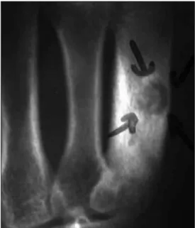
A 16 year-old female was admitted with noc-turnally aggravated local tenderness and swell-ing on her fifth metacarpal bone in February 2011. The complaints had been present for three months, and relieved with salicylates. The labo-ratory analysis was normal. In plane X-ray, in-creased sclerosis in the middle third of the fifth metacarpal bone extending from cortical area to the medulla was observed (Figure 1). Magnetic resonance imaging (MRI) showed intracortical lesion with surrounding sclerotic tissue.
Under general anesthesia, an arm tourni-quet was applied. A 3 cm dorsolateral incision
was used for exposure. A nidus was completely excised during surgery, but the defect left after tumor resection was filled with allograft since the patient did not approve autografting. Histo-pathologic evaluation confirmed the diagnosis of osteoid osteoma. The patient’s complaints were relieved in the immediate postoperative period, and during the four-year follow-up she did not express any complaints.
Case 2
An 18-year-old male was admitted to our clinic in January 2013 due to the pain in his left hand for one year. He was given rest and analgesics in the previous medical center but his
com-Figure 1. Plain radiograph of the first patient shows
ni-dus and surrounding sclerotic bone
Correspondence to:
Ali ŞEKER
Istanbul Medipol University Department of Orthopaedics and Traumatology
Istanbul Turkey
554
doi: 10.2298/SARH1610553S
Şeker A. et al. A rare localization of osteoid osteoma – Presentation of two cases
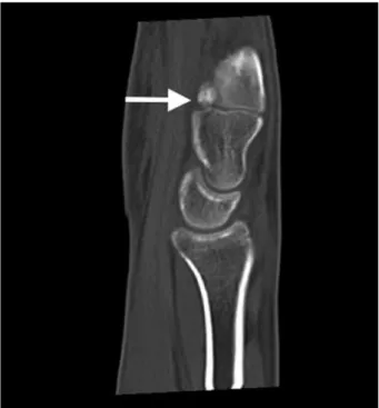
plaints did not disappear. His pain was disturbing espe-cially during nighttime and persisted during the day. The pain was reduced with non-steroidal anti-inflammatory drugs. On physical examination, tenderness was observed at the level of the proximal portion of his third metacarpal on palmar side of the left hand, but the finger and wrist range of motion were normal. A lesion which was 3 × 3 mm in diameter with well-defined margins in the proxi-mal volar side of the third metacarpal was detected in the computed tomography (CT) sections (Figure 2). The MRI revealed the lesion and edema in the surrounding bone (Figure 3). The mass was accepted as osteoid osteoma, and it was decided to perform an excision.
Under general anesthesia, an arm tourniquet was ap-plied. A 4 cm longitudinal incision parallel to the thenar crease was made. The transverse carpal ligament was cut. The median nerve and flexor tendons were retracted in order to reach the carpometacarpal joint. The joint capsule was opened and the lesion was exposed. The nidus was excised and the cavity was debrided (Figure 4). The skin
and subcutaneous tissues were closed and compressive bandage was applied.
The histopathologic examination confirmed the diag-nosis of osteoid osteoma (Figure 5). The pain disappeared after the excision. At the last control, the patient was pain free and working without limitation 22 months after the surgery.
DISCUSSION
Six to 13% of osteoid osteomas are localized in the hand. The phalanges are the most frequent sites for osteoid os-teoma in the hand. The carpal bones and metacarpals are rarely affected [1, 2, 4]. In our patients, the lesions were located in the metacarpal bones. In their series of nine-teen patients, Ambrosia et al. [5] reported only one patient whose metacarpal bone was affected.
The etiology of osteoid osteoma is unknown. Chro-mosomal abnormalities are suspected but have not been proven yet [6]. Kalil and Antunes [7] reported two broth-ers that had osteoid osteoma with similar properties, like onset of symptoms and localization. Another suspect for etiology of osteoid osteoma is trauma [1, 2]. Uda et al. [8] and Baron et al. [9] reported their cases of osteoid osteoma and claimed that the lesions were presented after injury. Our patient had neither family history nor trauma.
Pain is the main complaint in patients with osteoid os-teoma. It’s nocturnal and increases with activity. Salicylates
Figure 2. In the computed tomography section of the second patient,
a sclerotic lesion was observed at the proximal palmar side of the third metacarpal
Figure 3. Magnetic resonance imaging of the second patient
Figure 4. Intraoperative view of the lesion
Figure 5. Histopathologic examination of the lesion in Case 2 – low
555
Srp Arh Celok Lek. 2016 Sep-Oct;144(9-10):553-556
www.srpskiarhiv.rs
or other non-steroidal anti-inflammatory agents typically cause relief of the symptoms [10]. Basu et al. [11] reported a metacarpal osteoid osteoma without pain. Swelling can also be observed in superficially localized lesions [12]. Our patients were admitted to our clinic due to nocturnal pain which was being relieved with non-steroidal anti-inflammatory agents. The patient from the first case had a swelling on the dorsal side of her hand, but we did not observe any swelling in the second case. This may be re-lated to the deep localization of the osteoid osteoma in the second case. In our opinion, the nocturnal pain is the most important clinical symptom for osteoid osteoma. Patients’ response to painkillers should also be noted.
The diagnosis of a metacarpal osteoid osteoma can be challenging and can be made using clinical and radiologic findings. In plain radiographs, a radiolucent area sur-rounded with a sclerotic rim can be observed. However, 15–25% of lesions cannot be detected in plain radiographs [13, 14]. For the lesions located in the hand, due to small diameters, central radiolucent nidus may not be seen. Instead, a sclerotic area can draw attention [1]. On the other hand, in some cases neither lytic nidus nor reactive sclerosis can be seen [15]. In case of suspicion, CT, bone scintigraphy or MRI may be helpful [1, 2, 15]. In Case 1, the lesion was obvious in plain radiographs, but in Case 2 the X-rays were not helpful. Therefore, MRI and CT were
ordered. We prefer to obtain MRI or CT in addition to plain X-rays despite obvious radiographic findings. This helps surgeons not only in reaching diagnosis, but also during surgical planning.
Although excision of the nidus is the gold standard treatment modality for osteoid osteoma, there are few reports of spontaneous regression and good results with medical treatment [16]. Radio-frequency percutaneous ablation is a popularized method with very low morbidity [1]. Schmidt et al. [17] reported their series of 23 patients and gave the success rate as 100%. Mylona et al. [18] re-ported their cases who had been operated on for osteoid osteoma localized at technically challenging locations such as spine or intraarticular positions. The primary clinical success was 91.3%, and 100% for the second procedures. In our opinion, this method can be preferred if it is available in a healthcare institution, as it has lower morbidity com-pared to surgical excision. The success rate is high, but the traditional method also works well if it is perfomed right.
Despite its rare localization in the hand, osteoid oste-oma has to be considered in the differential diagnosis. In the case of high suspicion, we advise obtaining CT or MRI examinations due to the risk of negative radiographic find-ings. In our opinion, surgical excision is curative and a safe method of treatment, but radio-frequency percutaneous ablation would be an alternative modality in such cases.
1. Themistocleous GS, Chloros GD, Benetos IS, Efstathopoulos DG, Gerostathopoulos NE, Soucacos PN. Osteoid osteoma of the upper extremity. A diagnostic challenge. Chir Main. 2006; 25(2):69–76. [PMID: 16841767]
2. Chronopoulos E, Xypnitos FN, Nikolaou VS, Efstathopoulos N, Korres D. Osteoid osteoma of a metacarpal bone: a case report and review of the literature. J Med Case Rep. 2008; 2:285.
[DOI: 10.1186/1752-1947-2-285] [PMID: 18752665] 3. Herring JA. Benign Musculoskaletal Tumors. In: Herring JA.
Tachdjian’s Pediatric Orthopaedics. 5th Edition. Philadelphia: Elsevier Saunders; 2014. p. 1113–6.
4. Weber K. Benign Bone Tumors and Reactive Lesions. In: Lieberman JR. AAOS Comprehensive Orthopaedic Review. Rosemont: American Academy of Orthopaedic Surgeons; 2009. p. 391–5. 5. Ambrosia JM, Wold LE, Amadio PC. Osteoid osteoma of the hand
and wrist. J Hand Surg [Am]. 1987; 12:794–800. [DOI: 10.1016/S0363-5023(87)80072-2] [PMID: 3655246] 6. Muren C, Hoglund M, Engkvist O, Juhlin L. Osteoid osteomas of
the hand. Report of three cases and review of the literature. Acta Radiol. 1991; 32:62–6. [PMID: 2012734]
7. Kalil RK, Antunes JS. Familial occurrence of osteoid osteoma. Skeletal Radiol. 2003; 32:416–9.
[DOI: 10.1007/s00256-003-0660-y] [PMID: 12802523]
8. Uda H, Mizuzeki T, Tsuge K. Osteoid osteoma of the metacarpal bone presenting after an injury. Scand J Plast Reconstr Surg Hand Surg. 2002; 36:238–42. [PMID: 12427001]
9. Baron D, Soulier C, Kermabon C, Leroy JP, Le Goff P. Post-traumatic osteoid osteoma. Apropos of 2 cases and review of the literature. Rev Rhum Mal Osteoartic. 1992; 59:271–5. [PMID: 1496275]
10. Ilyas I, Younge DA. Medical management of osteoid osteoma. Can J Surg. 2002; 45:435–7. [PMID: 12500919]
11. Basu S, Basu P, Dowell JK. Painless osteoid osteoma in a meta-carpal. J Hand Surg. 1999; 24:133–4.
[DOI: 10.1054/jhsb.1998.0048] [PMID: 10190628]
12. Bilgin SS, Yildiz Y, Güçlü B, Sağlik Y. [Osteoid osteoma in the hand: an evaluation of eight patients]. Acta Orthop Traumatol Turc. 2004; 38(3):206–11. [PMID: 15347921]
13. Kreitner KF, Low R, Mayer A. Unusual manifestation of an osteoid osteoma of the capitate. Eur Radiol. 1999; 9(6):1098–100. [DOI: 10.1007/s003300050797] [PMID: 10415241]
14. Papathanassiou ZG, Megas P, Petsas T, Papachristou DJ, Nilas J, Siablis D. Osteoid osteoma: diagnosis and treatment. Orthopedics. 2008; 31(11):1118. [PMID: 19226086]
15. Burger IM, McCarthy EF. Phalangeal Osteoid Osteomas in the Hand: A Diagnostic Problem. Clin Orthop Relat Res. 2004; 1(427):198–203. [PMID: 15552158]
16. Aiba H, Hayashi K, Inatani H, Satoshi Y, Watanabe N, Sakurai H, et al. Conservative treatment for patients with osteoid osteoma: a case series. Anticancer Res. 2014; 34:3721–5. [PMID: 24982393] 17. Schmidt D, Clasen S, Schaefer JF, Rempp H, Duda S, Trübenbach J,
et al. CT-guided radiofrequency (RF) ablation of osteoid osteoma: clinical long-term results. Rofo. 2011; 183(4):381–7.
[DOI: 10.1055/s-0029-1245998] [PMID: 21246475] 18. Mylona S, Patsoura S, Galani P, Karapostolakis G, Pomoni A,
Thanos L. Osteoid osteomas in common and in technically challenging locations treated with computed tomography-guided percutaneous radiofrequency ablation. Skeletal Radiol. 2010; 39(5):443–9. [DOI: 10.1007/s00256-009-0859-7] [PMID: 20066410]
556 doi: 10.2298/SARH1610553S КРАТАК САДРжАЈ Увод Остеоидни остеом (ОО) јесте бенигни тумор који фор-мира остеоид, који се најчешће јавља на дугим костима. Ло-кализација на шаци је ретка и главни симптом је повремени бол. Циљ овог рада је приказ два болесника над којима је из-вршена операција услед метакарпалних остеоидних остеома. Прикази болесника Шеснаестогодишња болесница и осамнаестогодишњи болесник оперисани су услед мета-карпалних остеоидних остеома. Код оба пацијента је главни симптом био повремени бол. У оба случаја су се симптоми повукли после хируршке ексцизије нидуса. Закључак У случају изражене сумње, потребно је извршити снимање компјутеризованом томографијом или магнетном резонанцом због ризика да радиографски налази буду не-гативни. Хируршка ексцизија је куратива и безбедан метод лечења. Кључне речи: остеоидни остеом; метакарпална кост; хи-руршко лечење
Ретка локализација остеоидног остеома – приказ два болесника
Али Шекер1, Мехмет Бекир Унал2, Мелих Малкоч1, Аднан Кара1, Илкер Абдулах Сарикаја3, Ахмет Мурат Булбул1 1Истанбулски универзитет „Медипол“, Катедра за ортопедију и трауматологију, Истанбул, Турска; 2Универзитет „Бахчешехир“, Катедра за ортопедију и трауматологију, Истанбул, Турска; 3Дечја ортопедска клиника, Истанбул, ТурскаПримљен • Received: 11/12/2015 Ревизија • Revision: 14/04/2016 Прихваћен • Accepted: 18/04/2016