ABSTRACT
BACKGROUND: The aim of the present study was to present characteristic features and risk factors of paint thinner burns in order to raise awareness and help prevent these injuries.
METHODS: Records of patients admitted to the burn unit due to paint thinner burns were retrospectively reviewed, and patients with comprehensive data available were included in the study. Total of 48 patients (3 female and 45 male) with mean age of 27.79±11.49 years (range: 4–58 years) were included in the study.
RESULTS: Mean total hospitalization period was 30.25±27.11 days (range: 3–110 days), and mean total burn surface area was 32.53±24.06% (range: 3.0–90.0%). In 31 cases (64.6%), intensive care unit admission was required. Among all 48 patients, 9 (18.8%) died in hospital and remaining 38 were discharged after treatment. Primary cause of death was septicemia (n=7) or respiratory failure (n=6). Inhalation injury was present in 12 of the patients, 6 of whom died (50%). Statistically significant differences were found between expired and discharged patients when compared for presence of inhalation injury (p=0.01) and septicemia (p=0.031).
CONCLUSION: Ignition of paint thinner is an important cause of burn injuries that may result in very severe clinical picture. Patients require prompt and careful treatment. Clinicians should be aware that inhalation injury and sepsis are the 2 main factors affecting mortality rate in this group of patients. With increased awareness, preventive measures may be defined. Further studies are warranted to decrease mortality rate in this subgroup of burn patients.
Keywords: Inhalation injury; mortality; paint thinner burn.
all systems of the body, including the central nervous system, the lungs, the heart, the liver and the kidneys.[3]
Although data in the literature about paint thinner burns are limited, it has been associated with high total burn surface area percentage (%TBSA), and with resulting high mortality and morbidity rates. Furthermore, concomitant inhalation in-jury may also worsen clinical picture of the victims.[4] The aim
of the present study was to present the characteristic fea-tures and risk factors related to paint thinner burns in order to help prevent them.
MATERIALS AND METHODS
After receiving approval from the Kocaeli University School of Medicine ethics committee, this study was conducted at Kocaeli Derince Training and Research Hospital between January 2012 and December 2015. Patient records of those Address for correspondence: Murat Burç Yazıcıoğlu, M.D.
Derince Eğitim ve Araştırma Hastanesi, Genel Cerrahi Kliniği, Kocaeli, Turkey
Tel: +90 262 - 317 80 00 E-mail: [email protected] Qucik Response Code Ulus Travma Acil Cerrahi Derg
2017;23(1):51–55
doi: 10.5505/tjtes.2016.66178 Copyright 2017
TJTES
INTRODUCTION
Unfortunately, especially in developing countries, paint thin-ner is an easily accessible, common household product, and for that reason, paint thinner-associated accidents, including burns, are not rare.[1] Paint thinner is highly inflammable liquid
admitted to burn unit due to paint thinner burns were ret-rospectively reviewed and patients with comprehensive data were included in the study. During this period, total of 630 major burn patients were admitted and hospitalized in burn center, and among those, 48 (7.6%) were paint thinner burns. After initial assessment, intravenous fluid resuscitation was initiated in all cases with monitoring of urine output. Surgi-cal interventions, including debridement, escharatomy, fasci-otomy, and flap coverage, were performed as needed. Eryth-rocyte or fresh frozen plasma was transfused when required. Demographic details; routine laboratory data, including complete blood count, renal and liver function tests, and C-reactive protein (CRP) and serum electrolyte levels; intensive care unit (ICU) requirements; total hospitalization period; and presence of blood culture positivity were recorded. Rule of Nines was used to calculate %TBSA. Most affect-ed region and depth of burn were also recordaffect-ed for each patient using burn grading scale: Grade 1, superficial thick-ness of skin is involved; Grade 2, full thickthick-ness of skin is de-stroyed; Grade 3, skin, subcutaneous tissue, fat, and muscle
are destroyed; Grade 4, skin, subcutaneous tissue, and bone are destroyed.
Statistical Analysis
Statistical analysis of the results was conducted using SPSS software (version 21; IBM Corp., Armonk, NY, USA). Results were presented as mean±SD for continuous variables and as number and proportion (percentage) for categorical vari-ables. Descriptive statistics were used for analyses. P value of less than 0.05 was considered statistically significant.
RESULTS
Total of 48 patients (3 female and 45 male) with mean age of 27.79±11.49 years (range: 16–58 years) were included in the study. Burn took place at home in 14 cases, at work in 25 cases, and in another location in 9 cases. The patients arrived at the hospital by ambulance (n=36), with their own vehicle (n=7), or by air ambulance (n=5). For 25 patients (1 female, 24 male), paint thinner burn was due to work-related accident, and in remaining 23 cases (2 female, 21 male) it was
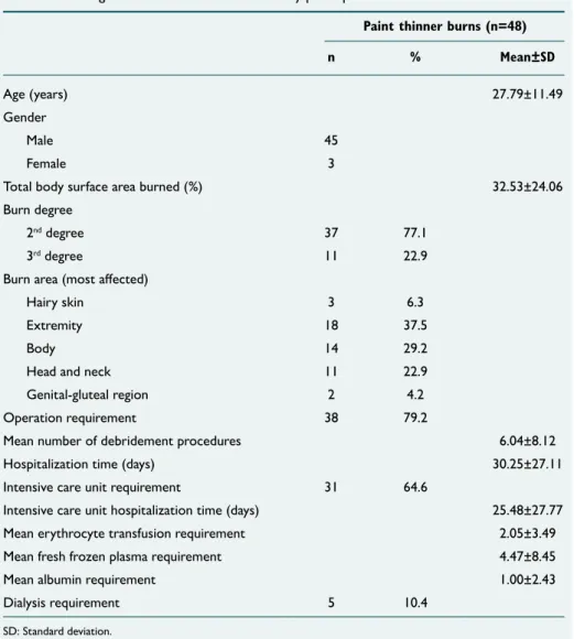
Table 1. The general characteristics of the study participants
Paint thinner burns (n=48)
n % Mean±SD
Age (years) 27.79±11.49
Gender
Male 45
Female 3
Total body surface area burned (%) 32.53±24.06
Burn degree
2nd degree 37 77.1
3rd degree 11 22.9
Burn area (most affected)
Hairy skin 3 6.3
Extremity 18 37.5
Body 14 29.2
Head and neck 11 22.9
Genital-gluteal region 2 4.2
Operation requirement 38 79.2
Mean number of debridement procedures 6.04±8.12
Hospitalization time (days) 30.25±27.11
Intensive care unit requirement 31 64.6
Intensive care unit hospitalization time (days) 25.48±27.77
Mean erythrocyte transfusion requirement 2.05±3.49
Mean fresh frozen plasma requirement 4.47±8.45
Mean albumin requirement 1.00±2.43
Dialysis requirement 5 10.4
son of patients who expired and those who were discharged, hemodialysis requirement did not yield statistically significant difference (p=0.10).
In all, 9 (18.8%) patients in the group expired and remaining 38 were discharged after treatment. Principal cause of death was septicemia (n=7) or respiratory failure (n=6). Septicemia was statistically significantly more common in expired group (p=0.031). When odds ratio (OR) was calculated, presence of septicemia increased mortality ratio 2.19 times (1.21–3.95). In comparison of expired and discharged patients regard-ing presence of respiratory failure requirregard-ing mechanic ven-tilation, respiratory failure was also statistically significantly more common in expired group (p=0.001). OR calculation
rized in Table 2. Interestingly, at admission, 41 (85.4%) pa-tients had CRP level higher than normal value, and 44 (91.7%) had the white blood cell and neutrophil count above normal values. In 39 (81.3%) of the patients, antibiotherapy was re-quired, and blood cultures were positive in 34 (70.8%) cases. In 7 of the 9 expired patients, blood cultures were positive and all those who died were receiving antibiotherapy.
DISCUSSION
The present study is retrospective assessment of patients with paint thinner burns conducted to determine general characteristics and outcomes. To the best of our knowledge, this is the largest study in the current literature evaluat-ing paint thinner burns. We determined that the victims of paint thinner burns were generally young males, %TBSA was high (32.53±24.06%), along with requirement for operation (79.2%) and ICU (64.6%) care. Total mortality ratio deter-mined in this group was 18.8%, and main cause of death was septicemia or respiratory failure.
Even though paint thinner burns are not rare, data about such burns in the current literature is limited. Queiroz et al.[5] investigated 293 patients admitted to ICU of burn
cen-ter between 2010 and 2012 and reported that 3.4% (n=10) of cases were due to paint thinner burn. We have reported prevalence of paint thinner burn of 7.6% in this study. Kulahci et al.[6] investigated demographic features of 9 male patients
who were the victims of paint thinner burn and reported that mean %TBSA was as high as 67.7% with mortality rate of 33.3%. Benbrahim et al.[7] examined demographic features of
17 patients admitted with paint thinner flame burns. Mean age of the patients was 32 years and nearly all of them (16/17) were male. Mean %TBSA was 23% in that study. Haberal et al.[8] retrospectively evaluated epidemiology of 28 paint
thin-ner burn injuries (25 male, 3 female) from period of 8 years and reported that mean age of the patients was 27.88±14.74 years and mean %TBSA was 48.82±27.39%. They stated that %TBSA was significantly larger in cases of paint thinner burn when compared with other sources of flame burn, and as in our study, the most commonly affected site was the ex-tremities. In that study, overall mortality rate was reported as
Table 2. Laboratory data of study participants at admission to the hospital
Paint thinner burns (n=48) Mean±SD Creatinine (mg/dL) 0.83±0.30 Urea (mg/dL) 27.68±9.84 Glucose (mg/dL) 138.36±90.88 Total protein (mg/dL) 5.32±1.62 Albumin (mg/dL) 3.07±0.98 Uric acid (mg/dL) 4.46±1.37
Aspartate amino transferase (IU/l) 53.57±96.67
Alanine aminotransferase (IU/l) 36.85±86.07
Potassium (mEq/L) 4.33±0.76
Sodium (mEq/L) 136.45±2.87
C-reactive protein 80.08±101.85
Hemoglobin (g/dL) 11.75±4.28
Mean platelet volume (fL) 8.45±1.24
Neutrophil (%) 70.12±19.04
Platelet count 266.76±143.82
WBC (103/µL) 21.78±11.19
39.3% and main cause of death was sepsis. Ozgenel et al.[9]
re-ported demographic data of 32 patients (30 males, 2 females) with paint thinner burns and indicated that mean age of pa-tients was 25.9±11 years and mean %TBSA was 33.6±24%. Mortality ratio was 15.6% in that study. In the present study, mean age of patients (27.79±11.49 years) and male predomi-nance were similar to other results. Also consistent with these studies, mean %TBSA was greater than 30% and mor-tality rate was greater than 15%.
In this study, main cause of death was septicemia or respira-tory failure. Inhalation injury was determined to be statis-tically significantly more common in the expired group and presence of inhalation injury was associated with more than 3 times greater mortality. Recently, inhalation injury was deter-mined to be independently associated with mortality in adults with %TBSA of 20% or greater.[10] Similarly, Chen et al.[11] also
reported that inhalation injuries significantly reduced sur-vival rate in patients with mild or moderate burns (burn in-dex<50%). Aguayo-Becerra et al.[12] reported that mortality
was higher for burns caused by inhalation injury and burns associated with infection. However, de Campos et al.[13] and
Moore et al.[14] did not determine significant association
be-tween inhalation injury and hospital mortality in severe adult burn patients admitted to burn ICU.
In our study, we determined that respiratory failure requir-ing mechanical ventilation increased mortality rate about 9 times. Similarly, Rosanova et al. also determined that mechan-ical ventilation was an independent variable related to mor-tality in children with burns.[15] Queiroz et al. also reported
that mechanical ventilation requirement was associated with significantly increased mortality rate in burn victims.[5] These
results were also compatible with our findings. In the pres-ent study, septicemia was one of the most common causes of mortality, and increased mortality by more than double. In parallel with our results, Krishnan et al.[16] reported that
multi-organ failure was primary cause of death, with sepsis being primary trigger in acute burn patients. Elkafssaoui et al.[17] also reported association of sepsis with increased
mor-tality. Multi-organ failure triggered by sepsis-associated in-flammatory cytokines may play a role in this association.[18] In
a recent review, Stewart et al.[19] did not recommend
system-ic antibiotsystem-ic prophylaxis for burns in low- and middle-income countries; however, further studies about prophylactic antibi-otic treatment, especially for patients with large paint thinner burns accompanied by inhalation injury, are warranted. In a retrospective study, Coban et al. investigated 411 burn patients and reported mortality rate of 5.6% (n=23). Among that study group, only 6 patients (1.4%) with acute renal fail-ure responded to hemofiltration. They also determined most common cause of mortality to be septicemia or effects of inhalation injury.[20] Saracoglu et al. recently reported that
main cause of death was multiple organ failure or infection in patients with electrical burns. However, they reported that
renal injury requiring hemofiltration was associated with an almost 12-fold increased risk for mortality.[21] In our study, we
did not determine a significant difference between expired or discharged groups regarding presence of hemodialysis re-quirement.
There are some limitations to this study that should be men-tioned. Although this is one of the largest studies in the lit-erature about paint thinner burns, the number of patients is still low. Secondly, blood culture results and microorganisms found were not recorded in this study, which may be the topic of another study to define an appropriate prophylaxis protocol.
Conclusion
Paint thinner ignition is an important cause of burn injuries that may cause very severe clinical picture in patients that requires prompt and careful treatment. Clinicians should be aware that presence of inhalation injury or sepsis were the 2 main factors affecting mortality rate in this group of patients. With increased awareness, preventive measures can be de-fined. Further studies are warranted in order to decrease mortality rate in this subgroup of burn patients.
Conflict of interest: None declared.
REFERENCES
1. Anderson CE, Loomis GA. Recognition and prevention of inhalant abuse. Am Fam Physician 2003;68:869–74.
2. Saito J, Ikeda M. Solvent constituents in paint, glue and thinner for plas-tic miniature hobby. Tohoku J Exp Med 1988;155:275–83. Crossref
3. Carabez A, Sandoval F, Palma L. Ultrastructural changes of tissues pro-duced by inhalation of thinner in rats. Microsc Res Tech 1998;40:56–62. 4. Yabanoglu H, Aytac HO, Turk E, Karagulle E, Belli S, Sakallioglu AE,
et al. Evaluation of Demographic and Clinical Characteristics of Patients who Attempted Suicide by Self-Inflicted Burn Using Catalyzer. Int Surg 2015;100:304–8. Crossref
5. Queiroz LF, Anami EH, Zampar EF, Tanita MT, Cardoso LT, Grion CM. Epidemiology and outcome analysis of burn patients admitted to an Intensive Care Unit in a University Hospital. Burns 2016;42:655–62. 6. Kulahci Y, Sever C, Noyan N, Uygur F, Ates A, Evinc R, et al. Burn
as-sault with paint thinner ignition: an unexpected burn injury caused by street children addicted to paint thinner. J Burn Care Res 2011;32:399– 404. Crossref
7. Benbrahim A, Jerrah H, Diouri M, Bahechar N, Boukind EH. Burns caused by paint thinner. [Article in French] Ann Burns Fire Disasters 2009;22:185–8. [Abstract]
8. Haberal M, Kut A, Basaran O, Tarim A, Türk E, Sakallioglu E, et al. Pre-ventable thermal burns associated with the ignition of paint thinner: ex-perience of a burn care network in Turkey. Minerva Med 2007;98:653–9. 9. Ozgenel GY, Akin S, Ozbek S, Kahveci R, Ozcan M. Thermal injuries
due to paint thinner. Burns 2004;30:154–5. Crossref
10. Sood RF, Gibran NS, Arnoldo BD, Gamelli RL, Herndon DN, Tomp-kins RG; Inflammation the Host Response to Injury Investigators. Early leukocyte gene expression associated with age, burn size, and in-halation injury in severely burned adults. J Trauma Acute Care Surg
15. Rosanova MT, Stamboulian D, Lede R. Risk factors for mortality in burn children. Braz J Infect Dis 2014;18:144–9. Crossref
16. Krishnan P, Frew Q, Green A, Martin R, Dziewulski P. Cause of
2010;16:353–6.
21. Saracoglu A, Kuzucuoglu T, Yakupoglu S, Kilavuz O, Tuncay E, Ersoy B, et al. Prognostic factors in electrical burns: a review of 101 patients. Burns 2014;40:702–7. Crossref
OLGU SUNUMU
Tiner yanıklarının genel özellikleri: Tek merkezli bir çalışma
Dr. Mustafa Celalettin Haksal,1 Dr. Cağrı Tiryaki,2 Dr. Murat Burç Yazıcıoğlu,2 Dr. Murat Güven,3
Dr. Ali Çiftci,2 Dr. Osman Esen,4 Dr. Hamdi Taner Turgut,2 Dr. Abdullah Yıldırım5 1Medipol Üniversitesi Tıp Fakültesi, Genel Cerrahi Anabilim Dalı, İstanbul
2Kocaeli Derince Eğitim ve Araştırma Hastanesi, Genel Cerrahi Kliniği, Kocaeli 3Kocaeli Derince Eğitim ve Araştırma Hastanesi, Yanık Tedavi Merkezi, Kocaeli 4Kocaeli Derince Eğitim ve Araştırma Hastanesi, Anestezi ve Reanimasyon Kliniği, Kocaeli 5Kocaeli Derince Eğitim ve Araştırma Hastanesi, Plastik ve Rekonstrüktif Cerrahi Kliniği, Kocaeli
AMAÇ: Bu çalışmanın amacı tinere bağlı yanıkları önlemek için bu yanıkların karakteristik özelliklerini incelemek ve risk faktörlerine olan farkındalığı artırmaktır.
GEREÇ VE YÖNTEM: Tiner yanığı nedeniyle yanık ünitesine kabul edilen hastalar geriye dönük olarak tarandı, hastaların klinik kayıtları kapsamlı bir şekilde incelendi. Ortalama yaşları 27.79±11.49 (dağılım, 16–58 yaş) olan toplam 48 hasta (3 kadın, 45 erkek) çalışmaya alındı.
BULGULAR: Ortalama hastanede kalış süresi 30.25±27.11 (dağılım, 3–110) gündü, ortalama toplam yanık yüzey alanı %32.53±24.06 (dağılım, %3.0–90.0). Toplam 31 hastada yoğun bakım ünitesi ihtiyacı oldu. Tiner yanığı olan hastaların dokuzu kaybedildi (%18.8), geriye kalan 38 hasta tedavileri sonrasında taburcu edildi. Ana ölüm nedeni septisemi (n=7) ve respiratuvar yetersizlikti (n=6). Hastaların 12’sinde inhalasyon yanığı eşlik ediyordu, bunlardan altısı kaybedilmişti (%50). Septisemi (p=0.031) ve inhalasyon hasarı (p=0.01) varlığı açısından karşılaştırıldığında, kaybedilen veya taburcu edilen hastalar arasındaki farklar anlamlı idi.
TARTIŞMA: Tinerle temas, hızlı ve dikkatli tedaviler gerektiren çok ciddi klinik tablolara neden olabilen önemli bir yanık nedenidir. Klinisyenlerin bunun bilincinde olmalı, bu hasta grubunda inhalasyon yanığı ve sepsisin mortalitenin iki önemli nedeni olduğunu bilmelidir. Artan bilinçle önleyici tedbirler tanımlanabilir. Bu hasta grubunda mortalitenin azaltılması için daha fazla çalışma yapılmasına ihtiyaç vardır.
Anahtar sözcükler: İnhalasyon hasarı; mortalite; tiner yanıkları.
Ulus Travma Acil Cerrahi Derg 2017;23(1):51–55 doi: 10.5505/tjtes.2016.66178