Early- and mid-term effects of percutaneous mitral balloon
valvuloplasty on left atrial mechanical functions in mitral stenosis
Mitral darlığında perkütan mitral balon valvüloplastinin kısa ve orta dönemde
sol atriyumun mekanik fonksiyonları üzerine etkisi
Department of Cardiology, Aksaray State Hospital, Aksaray;
*Department of Cardiology, Istanbul Kemerburgaz University Faculty of Medicine, İstanbul; #Department of Cardiology, Ataturk University Faculty of Medicine, Erzurum Sinan İnci, M.D., Mustafa Kemal Erol, M.D.,* Muhammet Hakan Taş, M.D.,# Eftal Murat Bakırcı, M.D.,# Hikmet Hamur, M.D.,# Şule Karakelleoğlu, M.D.#
Objectives: The aim of the study was to evaluate left atrial (LA) mechanical functions in MS before and after percutane-ous mitral balloon valvuloplasty (PMBV) and to follow it up in short- and mid-term.
Study design: We carried out a prospective study of 49 pa-tients with critical mitral stenosis (MS) who had normal sinus rhythm. LA mechanical functions were evaluated before and 24-48 h, 3 months, and 1 year after PMBV, which included LA passive emptying volume (LAPEV), LA active emptying volume (LAAEV), LA total emptying volume (LATEV), LA pas-sive emptying fraction (LAPEF), LA active emptying fraction (LAAEF), LA total emptying fraction (LATEF), and conduit vol-ume.
Results: The transthoracic echocardiography parameters of the MS patients before and 24-48 h, 3 months, and 1 year after PMBV were as follows: (a) mitral valve area 1.1 cm² (0.9-1.6); 2.2 cm² (1.8-2.8) (p<0.001); 2.2 cm² (1.7-2.9) (NS); 2.1 cm² (1.8-2.7) (p<0.001); (b) LAPEV 13 ml/m² (9-27); 11 ml/m² (8-19) (p<0.001); 10 ml/m² (7-19) (p<0.001); 10 ml/m² (6-18) (p<0.001); (c) LATEV 26 ml/m² (19-50); 21 ml/m² (16-40) (p<0.001); 20 ml/m² (15-36) (p<0.001); 19 ml/m² (15-34) (p<0.001); (d) Conduit volume 30 ml/m² (22-44); 33 ml/m² (26-46) (p<0.001); 34 ml/m² (30-42) (p<0.001); 36 ml/m² (31-42) (p<0.001), respectively. However, LAAEV, LAPEF, LAAEF, and LATEF were not altered after PMBV.
Conclusion: The findings of this study demonstrated an improvement of LA mechanical functions, which continued to improve for 1 year, after successful treatment of MS by PMBV.
Amaç: Çalışmanın amacı, mitral darlığı olan hastalarda per-kütan mitral balon valvuloplastinin (PMBV) sol atriyumun me-kanik fonksiyonları üzerine olan etkisini kısa ve orta dönemde değerlendirmektir.
Çalışma planı: Çalışmaya sinüs ritminde orta-ciddi mitral darlığı olan 49 hasta alındı. Sol atriyum pasif boşalma hacmi (LAPEH), sol atriyum aktif boşalma hacmi (LAAEH), sol atri-yum toplam boşalma hacmi (LATEH), sol atriatri-yum pasif boşal-ma fraksiyonu (LAPEF), sol atriyum aktif boşalboşal-ma fraksiyonu (LAAEF), sol atriyum toplam boşalma fraksiyonu (LATEF) ve konduit hacmi (KH) içeren sol atriyumun mekanik fonksiyon-ları, PMBV öncesi, işlem sonrası, işlemden 3 ay sonra ve iş-lemden 1 yıl sonra ölçüldü.
Bulgular: Mitral darlığı olan hastaların transtorasik ekokar-diyografi verileri PMBV öncesi, işlem sonrası, işlemden 3 ay sonra ve işlemden 1 yıl sonra olacak şekilde sıralandı: a) Mit-ral kapak alanı 1.1 cm² (0.9-1.6); 2.2 cm² (1.8-2.8) (p<0.001); 2.2 cm² (1.7-2.9) (NS); 2.1 cm² (1.8-2.7) (p<0.001); b) LAPEH 13 ml/m² (9-27); 11 ml/m² (8-19) (p<0.001); 10 ml/m² (7-19) (p<0.001); 10 ml/m² (6-18) (p<0.001); c) LATEH 26 ml/m² (19-50); 21 ml/m² (16-40) (p<0.001); 20 ml/m² (15-36) (p<0.001); 19 ml/m² (15-34) (p<0.001); d) KH 30 ml/m² (22-44); 33 ml/m² (26-46) (p<0.001); 34 ml/m² (30-42) (p<0.001); 36 ml/m² (31-42) (p<0.001). İşlem sonrası ve takiplerde LAAEH, LAPEF, LAAEF ve LATEF’de değişiklik izlenmedi.
Sonuç: Bulgularımız PMBV uygulanan mitral darlıklı hasta-larda başarılı tedavi sonrası sol atriyumun mekanik fonksi-yonlarının kısmen iyileştiğini ve bu iyileşmenin bir yıl boyunca devam ettiğini gösterdi.
Received:September 13, 2013 Accepted:February 18, 2014
Correspondence: Dr. Sinan İnci. Aksaray Devlet Hastanesi, Kardiyoloji Kliniği, Aksaray, Turkey. Tel: +90 382 - 212 91 00 e-mail: [email protected]
© 2014 Turkish Society of Cardiology
L
eft atrial (LA) functions consist of conduit, res-ervoir, and pump function.[1,2] Reservoir function occurs during left ventricular (LV) systole when the mitral valve is closed, the LA is relaxed, and the mi-tral annulus is temporarily displaced toward the apex. [3] The LA acts as a conduit in diastole, when the mi-tral leaflets open and allow blood to enter the left ven-tricle (LV). At end-diastole, the LA contracts, and the pump function occurs.[4]Several diseases have been acknowledged as pathological causes for mitral valve stenosis, among them rheumatic heart disease is the most prevalent. Rheumatic heart disease is a manifestation of rheu-matic carditis, which occurs in 60% to 90% cases of rheumatic fever.[5] Rheumatic mitral stenosis (MS) is associated with a considerable fusion of the com-missures, reduction of the mitral valve apparatus, and leaflet mobility. In MS, LA dimension increases and LA pump function deteriorates as time elapsed.[6-8]
Percutaneous mitral balloon valvuloplasty (PMBV) is a procedure that has been revealed to be an alternative to open and closed commissurotomy in the management of MS, currently the most common valvular disease. Short-, mid-, and long-term results obtained from management with PMBV in patients with appropriate symptomatic and mitral morphol-ogy demonstrated that this treatment modality is as effective as surgical procedures.[9] Successful PMBV decreases the intensity of spontaneous LA contrast, reduces the size of the LA, and improves LA function. [10] Improvements in LA mechanical functions after PMBV have been demonstrated in few clinical stud-ies. The studies in the literature have evaluated only the mechanical functions of the LA in the early period following PMBV, and no improvement has been not-ed in the pump functions of the LA in the acute phase.
The aim of the present study was to evaluate short- and mid-term effects of PMBV on the mechanical functions of LA. In addition to other mechanical functions, the present study particularly evaluated the pump functions in mid-term, which do not recover in the acute phase.
PATIENTS AND METHODS
We designed a prospective observational study to in-vestigate post-PMBV impact on LA mechanical func-tions in MS.
Subject
A total of 49 consecu-tive patients (male/ female 14/35; mean age 42±11 years) with symptomatic MS after successful PMBV were included in this study. Indications for PMBV were New York Heart class ≥II, ≤IV, planim-etered MVA, ≤1.5 cm²,
mitral regurgitation ≤2+, suitable valve morphology, and the absence of concomitant cardiovascular disease requiring surgical correction. All patients had sinus rhythm. A detailed written informed consent was ob-tained from all the patients. Approval of the study was obtained from the Local Ethics Committee. Exclusion criteria were as follows: left ventricular ejection frac-tion (LVEF) <50%, aortic regurgitafrac-tion greater than a mild degree or aortic stenosis, mitral regurgitation greater than a mild degree, tricuspid stenosis, clinical, echocardiographic or angiographic evidence of coro-nary artery disease, hypertension, diabetes mellitus, severe calcification of mitral valve annulus, clinical or laboratorical evidence of active rheumatic disease, chronic obstructive or restrictive lung disease, chronic pulmonary thromboembolism, and low-quality echo-cardiographic images for tissue Doppler imaging or velocity vector imaging analyses.
Transthoracic echocardiography and LA mechanical functions
In this study, a Vingmed system five Doppler echo-cardiographic (GE Vingmed ultrasound, Horten, Nor-way) unit with a 2.5 MHz FPA probe was used. All echocardiograms were recorded in left lateral decu-bitus with the parasternal long and apical 2-chamber and 4-chamber views. Mitral valve area (MVA) was calculated using the equation of continuity and plani-metric methods, and the average of the two methods was included in the analysis.
LA volumes were measured echocardiographically at the time of mitral valve opening (maximal, LA max volume), at the onset of atrial systole (p wave in the electrocardiography: LA volume before atrial systo-le), and in the mitral valve closure (minimal, LA min volume) according to the biplane area-length method
Abbreviations: LA Left atrial
LAAEF LA active emptying fraction LAAEV LA active emptying volume LAPEF LA passive emptying fraction LAPEV LA passive emptying volume LATEF LA total emptying fraction LATEV LA total emptying volume LV Left ventricular MS Mitral stenosis MVA Mitral valve area PAP Pulmonary artery pressure PMBV Percutaneous mitral balloon valvuloplasty
in the apical 4-chamber and 2-chamber views. All volumes were corrected for body surface area, and the following LA emptying functions were calculated:[11] 1. LA passive emptying volume = LA max volume
−LA volume before atrial systole;
2. LA passive emptying fraction = LA passive emp-tying volume/LA max volume × 100;
3. LA conduit volume = LV stroke volume (LA max volume − LA min volume);
4. LA active emptying volume = LA volume before atrial systole − LA min volume;
5. LA active emptying fraction = LA active emptying volume/LA volume before atrial systole × 100; 6. LA total emptying volume = (LA max volume −
LA min volume); and
7. LA total emptying fraction = LA total emptying volume/LA max volume × 100.
All measurements were averaged over three cardiac cycles.
Percutaneous balloon valvuloplasty
MS patients underwent PMBV, which was performed by three investigators. The MVA was calculated us-ing the Gorlin equation. Balloon dilation of the mitral valve was performed using a single balloon dilating technique.
Follow-up
Clinical and echocardiographic evaluations were per-formed before, and 24-48 h, 3 months, and 1 year after the PMBV. Recurrent stenosis was defined as >50%
loss of MVA calculated after the procedure and/or a valve area of 1.5 cm². A major cardiovascular event was defined as death, repeat of balloon valvuloplasty, or the need for mitral valve replacement during the follow-up period.
Statistical analysis
Statistical evaluation was performed using SPSS 15.0 (Statistical Package for the Social Sciences, Chica-go, IL, USA). Continuous variables were expressed as mean±standard deviation. The Friedman test was used to compare the consecutive measurements, and the Wilcoxon test was used for the post-hoc analysis. A value of p<0.05 was considered significant.
RESULTS
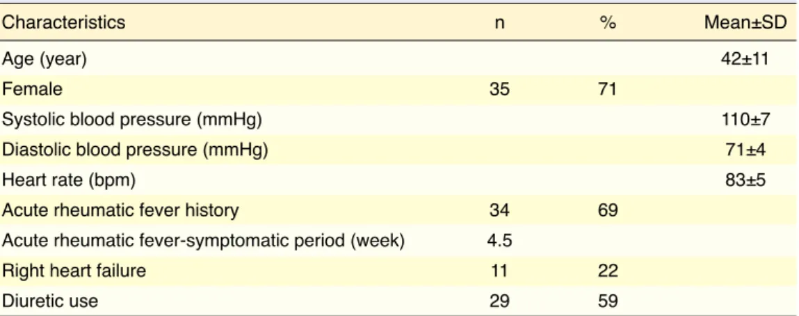
The baseline clinical and demographic properties of all study subjects are presented in Table 1. Thirty-five of the 49 patients that were enrolled in the study were female (71%). The average age of the patients was 42±11 years. Of the patients, only 34 had a past history of ARA, and 11 developed right heart failure. Major cardiovascular events (death, repeat balloon valvuloplasty, or mitral valve replacement) and re-current stenosis were not observed during the follow-up period. The results of the invasive hemodynamic measurements are shown in Table 2. No significant change was observed in the cardiac output following PMBV. Pulmonary capillary wedge pressure, pulmo-nary artery pressure (PAP), and mean gradient de-creased significantly following PMBV, whereas the MVA significantly increased.
Table 1. Clinical characteristics of study patients
Characteristics n % Mean±SD
Age (year) 42±11
Female 35 71
Systolic blood pressure (mmHg) 110±7
Diastolic blood pressure (mmHg) 71±4
Heart rate (bpm) 83±5
Acute rheumatic fever history 34 69
Acute rheumatic fever-symptomatic period (week) 4.5
Right heart failure 11 22
Diuretic use 29 59
increased after successful PMBV and in the follow-up period (p<0.01).
DISCUSSION
The present study showed that in patients with MS, successful PMBV improved LA reservoir and conduit functions, but not pump function, in both acute and chronic periods. Similar results were reported in a few clinical investigations. The current study demonstrat-ed incompletely improvdemonstrat-ed LA functions, 24-48 h, 3 months, and 1 year after PMBV. This is the first study indicating that LA functions continue to improve in the short- and mid-term after PMBV.
After PMBV, a substantial decline in transmitral gradient is usually observed. In the present study, a substantial decrease in transmitral gradient after PMBV, followed by a decrease in LA volume and an increase in the conduit volume, was observed, which may represent a close relationship among pressure, A comparison of the evaluated parameters before
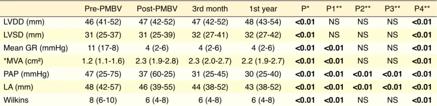
and after PMBV, measured by transthoracic echocar-diography, is presented in Table 3. The measured MVA had a significant increase after successful PMBV and in the follow-up period (p<0.01). The mean gradient value, PAP, LA diameter, and Wilkins the score sig-nificantly decreased after successful PMBV and in the follow-up period (p<0.01). A comparison of PMBV LA mechanic functional parameters before and after PMBV, measured by transthoracic echocardiography, is given in Table 4. LA maximum volume, minimum volume, atrial presystolic volume, LA passive emp-tying volume (LAPEV), and LA total empemp-tying vol-ume (LATEV) significantly decreased after successful PMBV and in the follow-up period (p<0.01). How-ever, LA passive emptying fraction (LAPEF), LA ac-tive emptying fraction (LAAEF), LA acac-tive emptying volume (LAAEV), and LA total emptying fraction (LATEF) were not altered after successful PMBV and in the follow-up period. Conduit volume significantly
Table 2. Pre- and post-PMBV invasive hemodynamic measurements in mitral valve stenosis patients
Pre-PMBV Post-PMBV p
Cardiac output (L/min) 4.4±0.9 4.6±0.9 NS
Left atrium-Left ventricle gradient (mmHg) 11.1±3.2 4.2±0.9 <0.01 Mitral valve area measured using the Gorlin equation (cm²) 1.1±0.2 2.1±0.4 <0.01 Pulmonary capillary wedge pressure (mmHg) 20.6±4.5 9.2±2.4 <0.01 Mean pulmonary artery pressure (mmHg) 34.4±5.7 26.5±4.9 <0.01
Pre-PMBV: Pre-percutaneous balloon valvuloplasty, Post-PMBV: Post-percutaneous mitral balloon valvuloplasty, NS: Not significant, SD: Standard devia-tion. The values are shown as the mean±SD, Student’s t-test.
Table 3. Comparison before and after PMBV values measured by transthoracic echocardiography
Pre-PMBV Post-PMBV 3rd month 1st year P* P1** P2** P3** P4** LVDD (mm) 46 (41-52) 47 (42-52) 47 (42-52) 48 (43-54) <0.01 NS NS NS <0.01 LVSD (mm) 31 (25-37) 31 (25-39) 32 (27-41) 32 (27-42) <0.01 NS NS NS <0.01 Mean GR (mmHg) 11 (17-8) 4 (2-6) 4 (2-6) 4 (2-6) <0.01 <0.01 NS NS <0.01 *MVA (cm²) 1.2 (1.1-1.6) 2.3 (1.9-2.8) 2.3 (2.0-2.7) 2.2 (1.9-2.7) <0.01 <0.01 NS NS <0.01 PAP (mmHg) 47 (25-75) 37 (60-25) 31 (25-45) 30 (25-40) <0.01 <0.01 <0.01 <0.01 <0.01 LA (mm) 48 (42-57) 46 (39-55) 44 (38-52) 43 (38-52) <0.01 <0.01 <0.01 <0.01 <0.01 Wilkins 8 (6-10) 6 (4-8) 6 (4-8) 6 (4-8) <0.01 <0.01 NS NS <0.01
Results are shown as mean±SD, rank (min-max) values. *Friedman test, **Wilcoxon test. *The average of the continuity equation and planimetric measure-ments were included. P1 - Pre-PMBV versus post-PMBV, P2 - Post-PMBV versus 3rd month, P3 - 3rd month versus 1st year, P4 - Pre-PMBV versus 1st year, LA: Left atrial diameter; LVDD: Left ventricular diastolic diameter; LVSD: Left ventricular systolic diameter; Mean GR: Mitral mean gradient; PMBV: Percutaneous balloon mitral valvuloplasty; MVA Pht: pressure half time mitral valve area; MVA plan: Planimetric mitral valve area; PAP: Pulmonary artery pressure; NS: Not significant; SD: Standard deviation.
volume, and function. These LA changes were mainly detected after 24-48 h of PMBV but not as marked after the 12 months, which could be explained by the substantial decrease in mitral valve afterload imme-diately after PMBV. The improvement in the Wilkins score in the acute phase was caused by increased valve mobility following PMBV. No changes were noted in the other parameters (thickening, calcifica-tion, or infravalvular structures).
LA functions have a prognostic clinical impact in several different scenarios. Stefanadis et al. found that PMV significantly decreases LA stiffness and in-creases LA systolic emptying index and LA stroke. Following PMBV, reservoir function increased in pa-tients with atrial fibrillation, but not in papa-tients with sinus rhythm.[10] In a study of 337 patients with dilated cardiomyopathy, a 3.8-times higher risk for death and the need for transplantation were observed when LA maximum volume was >68.5 mL/m2.[12] LA has prog-nostic implications in Chagas heart disease patients[13] and in sinus rhythm patients.[14] Vieira et al.[15] dem-onstrated improvements in LA reverse remodeling, in symptomatic MS patients undergoing PMBV, as analyzed by 2D and 3D echocardiography; and LAEF was observed after 72 h and 12 months of the
pro-cedure.Bitigen et al.[10] evaluated the LA mechanical functions before and after PMBV. Despite their in-dication of an improvement on conduit volume and reservoir functions, LA functions did not completely recover. In the current study, PMBV enabled a contri-bution to cardiac functions by decreasing LA volume both in the acute and long term. The researchers ob-served that LAVmax, LAmin, LA presystolic volume, LATEV, and LAPEV improved, and conduit volume increased following successful PMBV and during the follow-up period. However, LATEF, LAAEV, LAPEF, and LAAEF did not improve following suc-cessful PMBV and during the follow-up period. In spite of the clear improvement on the atrial volume function rates after a successful PMBV and 1 year of follow-up, the fact that fractional volume rates did not improve may be thought to be due to the late improve-ments in anatomical changes such as LA dilatation, fibrosis of the atrial wall, and disorganization of atrial muscle bundles.[16] Such improvements may not have been found because only mid-term follow-up results were available. Improvement in the fractions of the atrial volume can be anticipated in studies evaluating long-term results.
One of the most interesting points in this study
Table 4. Comparison of pre- and post-PMBV LA mechanic functional parameters measured by transthoracic echocardiography
Pre-PMBV Post-PMBV 3rd month 1st year P* P1** P2** P3** P4** LAV max (ml/m²) 59 (42-76) 48 (34-62) 44 (29-59) 43 (28-57) <0.01 <0.01 <0.01 <0.01 <0.01 LAV min (ml/m²) 33 (20-45) 27 (14-38) 24 (11-36) 23 (10-35) <0.01 <0.01 <0.01 <0.01 <0.01 LATEF (%) 0.43 (0.32-0.69) 0.45 (0.35-0.66) 0.45 (0.33-0.65) 0.45 (0.31-0.66) 0.05 0.04 NS NS 0.05 LATEV (ml/m²) 26 (19-50) 21 (16-40) 20 (15-36) 19 (15-34) <0.01 <0.01 <0.01 <0.01 <0.01 Reservoir function LAV p 46 (29-61) 37 (21-49) 34 (20-47) 32 (18-45) <0.01 <0.01 <0.01 <0.01 <0.01 Conduit function LAPEV (ml/m²) 13 (9-27) 11 (8-19) 10 (7-19) 10 (6-18) <0.01 <0.01 NS 0.03 <0.01 LAPEF (%) 0.21 (0.15-0.37) 0.22 (0.15-0.35) 0.23 (0.14-0.38) 0.24 (0.14-0.41) NS NS NS NS NS CV 30 (22-44) 33 (26-46) 34 (30-42) 36 (31-42) <0.01 <0.01 <0.01 <0.01 <0.01 Pump function LAAEV (ml/m²) 10 (8-23) 10 (6-21) 9 (5-17) 9 (4-16) NS NS NS NS NS LAAEF (%) 0.27 (0.20-0.51) 0.28 (0.20-0.51) 0.28 (0.20-0.51) 0.29 (0.16-0.47) NS NS NS NS NS
Results are shown as mean±SD, rank (min-max) values. *Friedman test, **Wilcoxon test. P1 - Pre-PMBV versus post-PMBV, P2 - Post-PMBV versus 3rd month, P3 - 3rd month versus 1st year, P4 - Pre-PMBV versus 1st year. LAV max: Left atrial maximal volume (cm³/m²); PMBV: Percutaneous balloon mitral valvuloplasty; CV: Conduit volume; LAV min: Left atrial minimal volume (cm³/m²); LAV p: Left atrial presystolic volume (cm³/m²); LAPEV: LA passive emptying volume (cm³/m²); LAPEF: LA passive emptying fraction (%); LAAEV: LA active emptying volume (cm³/m²); LAAEF: LA active emptying fraction (%); LATEV: LA total emptying volume (cm³/m²); LATEF: LA total emptying fraction (%); NS: Not significant; SD: Standard deviation.
The present study indicated that PMBV improved the LA mechanical functions including conduit and reservoir functions in the acute phase, and this favor-able effect lasted for 1 year. No improvement was not-ed in the LA pump functions, and long-term studies are warranted to elucidate long-term effects.
Conflict-of-interest issues regarding the authorship or article: None declared
REFERENCES
1. Inoue K, Owaki T, Nakamura T, Kitamura F, Miyamoto N. Clinical application of transvenous mitral commissur-otomy by a new balloon catheter. J Thorac Cardiovasc Surg 1984;87:394-402.
2. Nobuyoshi M, Hamasaki N, Kimura T, Nosaka H, Yokoi H, Yasumoto H, et al. Indications, complications, and short-term clinical outcome of percutaneous transvenous mitral commis-surotomy. Circulation 1989;80:782-92. CrossRef
3. Tuzcu EM, Block PC, Palacios IF. Comparison of early versus late experience with percutaneous mitral balloon valvuloplas-ty. J Am Coll Cardiol 1991;17:1121-4. CrossRef
4. Vahanian A, Michel PL, Cormier B, Ghanem G, Vitoux B, Maroni JP, et al. Immediate and mid-term results of percu-taneous mitral commissurotomy. Eur Heart J 1991;12 Suppl B:84-9.
5. Kawakita S. Rheumatic fever and rheumatic heart disease in Japan. Jpn Circ J 1986;50:1241-5. CrossRef
6. Fukuchi M, Kumagai K, Sakuma M, Kagaya Y, Watanabe J, Tabayashi K, et al. Warfarin-intractable, intraatrial throm-bogenesis in a 52-year-old woman with mitral stenosis and chronic atrial fibrillation. Tohoku J Exp Med 2004;203:59-63. 7. Akdeniz B, Badak O, Bariş N, Aslan O, Kirimli O, Göldeli
O, et al. Left atrial appendage-flow velocity predicts car-dioversion success in atrial fibrillation. Tohoku J Exp Med 2006;208:243-50. CrossRef
8. Uslu N, Nurkalem Z, Orhan AL, Aksu H, Sari I, Soylu O, et al. Transthoracic echocardiographic predictors of the left atrial appendage contraction velocity in stroke patients with sinus rhythm. Tohoku J Exp Med 2006;208:291-8. CrossRef
9. Ben Farhat M, Ayari M, Maatouk F, Betbout F, Gamra H, Jar-ra M, et al. Percutaneous balloon versus surgical closed and open mitral commissurotomy: seven-year follow-up results of a randomized trial. Circulation 1998;97:245-50. CrossRef
10. Bitigen A, Türkmen M, Karakaya O, Saglam M, Barutcu I, Esen AM, et al. Early effects of percutaneous mitral valvulo-plasty on left atrial mechanical functions. Tohoku J Exp Med 2006;209:285-9. CrossRef
11. Erol MK, Yilmaz M, Acikel M, Karakelleoglu S. Left atrial mechanical function in patients with essential hypertension. Acta Cardiol 2002;57:323-7. CrossRef
12. Rossi A, Cicoira M, Zanolla L, Sandrini R, Golia G, Zardini P, was the fast improvement of LA mechanical function
even in the post-PMBV acute period. In mitral valve stenosis, LA dysfunction was found to be associated with increased LA afterload.[17] The rapid decline in the afterload, as well as acute hemodynamic changes occurring immediately after PMBV, can explain this rapid recovery. Similar results have been reported in studies with a follow-up period of 1 year, which sug-gest that favorable mid-term effects of PMBV on the structural alterations in the LA can be explained by the improvement in the LA contraction and LA chamber stiffness. According to Pamir et al.[18] after the PMBV, the left atrium pressure decreases; however, there was no change in the left ventricle’s early diastolic filling fraction; and following the PMBV, the LV-end dia-stolic pressure increases in an important level. Mohan et al.[19] found that the decrease in atrial contribution in younger patients with MS was lower than that in older patients. While the atrial contraction in young patients recovered, there was no recovery in the LA function in older patients.Younger patients achieved better atrial filling after PMBV. This response was due to the improvement in atrial function, which is better in younger patients.[20]
Over the past several years, PMBV has become an accepted alternative to surgery in the treatment of MS. This procedure is highly successful with a low complication rate and significant short- and long-term improvements in both hemodynamics and patient symptoms.[21-23] Abrupt changes in metabolic and he-modynamic parameters following PMBV have at-tracted attention of most clinicians, and this has led to a rise in the number of research in this field.
The current study found improvement of LA me-chanical functions after successful treatment of MS with PMBV, and the improvement continued for 1 year. MS should be treated not only to avoid pulmo-nary edema and dysrhythmia, but also to alleviate the negative effect of impaired LA functions.
Limitations
This study included a relatively small number of pa-tients with MS. The parameters used to predict LA mechanical functions were not independent param-eters. Imaging methods such as real time 3D echocar-diography or deformation analysis were not used. The effect of PMBV on late prognosis could not be evalu-ated because the follow-up period was only 1 year.
Cardiol 1991;67:1121-6. CrossRef
18. Pamir G, Ertaş F, Oral D, Gümüş H, Omürlü K, Karaoguz R. Left ventricular filling and ejection fraction after success-ful percutaneous balloon mitral valvuloplasty. Int J Cardiol 1997;59:243-6. CrossRef
19. Mohan JC, Agrawal R, Arora R, Khalilullah M. Atrial contri-bution to left ventricular filling in mitral stenosis: effects of balloon mitral valvuloplasty. Indian Heart J 1994;46:129-32. 20. Krishnamoorthy KM, Dash PK. Effect of age on atrial
contri-bution to ventricular filling after balloon mitral valvuloplasty in mitral stenosis. Int J Cardiol 2004;96:241-6. CrossRef
21. Fawzy ME. Percutaneous mitral balloon valvotomy. Catheter Cardiovasc Interv 2007;69:313-21. CrossRef
22. Guérios EE, Bueno R, Nercolini D, Tarastchuk J, Andrade P, Pacheco A, et al. Mitral stenosis and percutaneous mitral val-vuloplasty (part 1). J Invasive Cardiol 2005;17:382-6. 23. Hernandez R, Bañuelos C, Alfonso F, Goicolea J,
Fernández-Ortiz A, Escaned J, et al. Long-term clinical and echocardio-graphic follow-up after percutaneous mitral valvuloplasty with the Inoue balloon. Circulation 1999;99:1580-6. CrossRef
et al. Determinants and prognostic value of left atrial volume in patients with dilated cardiomyopathy. J Am Coll Cardiol 2002;40:1425. CrossRef
13. Nunes MC, Barbosa MM, Ribeiro AL, Colosimo EA, Rocha MO. Left atrial volume provides independent prognostic val-ue in patients with Chagas cardiomyopathy. J Am Soc Echo-cardiogr 2009;22:82-8. CrossRef
14. Tsang TS, Abhayaratna WP, Barnes ME, Miyasaka Y, Gersh BJ, Bailey KR, et al. Prediction of cardiovascular outcomes with left atrial size: is volume superior to area or diameter? J Am Coll Cardiol 2006;47:1018-23. CrossRef
15. Vieira ML, Silva MC, Wagner CR, Dallan LA, Kajita LJ, Oliveira WA, et al. Left atrium reverse remodeling in patients with mitral valve stenosis after percutaneous valvuloplasty: a 2- and 3-dimensional echocardiographic study. [Article in English, Spanish] Rev Esp Cardiol 2013;66:17-23. [Abstract] 16. Catherine M, Bonow O, Bonow RO. Valvular heart disease.
In: Bonow RO, Mann DL, Zipes DP, Libby P, Braunwald E, editors. Braunwald’s heart disease: a textbook of cardiovascu-lar medicine. 9 th ed. Philadelphia: Elselvier, Saunders; Chap-ter 66. 2012. p. 1468-1529.
17. Matsuzaki M, Tamitani M, Toma Y, Ogawa H, Katayama K, Matsuda Y, et al. Mechanism of augmented left atrial pump function in myocardial infarction and essential hypertension evaluated by left atrial pressure-dimension relation. Am J
Key words: Atrial function, left; balloon valvuloplasty; cardiac cath-eterization/methods; mitral valve stenosis/therapy.
Anahtar sözcükler: Atriyal fonksiyon, sol; balon valvuloplasti; kalp kateterizasyonu/yöntem; mitral kapak stenozu/tedavi.