An Evaluation of the Current Rehabilitation Methods for
Women with Breast Cancer
Corresponding Author Yazışma Adresi Sibel Kibar Ufuk Üniversitesi Tıp Fakültesi, Fiziksel Tıp ve Rehabilitasyon AD, Ankara, Turkey
E-mail: [email protected] Received/Geliş Tarihi: 07.10.2013 Accepted/Kabul Tarihi: 21.04.2014
Meme Kanseri Olan Kadınlar İçin Güncel Rehabilitasyon
Yöntemlerinin Değerlendirmesi
Meltem Dalyan Aras1, Sibel Kibar2, Sibel Ünsal Delialioğlu1
1Ankara Physical Medicine and Rehabilitation Education and Research Hospital, Department of Physical Medicine and Rehabilitation, Ankara, Turkey 2Ufuk University, Faculty of Medicine, Department of Physical Medicine and Rehabilitation, Ankara, Turkey
ABSTRACT
At present, breast cancer (BC) survival rates are increasing day by day; therefore, the disabilities associated with BC therapy and the disease itself require a variety of rehabilitation modalities frequently. After the initial treatment, breast cancer patients require a dynamic follow-up period to check for tumor recurrence, metastasis, as radiation therapy side effects as well as side effects of chemotherapy, and physiatric evaluations and rehabilitative interventions must be a part of this process. Functional problems which need rehabilitation and have unfavourable effect on quality of life, emerges after breast cancer therapy. For example, chemotherapy may induce neuropathy, impair concentration and memory, and cause fatigue, and when combined with radiotherapy after axillary lymph node dissection (ALND), it has been associated with chronic pain in the ipsilateral arm. Furthermore, axillary dissection and/or radiation therapy in conjunction with chemotherapy can cause limitations in shoulder range of motion, nociseptive and neuropathic pain, numbness, myofascial pain, loss of strength, pectoral tightness, axillary cord development, and lymphedema. In our opinion, specifying rehabilitation needs and early treatment may improve the quality of life for breast cancer patients and stimulate them to more readily join in normal social activities after breast cancer treatment. The aim of this study is to review the rehabilitation requeriments of women with breast cancer.
Keywords: Breast cancer, rehabilitation, functional problems, upper extremity impairment
ÖZET
Günümüzde, meme kanseri tedavisi sonrası sağkalım oranları gün geçtikçe artmaktadır, bu nedenle, takip sürecinde hastalığın kendisine ve tedavisine bağlı komplikasyonların farklı rehabilitasyon yöntemlerine ihtiyacı olmaktadır. Tedavi sürecinin sonrasında tümörün tekrarı ve ortaya çıkabilecek metastazlar, radyoterapi ve kemoterapinin yol açabileceği yan etkiler açısından hastaların aktif olarak takibi gerekmektedir. Fiziatrik değerlendirmeler ve rehabilitasyon yaklaşımları da bu sürecin parçası olmalıdır. Meme kanseri tedavisi sonrası hastaların günlük yaşam aktivitelerini kötü yönde etkileyen, rehabilitasyon gerektiren fonksiyonel problemler ortaya çıkmaktadır. Örneğin kemoterapi nöropati gelişimini uyarabilir, konsantrasyon ve hafızayı bozabilir ve yorgunluğa neden olabilir. Üstelik aksiller lenf nodu diseksiyonu sonrası yapılan radyoterapi aynı taraf üst ekstremitede eklem hareket kısıtlılığı, nöropatik ve nosiseptif ağrı, uyuşukluk, miyofasial ağrı, kas gücü kaybı, pektoral gerginlik, aksiller kord gelişimi ve lenfödem gibi omuz-kol problemlerine neden olabilir. Meme kanseri tedavisi sonrası rehabilitasyon ihtiyacı olabilecek durumların belirlenmesi ve zamanında müdahale edilmesinin hastaların yaşam kalitesini iyileştireceğini ve sosyal hayata katılımlarını hızlandıracağını düşünüyoruz. Bu derlemenin amacı meme kanseri olan kadınlarda rehabilitasyon gerektiren durumları gözden geçirmektir.
Introduction
Contemporary cancer rehabilitation is a concept that is defined by the patient and involves helping a person with this disease obtain maximum physical, social, psychological, and vocational functioning within the limits imposed by the disease and its treatment (1) with a goal of improving multiple dimensions of life satisfaction (2).
Currently more cancer patients are being diagnosed at early, curable stages, and the five-year survival rates are increasing due to early diagnosis, effective screening programs, and a broader selection of multimodal treatments along with better general medical management (3). There is often a variety of impairments related to tumor effects or cancer treatment from the time of diagnosis to palliation (2,4,5). Individuals with this disease need to maintain functional status while also receiving appropriate rehabilitation and adequate symptom control as needed.
Multiple studies have documented the rehabilitation needs among cancer patients, and the most common thread among these studies was that these needs were going unmet (6-9). In fact, there is an underuse of rehabilitation services for cancer patients across the world due to a variety of reasons that include the failure by acute care staff to identify functional impairments, the absence of appropriate rehabilitation referrals, the lack of awareness of rehabilitation services, and an inadequate knowledge about such services among patients and family members. One of the first studies on this topic was performed by Lehman et.al who studied 805 patients at cancer referral centers in 1978. They found that 438 patients of all tumor types had rehabilitation needs. Fifty-two percent had psychological problems, 35% had generalized weakness, 30% had problems associated with daily activities, 25% had difficulties with ambulation, 7% had deficits in transfers, and 7% had deficits in communication (7). Undertreatment was particularly prominent among minorities and the socioeconomically disadvantaged (10). The topic of unmet rehabilitation and physical needs of cancer patients is still being investigated (7,8), but some studies about breast cancer (BC) have demonstrated inadequate evaluation of rehabilitation needs (11).
Breast Cancer
Among women, BC is the most frequently diagnosed type of carcinoma, and it is the leading cause of cancer deaths worldwide (12,13). However, more than 85% of BC patients are alive five years after their diagnosis. For instance, in the United States, there are 11 million cancer
survivors, and there are more than 2.4 million who have survived BC; hence, this group makes up 22% of all cancer survivors. However, the increased survival rates have also meant an escalation in complications related to BC treatment, which has necessitated the need for more rehabilitation modalities (12,13).
Determination of operability is the initial step in the treatment of BC after it has been diagnosed. Clinical and pathological staging are performed to identify the prognostic factors and treatment options. These are determined according to the stage of the disease, tumor size, biological characteristics of the tumor, estrogen and progesterone receptor expressions, the risks and benefits of the treatment protocols, and the age and preferences of the patient. Treatment principles are based on the therapeutic goal of complete excision of the tumor and management of the axilla with surgery and/or radiation therapy. Adjuvant systemic therapy is applied to prevent the recurrence of BC and the occurrence of distant metastasis. Recently, more conservative surgical approaches for BC have been adapted. Breast conservation surgery and sentinel lymph node dissections (SLND) are now performed more often than modified radical mastectomies and axillary lymph node dissection (ALND). However, locally advanced BCs are treated with modified radical mastectomies along with chemotherapy. In addition, radiotherapy of the chest wall is considered if the chest wall or nodal recurrence is high, the tumor is large or multicentric, or four or more axillary nodes have metastasized (3,12,13).
Three systemic adjuvant therapy modalities are currently used in the treatment of BC. These include endocrine treatments, such as tamoxifen, aromatase inhibitors, or ovarian suppression, anti-human epidermal growth receptor factor-2 (anti-HER-2) therapy with the humanized monoclonal antibody trastuzumab, and chemotherapy. The selection of the adjuvant treatment is determined by the biological features and stage of the BC. Chemotherapy regimes of multiple cycles may be administered before, during, or after radiation treatment, with epirubicin, doxorubicin, cyclophosphamide, 5-fluorouracil (5-FU), and taxanes being the most commonly used agents. Tamoxifen and aromatase inhibitors, such as anastrozole and letrozole, are adjuvant endocrine therapy modalities, and trastuzumab is yet another adjuvant therapy for patients with positive HER-2 expression (3,1HER-2,13).
After the initial treatment, BC patients require a dynamic follow-up period for local and regional tumor recurrence, contralateral BC, distant metastasis, late or ongoing side effects of radiation therapy, chemotherapy, and anti-estrogen therapies. Physiatric evaluations and rehabilitative interventions must be a part of
this process as well as in the initial treatment period. Quality of life (QOL), functional status, and physiological integrity after treatment have emerged as important public health issues in BC survivors (14) since there are several functional problems and common BC sequelae that require rehabilitation (15-17). Table 1 shows a summary of these common functional problems. For example, chemotherapy may induce neuropathy, impair concentration and memory, and cause fatigue. Additionally, chemotherapy with concurrent radiotherapy after ALND has been associated with chronic pain in the ipsilateral arm. Moreover, ALND and/ or radiation therapy can cause regional upper extremity dysfunction that includes limited shoulder range of motion, chronic pain, numbness, neuropathic pain, myofascial pain syndrome, loss of strength, pectoral tightness, axillary cord development, and lymphedema. These impairments are significantly associated with poor QOL in BC survivors (15-17).
Breast Cancer Rehabilitation
Shoulder Dysfunction
Shoulder dysfunction and restriction of shoulder mobility is common following BC surgery (Figure 1), with incidence rates ranging from 17-70% (18). Maunsel et al. reported this impairment in 32% of the patients in their study, but only 13% had limitations below 90° (19). In another study, shoulder dysfunction was reported at a rate of 36% (20). There are also differences between SLND and ALND in terms of shoulder function. For example, one study indicated that at the postoperative second week, shoulder range of motion was limited in up to 45% of the patients who had SLND, whereas 86% of the patients who had ALND had this problem (21). On
the other hand, a three-year follow-up of a patient group showed that 34% of the patients who underwent ALND and 15% of the patients who underwent SLND reported limited shoulder movement (22). This impairment can be caused by axillary web syndrome, pectoral tightness, radiation fibrosis, nerve injury, pain, anxiety, lack of use, and overprotection of the extremity as well as the actual surgery itself. A preoperative evaluation of the patient in terms of range of motion, strength, sensation, posture, endurance, and general functionality is recommended, and the patient and caregivers should be instructed regarding range of motion exercises and precautions, breathing exercises, and initial mobility after surgery. While considering the principles of wound healing, shoulder and arm rehabilitation should start as soon as possible postoperatively, with patients being encouraged to begin shoulder range of motion and stretching exercises after a few days since early remobilization does not increase morbidity. The details of shoulder remobilization are given in Table 2 (3).Passive, active-assisted, and active exercises for the shoulder should also be incorporated in the care plan. Activities such as wall walking, pendulum exercises, and internal and external rotation with the use of a cane are some of the early remobilization efforts. Compliance with the home exercise program is very important, and ongoing shoulder exercises are recommended, particularly for preventing contracture during the radiation therapy period. Studies have demonstrated that a structured,
Figure 1. Restriction of shoulder mobility after breast cancer surgery.
Table 1. Rehabilitation needs of patients with BC.
Shoulder dysfunction, frozen shoulder Lymphedema of the limb
Axillar lymphedema Axillary web syndrome Pectoralis tightness and pain Myofascial dysfunction and pain Loss of sensation and numbness Upper extremity pain
Loss of strength in the upper extremity Tumor or treatment related pain syndromes Osteoporosis
Fatigue
Morbidity related with breast reconstruction
Table 2. Shoulder remobilization following breast surgery.
Postoperative day Flexion Abduction Rotation
1 through 3 40°-45° 40°-45° To tolerance 4 through 6 45°-90° 45° To tolerance 7 onward To tolerance To tolerance To tolerance
prospective physiotherapy model is associated with better upper extremity function (23-25). Manual therapy, mobilization techniques, stretching exercises, adequate pain management, subacromial or intra-articular injections of anesthetics and steroids, capsular distension, and intraoperative shoulder manipulation under anesthesia are the other interventional and pharmacological approaches in the management of refractory shoulder dysmobility (16).
Axillary Web Syndrome
Axillary web syndrome, also known as axillary cord, syndrome of axillary adhesion, and cording lymphedema, was first defined by Moskovitz et al. who determined that it generally develops from one to five weeks after surgery and is responsible for a significant decrease in shoulder mobility (Figure 2) (26). The palpable cords that are involved are the lymphatic vessels whose course has been interrupted or occluded during ALND. Pathological assessment found these cords to be sclerosed veins and lymphatic vessels with surrounding fibrosis. The cords might be found only in the axilla, or they could extend distally along the anterior, medial aspect of the arm, or even the palm. The incidence rate differs among authors, which is presumably due to the various diagnostic criteria used in the studies. In a cohort of 85 patients, axillary web syndrome developed in 20% of the patients after SLND and in 72% after ALND, (21) and in a retrospective study of 750 mixed cases, Moskovitz reported this syndrome had an incidence rate of 6%. However, 74% of the patients in that study had a shoulder abduction limitation of 90° (26). Although the clinical course is undefined, this is a benign and non-progressive situation. Home exercise consisting of wall walking, Codman’s exercises, stretching and active-assistive exercises, manuel release techniques,
topical heat, and appropriate analgesic medications are the stardard therapeutic approaches. However, topical heat treatments must be performed with caution because of the risk of lymphedema and burn. Interventional techniques are generally contraindicated (16).
Myofascial Pain Syndrome
Myofascial dysfunctionis characterized by painful trigger points with radiating pain. In BC patients, the scapular retractor muscles frequently develop trigger points. Anxiety, pain, and shoulder capsule tightness are the other common etiological factors for myofascial pain. Pectoral tightness resulting from surgery, radiation fibrosis, a posture characterized by truncal anteroflexion, and scapular protraction are the main causative factors for myofascial pain and trigger points. The scapular retractor muscles try to overcome these issues and eventually become overused and strained. Although the exact incidence rate is not known, a cohort of 163 stage IV BC patients showed that 21% developed moderate to severe myofascial pain (27).Myofascial release, stretching and relaxation techniques, posture exercises, analgesics, antispasmodics, and trigger point injections are the most frequent treatment options.
Breast Cancer-Related Lymphedema
Lymphedema is defined as an abnormal accumulation of protein-rich fluid in a body part secondary to the insufficiency of the lymphatic system (28). Resection or irradiation of lymph nodes and vessels may cause lymphatic congestion that causes lymphedema in the affected territories. This sequela is associated with cosmetic deformity, physical discomfort, disability, recurrent episodes of cellulitis and lymphangitis, and reduced QOL (2,17). Secondary lymphedema has been reported as a consequence of treatment for several solid tumors, for example BC, melanoma, and head and neck tumors along with gynecological and genitourinary malignancies. The overall incidence rate of lymphedema among cancer survivors is about 15.5%, and this is related to the type and extent of treatment, anatomic location, clinical definition, patient characteristics, assessment techniques, and length of the follow-up period (29).
Lymphedema is one of the most recognized complications after BC (Figure 3). A transient lymphostasis occurs immediately after surgery as well as during radiation and lymphatic restoration, and the occurrence of collateral lymph drainage pathways is common (29-32). Lymphostasis can become chronic, leading to the development of lymphedema in the arm, axilla, breast, and trunk. Physical morbidities associated with lymphedema include skin changes, a sense of heaviness
and tightness, and a loss of sensation, strength and functional ability along with pain and several psychosocial difficulties (29-32). Incidence rates of lymphedema after BC have been recorded as low as 6% and as high as 67%, but they are generally between 20% and 30% (32). The risk factors are grouped in three main categories (33-37). Disease-related factors are tumor stage, nodal status, and tumor location. Patient-related factors include age, obesity, hypertension, infection, cellulitis, and elapsed time since treatment. Treatment-related etiologies are connected with the type of surgery, radiotherapy, and other adjuvant therapies. The risk of lyphedema increases after more extensive resections, such as those that occur with level-2 axillary clearance and mastectomies. Patients undergoing SLND and breast-conserving treatment are less likely to develop lymphedema, but the risk is not completely eliminated. One study found 17% of women with SLND alone (no follow-up axillary dissections) developed grade 1 or higher lymphedema (38), and several other studies agree that SLND decreases the risk of lymphedema (39-41).
The diagnosis of lymphedema is traditionally made by a comparison of limb sizes. Most clinicians agree that a difference of more than 2 cm or a 5% interlimb volume difference is clinically significant. Even when diagnostic thresholds are not reached, the clinical signs and symptoms might be meaningful because of the frequent insidious onset of lymphedema. There are several established lymphedema rating systems, (42,43) but in daily practice, circumference measurements and staging are preferable. In stage 1, the limb swells and pits with pressure. Elevation alone may relieve the swelling. In stage 2, the limb becomes firmer, and it may not pit with pressure. Skin changes may be noted. Finally, in stage 3, very thick skin and large skinfolds develop. Clinical
assessment includes ruling out infection (or treating it if present) and determining whether there is venous thrombosis or cancer recurrence. Serial measurements should be performed during follow-up visits (42,43).
Awareness of BC-related lymphedema has increased both in oncology units and among cancer survivors themselves. Several therapeutic approaches with different evidence levels for the treatment of lymphedema disease along with patient- and treatment-related factors can be seen in Table 3 (3,16,30,42,44). Their objectives are to decrease swelling, control the symptoms, and prevent complications. Decongesting the lymphatic pathways, maintaining collateral drainage, and stimulating the healthy lymphatics can be used in an effort to decrease the swelling in the extremity (3,16,30,42,44).
The management of lymphedema begins with obtaining information regarding the lymphatic circulation and the pathology that is causing the lymphedema as well as its signs, symptoms, and complications. The patients are then instructed about the self-care and skin maintenance, balanced nutrition, including an increase in protein and decrease in salt intake, weight management, and physical activity. The most widely accepted approach for lymphedema management includes the combination of multiple elements.
Complex physical therapy, also called complex decongestive therapy (CDT), is a treatment regimen for lymphedema that involves manual lymph drainage (MLD), bandaging, exercises, support garments and skin hygiene (45).CDT occurs in two phases (16,31,42).
Figure 3. Lymphedema in left hand and forearm.
Table 3. Treatment of BC- related lymphedema.
Skin care
Education for hygiene and infection prevention Weight control
Elevation
Compression garments Compression bandages Manual lymphatic drainage Self lymphatic drainage
Complex decongestive treatment Pneumatic pumps
Electrophysiological modalities (LLLT,ES) Oral medication
Exercise (remedial, aerobic, ROM, breathing, PRE)
LLLT low: Level laser therapy, ES: Electirical stimulation, ROM:
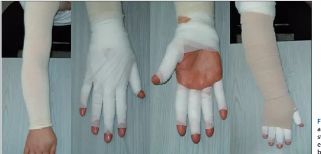
The initial phase focuses on reducing limb volume and the symptoms and consists of a certified therapist who performs MLD, applies multi-layered, short-stretch compressive bandages, and aids the patient as they do remedial exercises. Treatment can last from one to four weeks and may involve from two to 20 sessions depending on the severity of the condition. Afterwards, the volume reduction patients are transitioned to phase II of CDT, which is a long-term maintenance program. In this phase, the patients wear compression sleeves and gloves during the day and apply compression bandages at night. They also perform remedial exercises daily and receive additional MLD sessions as needed. Phase II continues indefinitely, and compliance is important for long-term lymphedema control. Manual lymphatic drainage is a specific massage technique designed to enhance sequestration and lymph transport. It differs from classical massage in terms of stroke duration, orientation, pressure, and sequence and is administered with light finger or hand pressures of 30mm Hg. In addition, this technique allows for the elimination of the congested truncal lymph by shifting it to lymphotomes with preserved drainage. Acute cellulitis, uncontrolled bacterial infection, arterial or venous occlusion, and renal dysfunction are relative contraindications of MLD. Compression bandages that are to be used are short-stretch and non-elastic (Figure 4). More layers of bandages can also be applied distally so that a distal-to-proximal compression gradient is achieved since the bandages have a high working pressure and exert force on the underlying contracting muscles. In addition, they exert low resting pressure when the muscles relax. Recently, there have been a few studies about kinesiotape applications as a compression therapy. While Tsai et al reported that kinesiotaping could be an alternative choice for compression bandages, another survey results
suggest that it could not replace the bandage(46, 47). Compression garments (Figure 5) help to maintain lymphatic flow, reduce accumulated protein, improve venous return, and properly shape and reduce the size of the limb. They also protect the limb from potential trauma. The garments should be class II and be replaced every six to nine months (16,31,42).
Exercises and physical activities are integral parts of lymphedema management (31,42). Remedial, aerobic, strength, and endurance training, range of motion activities, and posture and pulmonary exercise programs should be integrated into a comprehensive regimen of BC rehabilitation. Remedial exercises are a specific group of repetitive movements designed to encourage repeated muscle contractions within the lymphedematous territory. Rhythmic contraction and relaxation trigger the smooth muscles within the lymphatic system, and these exercises should always be performed while wearing the compression garments or bandages. Aerobic conditioning also has a positive effect in the transport of lymph through the elevated sympathetic tonus. It can also be utilized in the management of cancer-related fatigue. Flexibility exercises enhance posture, normalize biomechanics, and possibly facilitate lymph flow. Yoga and pilates might also be beneficial, but there is a need for research in terms of safety and the proper timing to start these exercises after surgery. In the past, most of the recommendations for BC survivors included avoiding the use of the affected arm. This approach usually was misguided and left the patients with the risk of deconditioning the arm. Weightlifting exercises were not prescribed for BC survivors in the past, whether they had lymphedema or not, but the recent findings of several randomized controlled trials have removed the concerns that these exercises increase the risk of lymphedema
Figure 4. The
application of short-stretch and non-elastic compression bandages.
[45-48]. Strength training should be done progressively. Patients should start with light weights, and their activities should be controlled and supervised at the beginning. The same research that demonstrated that weightlifting was not harmful also showed that upper body exercise for BC survivors with lymphedema as well as those at risk for this condition was also safe. Evidence also exists that indicates that an exercise program for BC survivors consisting of slow progression reduces the symptoms, decreases the incidence of exacerbations of lymphedema, and increases the work capacity of the limb (48-51).
Pneumatic pump therapy and low level laser therapy (LLLT) are other physical treatment modalities for BC-related lymphedema (42). Contraindications for the use of pneumatic devices include acute deep vein thrombosis and inflammatory edema. When utilizing these devices, the therapeutic pressure should be between 30 to 60mm Hg, and the treatment time per session should range from 30 minutes to up to an eight-hour period, but this depends on the condition of the patient and the device. The isolated use of pneumatic devices is not currently advised, but they can be utilized as part of a comprehensive management program for lymphedema. There are also a few studies that have suggested that LLLT is an effective treatment modality for lymphedema, but the authors of these cohorts came to the conclusion that it should be considered as an adjuvant therapy that could be used in conjuction with other treatment methods (52,53).
There are also some surgical procedures for the treatment of primary lymphedema, but there is no
convincing data that surgery is a viable option for BC-related lymphedema. Additionally, medications such as diuretics have not been effective unless there is an origin of mixed edema like cardiogenic edema and lymphedema. Furthermore, benzopyrones and various flavanoids have also had no proven effect in these cases.
Pain
The prevalence of cancer-related pain is 28% among patients with newly diagnosed cancer, and 50-70% of these patients receive antineoplastic therapy. However, the rate increases to 64-80% among patients with advanced disease (3). There might be a variety of pain syndromes in cancer patients, but the majority of pain is due to the tumor effects. Cancer therapy or unrelated causes have also been noted as other sources of pain in patients with carcinoma. If the pain is due to direct tumor spread, antitumor therapy is critical for pain management. The differential diagnosis of acute and chronic pain as well as its location, severity, and quality should be evaluated, and the impact of pain on sleep and daily activities also needs to be assessed. Psychological factors such as depression, anxiety, and distress may increase the pain experience, so these conditions should be identified and treated appropriately. Pain treatment in cancer patients is built on the guidelines established by the World Health Organization (3). The first line in treatment is nonopioid analgesics, such as paracetamol and nonsteroidal anti-inflammatories. If these fail to control the pain, opioids (codeine, oxycodone, morphine, fentanyl, methadone, etc.) can be added, with the dosage being determined according to the severity of the patient’s symptoms. In some cases, adjuvant pain medications (antidepressants, benzodiazepines, corticosteroids, anti-epileptics, muscle relaxants, etc) might offer better results for pain control, and modalities such as heat, cold, electricity, transcutaneous electrical nerve stimulation (TENS), and acupuncture along with cognitive and behavioral strategies can also be tried for pain alleviation in cancer patients. Interventional approaches such as nerve blocks, intraspinal opioid administrations, and neurosurgical and neuroablative procedures may also be beneficial in the treatment of refractory cancer pain.
Persistent postsurgical pain has been reported in 10-50% in patients, including those who underwent BC surgery (54). This type of pain in BC survivors is often termed as postmastectomy pain syndrome, especially if there are neuropathic pain descriptors such as burning, tingling, and lancinating. Neuroma formation, sacrifice of the intercostobrachial nerve, fibrotic entrapment, and surgical compromise of cutaneous innervation have been suggested as relevant pathogenic mechanisms. Physical therapy consisting of desensitization and TENS along with range of motion to improve strength and flexibility
Figure 5. Compression garment for arm.
exercises can also be prescribed. Adjuvant analgesics such as gabapentin, pregabalin, paroxetine, and duloxetine are the preferred agents for this type of pain. Interventional pain management, including intercostal or paravertebral nerve blocks and radiofrequency ablation/cryoablation, might be necessary in refractory cases.
Fatigue
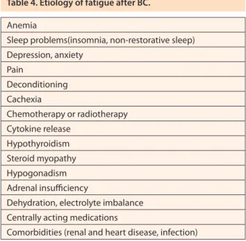
Almost 90% of cancer patients experience cancer-related fatigue at some point in their disease course (55,56). Fatigue is defined as an unusual, persistent, subjective sense of tiredness related to cancer or cancer treatment that interferes with normal functioning, and it is an important barrier to the rehabilitation of cancer patients. Pain, depression, anxiety, dyspnea, and insomnia are common symptoms associated with this condition. Although the pathophysiology of fatigue is not completely understood, a group of possible mechanisms have been proposed that includes cytokines, serotonin dysregulation, hypothalamic-pituitary-adrenal (HPA) axis dysfunction, and abnormal sleep-wake cycles. Certain conditions (Table 4)contribute to fatigue, and these should be examined and ruled out if possible. Those that are present should then be treated in a relevant manner (3,55,56). Medications such as methylphenidate, modafinil, paroxetine, prednisone, and L-carnitine have been proven to be beneficial in alleviating fatigue. For some patients, psychosocial interventions (stress management, support groups, energy conservation, etc) and improved sleep hygiene can also be effective. As there is growing evidence that aerobic and resistive types of exercises have a positive effect on cancer-related fatigue, patients should be encouraged to begin an exercise regimen and be provided with a detailed recommendation of an exercise program to fit their needs.
Rehabilitation After Breast Reconstruction
Breast reconstruction is a part of BC treatment that is pursued by many BCpatients (3). The advantages of this procedure are improved QOL and a better self-image. Studies have shown that breast reconstruction does not alter the biological behavior of BC. In addition, it does not interfere with adjuvant chemotherapy and is not associated with a delay in the diagnosis of BC recurrence. Reconstruction can be performed any time after surgery (57,58), and saline or silicone implants and autolog procedures are the current options of choice. The most common breast reconstruction procedures are a tranverse rectus abdominis myocutaneous flap (TRAM), lattisumus dorsi flap, a gluteus maximus flap, and a distal inferior epigastric perforator flap, but the TRAM procedure is the most common (3,57,58). Rehabilitation consultations can be useful for preventing musculoskeletal sequelae in flap procedures.
Rehabilitation after TRAM procedure is an important issue. The patients should be trained in proper body movements to reduce stress on the abdominal wall such as positioning the head of the bed 45-degree angle and instructing the patient to lie on the uninvolved side in a fetal position. The patients should be adviced to maintain trunk flexion during transfers and ambulation in their usual life. Active and active assistive exercises of the upper extremities should be given in the first week after surgery. Abdominal strengthening exercises should begin a few weeks aftersurgery. Lifting and sit-ups are not permitted until 6 weeks after surgery. Lumbar stabilization (core) exercises are the most efficient exercises for trunk strengthening. The resistance exercise intensity may be increased due to patients’ tolerance day by day (59,60).
Shoulder dysfunction might occur after the latissumus dorsi flap procedure as this muscle is an important shoulder adductor, internal rotator, and depressor. Shoulder range of motion and strength, scapulothoracic joint function, and glenohumeral joint mechanics should be restored in these patients.
Special Considerations After Adjuvant Medical and Radiation Treatments
Chemotherapy is usually administered in four to six cycles of three to four weeks in length, and hormonal treatment is prescribed over a period of years. Continuation of regular activities during these treatments is recommended. The following consequences of adjuvant treatments require monitoring: weight gain, loss
Table 4. Etiology of fatigue after BC.
Anemia
Sleep problems(insomnia, non-restorative sleep) Depression, anxiety Pain Deconditioning Cachexia Chemotherapy or radiotherapy Cytokine release Hypothyroidism Steroid myopathy Hypogonadism Adrenal insufficiency
Dehydration, electrolyte imbalance Centrally acting medications
of muscle tissue, fatigue, lifestyle changes, depression, premature ovarian failure resulting in osteoporosis (e.g,. estrogen deprivation caused by aromatase inhibitors and choosing surgery over ablation), serious cardiac side effects like cardiomyopathy after anthracycline chemotherapy, and neurological side effects of certain chemotherapy agents such as neuropathy and impairment in concentration and memory. Rehabilitation activities must be considerd with these in mind and should be managed in order to increase the patient’s QOL and functional ability (3,4,30,61).
Radiation therapy-related impairments include the following: increased obstruction of arm lymphatics, tightness of the chest wall and pectoralis that decreases shoulder mobility, pain in the subcutaneous tissue, intercostal muscles and ribs, reduced pulmonary capacity, rib fractures, brachial plexopathy, myelopathy, encephalopathy, and cerebrovascular ischemia. These conditions should also be evaluated at physical medicine and rehabilitation clinics (3,4,30,61).
Rehabilitation After Systemic Complications
Metastatic (stage IV) BC is defined by the spread of the tumor beyond the breast, chest wall, and regional lymph nodes. The most common sites include the bones (vertebrae, pelvis, ribs, and proximal long bones), lungs, liver, lymph nodes, and brain. Therapy is not generally curative in advanced BC. Treatment goals include the prolongation of life, reduction and palliation of cancer-related symptoms, and maintenance ofQOL and function. The symptoms are related to the location and extent of the tumor, but bone discomfort, fracture, neurological compromise, metabolic complications, lymphadenopathy, skin changes, cough, and dyspnea are commonly seen (3,61). Moreover, these symptoms are often accompanied by pain, fatigue, mobility and self-care deficits, vocational problems, and depression. Rehabilitation goals during this period of the disease include the following: 1- To achieve good pain relief and comfort, 2- To focus on maximizing independence with regard to mobility, ambulation, and self care, 3-To maintain the stability of the spine and long bones, 4- To offer psychosocial support and prevent progressive isolation, 5- To provide education for caregivers, 6- To develop public health strategies for terminally ill patients (61).
Conclusion
After BC treatment in the follow-up period, evaluating the patients in terms of the clinical features of the disease and the complications of related treatment modalities has become more necessary. We suggest that by specifying the rehabilitation needs (shoulder
dysfunction, lymphedema, pain, neuropathy, faigue, etc.) and providing extensive counseling to these patients, their QOL may improve, which could motiviate them to participate more readily in normal social activities.
References
1. Ganz PA. The status of cancer rehabilitation in the late 1990s. Mayo Clin Proc 1999;74:939-40.
2. Cheville AL. Cancer rehabilitation. Semin Oncol 2005;32:219-24.
3. Cheville A. Cancer rehabilitation. In: Bradom RL, editor. Physical Medicine & Rehabilitation. 3rd ed. Philadelphia: Saunders Elsevier; 2007: p 1369-96.
4. Aras M, Ünsal S. The essence of rehabilitation of patients with cancer. Turk J Phys Med Rehab 2007; 53:74-7.
5. Vargo MM. The oncology-rehabilitation interface: Better systems needed. J Clin Oncol 2008;26:2610-1.
6. Lehmann JF, DeLisa JA, Warrren CG, DeLateur BJ, Bryant PL, Nicholson CH. Cancer rehabilitation: assessment of need, development and evaluation of model care. Arch Phys Med Rehabil 1978;59:410-9.
7. Veloso AG, Sperling C, Holm LV, Nicolaisen A, Rottmann N, Thayssen S, Christensen RD, Lehmann Knudsen J, Hansen DG. Unmet needs in cancer rehabilitation during the early cancer trajectory--a nationwide patient survey. Acta Oncol 2013;52:372-81.
8. Ng R, Verkooijen HM, Ooi LL, Koh WP. Unmet psychosocial needs among cancer patients undergoing ambulatory care in Singapore. Support Care Cancer 2012;20:1049-56. 9. Aras M, Unsal S, Atalay N, Taflan-Selçuk S. Rehabilitation
needs of patients with cancer. Turk J Phys Med Rehab 2009;55:25-9.
10. Cheville AL, Troxel AB, Basford JR, Kornblith AB. Prevalence and treatment of patterns of physical impairments in patients with metastatic breast cancer. J Clin Oncol 2008;26:2621-9
11. von Heymann-Horan AB, Dalton SO, Dziekanska A, Christensen J, Andersen I, Mertz BG, Olsen MH, Johansen C, Bidstrup PE. Unmet needs of women with breast cancer during and after primary treatment: a prospective study in Denmark. Acta Oncol. 2013;52:382-90.
12. Epidemiology of cancer. In: Principles and Practice of Oncology. 8th ed. Philadelphia: Lippincott Williams & Wilkins; 2008.
13. World Health Organisation [internet]. Available from: http:// www.who.int/mediacentre / factsheets/fs297/en/index. html
14. Pockaj BA, Degnim AC, Boughey JC, Gray RJ, McLaughlin SA, Dueck AC, Perez EA, Halyard MY, Frost MH, Cheville AL, Sloan JA. Quality of life after breast cancer surgery: What have we learned and wher should we go next? J Surg Oncol 2009;99:447-55.
15. Smoot B, Wong J, Cooper B, Wanek L, Topp K, Byl N, Dodd M. Upper extremity impairments in women with and without lymphedema following breast cancer treatment. J Cancer Surviv 2010;4:167-178.
16. Cheville AL, Tchou J. Barriers to rehabilitation following surgery for primary breast cancer. J Surg Oncol 2007;95:409-18.
17. Unsal S, Aras M, Kurt EE, Özel S. Demographic and clinical characteristics of patients with breast cancer-related lymphedema. Turk J Phys Med Rehab 2009;55:25-9.
18. Dinçer U, Kaya E, Çakar E, Kıralp MZ, Dursun H. Effectiveness of comprehensive rehabilitation program and home-based exercise in middle and long term mastectomy related disability. Turk J Phys Med Rehab 2007;53:138-43.
19. Maunsell E, Brisson J, Deschenes L. Arm problems and physiological distress after surgery for breast cancer. Can J Surg 1993; 36:315-20.
20. Rietman JS, Dijkstra PU, Debreczeni R, Geertzen JH, Robinson DP, De Vries J. Impairments, disabilities and health related quality of life after treatment for breast cancer: a follow-up study 2.7 years after surgery. Disabil Rehabil 2004;26:78-84. 21. Leidenius M, Leppänen E, Krogerus L, von Smitten K. Motion
restriction and axillary web syndrome after sentinel node biopsy and axillary clearance in breast cancer. Amer J Surg 2003;185:127-130.
22. Leidenius M, Leivonen M, Vironen J, von Smitten K. The consequences of long-time arm morbidity in node negative breast cancer patients with sentinel node biopsy or axillary clearance. J Surg Oncol 2005;92:23-31.
23. Springer BA, Levy E, McGarvey C, Pfalzer LA, Stout NL, Gerber LH, Soballe PW, Danoff J. Pre-operative assessment enables early diagnosis and recovery of shoulder function in patients with breast cancer. Breast Cancer Res Treat 2010;120:135-147.
24. Box RC, Reul-Hirche HM, Bullock-Saxton JE, Furnival CM. Shoulder movement after breast cancer surgery: results of a randomized controlled study of postoperative pysiotherapy. Breast Cancer Res Treat 2002;75:35-50.
25. McNeely ML, Campbell K, Ospina M, Rowe BH, Dabbs K, Klassen TP, Mackey J, Courneya K. Exercise interventions for upper-limb dysfunction due to breast cancer treatment (review). Cochrane Database Syst Rev. 2010 16;(6):CD005211. 26. Moskovitz AH, Anderson BO, Yeung RS, Byrd DR, Lawton
TJ, Moe RE. Axillary web syndrome after axillary dissection. Amer J Surg 2001;181:434-439.
27. Cheville AL, Troxel AB, Basford JR, et al. Rehabilitation needs and service utilization in patients with stage IV breast cancer. ECCO 14th Annual Assembly; 2007 September Barcelona, 2007.
28. Harwood CA, Mortimer PS. Causes and clinical manifestations of lymphatic failure. Clin Dermatol 1995;13:459-471. 29. Cormier JN, Askew RL, Mungovan KS, Xing Y, Ross MI,
Armer JM. Lymphedema beyond breast cancer. Cancer 2010;116:5138-49.
30. Chalasani P, Downey L, Stopeck AT. Caring for the breast cancer survivor: A guide for primary care physician. Am J Med 2010;123:489-495.
31. Smoot B, Wong J, Cooper B, Wanek L, Topp K, Byl N, Dodd M. Upper extremity impairments in women with or without lymphedema following breast cancer treatment. Cancer Surviv 2010;4:167-178.
32. Schmitz KH. Balancing lymphedema risk: Exercise versus deconditioning for breast cancer survivors. Exerc Sport Sci Rev 2010;38:17-24.
33. Cheville AL. Current and future trends in lymphedema management: Implications for women’s health. Phys Med Rehabil Clin Am 2007;18:539-553.
34. Soran A, Aydın C, Harlak A, Johnson R. Is sentinel node biopsy a real hope in the prevention of breast cancer-related lymphedema. J Breast Health 2006;2:1-6.
35. Tsai RJ, Dennis LK, Lynch CF, Snetselaar LG, Zamba GK, Scott-Conner C. The risk of developing arm lymphedema among breast cancer survivors: a meta- analysis of treatment factors. Ann Surg Oncol 2009;16:1959-1972.
36. Gärtner R, Jensen MB, Kronborg L, Ewertz M, Kehlet H, Kroman N. Self-reported arm-lymphedema and functional impairment after breast cancer treatment- A nationwide study of prevalence and associate factors. The Breast 2010;19:506-515.
37. Petrek JA, Senie RT, Peters M, Rosen PP. Lymphedema in a cohort of breast carcinoma survivors 20 years after diagnosis. Cancer 2001;92:1369-1377.
38. Francis WP, Abghari P, Du W, Rymal C, Suna M, Kosir MA. Improving surgical outcomes: standardizing the reporting of incidence and severity of acute lymphedema after sentinel node biopsy and axillary lymph node dissection. Am J Surg 2006;192:636-639.
39. Sener SF, Winchester DJ, Martz CH, Feldman JL, Cavanaugh JA, Winchester DP, Weigel B, Bonnefoi K, Kirby K, Morehead C. Lymphedema after sentinel lymphadenectomy for breast carcinoma. Cancer 2001;92:748-752.
40. Burak WE, Hollenbeck ST, Zervos EE, Hock KL, Kemp LC, Young DC. Sentinel lymph node biopsy results in less postoperative morbidity compared with axillary lymph node dissection for breast cancer. Am J Surg 2002;183:23-27.
41. Golshan M, Martin WJ, Dowlatshahi K. Sentinel lymph node biopsy lowers the rate of lymphedema whan compared with standart axillary lymph node dissection. Am Surg 2003;69:209-211.
42. Cheville AL, McGarvey CL, Petrek JA, Russo SA, Taylor ME, Thiadens SR. Lymphedema management. Semin Radiation Oncol 2003;13:290-301.
43. Cheville AL, McGarvey CL, Petrek JA, Russo SA, Thiadens SR, Taylor ME. The grading of lymphedema in oncology clinical trials. Semin Radiation Oncol 2003;13:214-225.
44. McNeely ML, Peddle CJ, Yurick JL, Dayes IS, Mackey JR. Conservative and dietary interventions for cancer-related lymphedema. Cancer 2010;0:1-13.
45. Harris SR1, Hugi MR, Olivotto IA, Levine M; Steering Committee for Clinical Practice Guidelines for the Care and Treatment of Breast Cancer. Clinical practice guidelines for the care and treatment of breast cancer: 11. Lymphedema. CMAJ 2001;164:191-9.
46. Tsai HJ1, Hung HC, Yang JL, Huang CS, Tsauo JY. Could Kinesio tape replace the bandage in decongestive lymphatic therapy for breast-cancer-related lymphedema? A pilot study. Support Care Cancer 2009;17:1353-60. 47. Smykla A, Walewicz K, Trybulski R, Halski T, Kucharzewski
M, Kucio C, Mikusek W, Klakla K ,Taradaj J.Effect of Kinesiology Taping on breast cancer-related lymphedema: arandomized single-blind controlled pilot study.Biomed Res Int 2013;2013:767106.
48. Schmitz KH, Ahmed RL, Troxel A, Cheville A, Smith R, Lewis-Grant L, Bryan CJ, Williams-Smith CT, Greene QP. Weight lifting in women with breast-cancer-related lymphedema. N Engl J Med 2009;361:664-673.
49. Schmitz KH, Ahmed RL, Troxel AB, Cheville A, Lewis-Grant L, Smith R, Bryan CJ, Williams-Smith CT, Chittams J. Weight lifting for women at risk for breast cancer-related lymphedema. JAMA 2010;304:2699-2705.
50. Kim DS, Sim YJ, Jeong HJ, Kim GC. Effect of active resistive exercise on breast-cancer-related lymphedema: A randomized controlled trial. Arch Phys Med Rehabil 2010;91:1844-1848.
51. Schmitz KH, Troxel AB, Cheville A, Grant LL, Bryan CJ, Gross CR, Lytle LA, Ahmed RL. Physical activity and lymphedema (the PAL trial): assessing the safety of progressive strength training in breast cancer survivors. Contemporary Clinical Trials 2009;30:233-245.
52. Omar MTA, Ebid AAG, Morsy AME. Treatment of post-mastectomy lymphedema with laser therapy: Double blind placebo control randomized study. J Surg Res 2011;165:82-90.
53. Lau RWL, Cheing GLY. Managing postmastectomy lymphedema with low-level laser therapy. Photomedicine and Laser Surgery 2009;27:763-769.
54. Kehlet H, Jensen TS, Woolf CJ. Persistent postsurgical pain: risk factors and prevention. Lancet 2006;367:1618-1625. 55. Cheville AL. Cancer- related fatigue. Phys Med Rehabil Clin
N Am 2009;20:405-416
56. Schultz SL, Dalton SO, Christensen J, Carlsen K, Ross L, Johansen C. Factors correlated with fatigue in breast cancer survivors undergoing a rehabilitation course, Denmark, 2002-2005. Psycho-Oncology 2011;20:352-60.
57. Avraham T, Daluvoy SV, Riedel ER, Cordeiro PG, Van Zee KJ, Mehrara BJ. Tissue expander breast recontruction is not associated with an increased risk of lymphedema. Ann Surg Oncol 2010;17:2926-932.
58. Elder EE, Brandberg Y, Björklund T, Rylander R, Lagergren J, Jurell G, Wickman M, Sandelin K. Quality of life and patient satisfaction in breast cancer patients after immediate breast reconstruction: a prospective study. Breast 2005;14:201-208.
59. McAnaw MB, Harris KW. The role of physical therapy in the rehabilitation of patients with mastectomy and breast reconstruction. Breast Dis 2002;16:163-74.
60. Monteiro M. Physical therapy implications following the TRAM procedure. Phys Ther 1997;77:765-70.
61. Vargo MM, Gerber LH. Rehabilitation for patients with cancer diagnosis. In: DeLisa JA, editor. Physical Medicine & Rehabilitation. 4th ed. Philadelphia: Lippincott Williams & Wilkins; 2005. p 1770-1794.