| Journal of Clinical and Analytical Medicine
1
Total Pancreatic Fracture Due to Blunt Trauma
Total Pancreatic Fracture Due to Blunt Trauma:
Report of a Rare Case
Künt Karın Travmasına Bağlı Total Pancreas Kırılması:
Nadir Bir Vaka Sunumu
DOI: 10.4328/JCAM.1738 Received: 18.03.2013 Accepted: 03.04.2013 Printed: 01.04.2016 J Clin Anal Med 2016;7(suppl 2): 110-3 Corresponding Author: Kamil Gulpinar, Ufuk Universitesi Tip Fakultesi Genel Cerrahi A.B.D, Konya Yolu No: 86-88 Balgat 06520 Ankara, Turkey.
T.: +90 3122044306 F.: +90 3122026213 GSM: +905327470269 E-Mail: [email protected]
Özet
Künt karın travmasına bağlı olarak gelişen nadir bir pankreas fraktürü vakasını sunmaktayız. 70 yaşında erkek hasta araç içi trafik kazası geçirmiş, fizik muaye-ne, ultrasonografi ve bilgisayarlı tomografi incelemeleri sonucunda pankres trav-masından şüphelenilmiştir. Dalak yaralanması ve portal venin pankreasın hemen altından geçtiği bölgede total pankreatik fraktür tanısı magnetik rezonans kolan-jiyopankreatografi ile konuldu. Hasta acilen operasyona alınarak splenektomi ve distal pankreatektomi uygulandı. Sunduğumuz bu vakada çok sık olarak karşımıza çıkmayan künt travmaya bağlı pankreatik fraktürü, komplikasyonlarını ve tanısal ve tedavisel zorlukları tartıştık.
Anahtar Kelimeler
Pankreas; Abdominal Yaralanmalar; Cerrahi Prosedürler
Abstract
A rare case of pancreatic fracture due to blunt trauma was presented. The patient was 70 year old male who had a motor vehicle collision and was suspected a pan-creatic trauma due his examinations with ultrasound and computerized tomogra-phy. The diagnosis of splenic injury and pancreas body total fracture in the point where the portal vein crosses the pancreatic body was made with the help of magnetic resonance cholangiopancreatography. He was taken to emergency sur-gery where a splenectomy and a distal pancreatectomy were performed. We rep-resented this infrequent case of pancreatic fracture and its complications after blunt abdominal trauma and discuss the diagnostic and management practices.
Keywords
Pancreas; Abdominal Injuries; Surgical Procedures
Kamil Gulpinar, S. Erpulat Ozıs, Burak Konak, Suleyman Ozdemir Ufuk University Faculty of Medicine, Department of General Surgery, Ankara, Turkey
I Journal of Clinical and Analytical Medicine
| Journal of Clinical and Analytical Medicine Total Pancreatic Fracture Due to Blunt Trauma
2
Introduction
Blunt abdominal trauma can cause severe damages in all in-ternal organs. Trauma to the pancreas especially isolated pan-creatic trauma is a very uncommon incident in these injuries[1]. Pancreatic fracture due to abdominal blunt trauma reported to be 0.4 per 100 000 population [2] or 1 per 250 000 hos-pital admissions[3]. The diagnosis of a pancreatic fracture is challenging as the physical signs are often absent and labo-ratory parameters such as serum amylase are inaccurate for diagnosis[4]. As the diagnosis and the treatment are difficult, a combined morbidity and mortality rate of 50% has been re-ported[1]. It is critically important to provide early treatment in these patients, therefore; infrequent pancreatic injuries should certainly be considered in the differential diagnosis in blunt trauma cases.
Herein we represented a rare case of pancreatic fracture after blunt abdominal trauma and discuss the diagnostic and man-agement practices.
Case Report
A 70 year old male admitted to the emergency service with severe abdominal pain after having a motor vehicle collision. He stated that he hit his chest and upper abdominal area through the steering wheel hardly during the accident. In his initial physical assessment; he was stable with a blood pressure of 160/80mmHg, heart rate of 96 /min and fever of 36.4 C. He had an abdominal tenderness especially in upper quadrants. A total blood count revealed a white blood cell count of 15000/mm3, hemoglobin of 12.6 g/dl and serum amylase of 800 iu /i. An ultrasound examination in the emergency service was initiated. A large hypo-echogenic area with dimensions of 10x8 cm was observed around the liver and spleen and interpreted as hema-toma secondary to solid organ trauma. For further investiga-tion a computed tomography (CT) scan was performed. Axial contrast-enhanced CT scans showed extensive spleen lacera-tions and a fracture line at the pancreatic body surrounded with fluid and hemorrhage between the pancreatic fragments. De-pending on the CT findings magnetic resonance cholangiopan-creatography (MRCP) was initiated and axial T2 weighted fat suppressed magnetic resonance (MR) image showed a fracture of the pancreatic body with associated fluid collections. Preop-erative diagnosis of an acute abdomen due pancreatic fracture was decided and the patient underwent emergency surgery. In the exploration of the abdomen; a spleen rupture was seen with an intra-abdominal bleeding of approximately 2 liters of blood was suctioned. In addition, the mesentery of small and large intestine was found to be filled with hematoma due pancre-atic fracture. A retroperitoneal dissection were performed and found that the pancreas body was fractured totally in the point where the portal vein crosses the pancreatic body. Pancreatic fluid leakage was also inspected. After the exploration; a sple-nectomy and a distal pancreatectomy were performed. A silicon drainage tube was placed in the resection area of the pancreas before the anatomical closure of the patient.
The patient follow-up postoperatively was uneventful and he was discharged from the hospital on day 4.
After one week of the discharge, he re-admitted to our clinic with severe pain of the abdomen and high fever of 39 C. He had
a white blood cell count of 20000/mm3. CT scan examination showed multi centered fluid and abscess like fluid, and there-fore the patient underwent relaparotomy. In the intraabdominal exploration; a close small intestine perforation adjacent to the resection area (due pancreatic fluid digestion) were found with intraabdominal multiple abscess foci. The perforated intestinal section was resected, the abscesses were drained as much as possible and the operation was ended with a replacement of silicon drainage. In the first postoperative day, the drainage fluid laboratory assessment showed very high amylase levels and an MRCP was planned. MRCP showed pancreatic fluid leak-age from the resection area. An ERCP-guided stent placement is considered and nasobilliary drainage was obtained. After one week from the replacement of drainage and appropriate antibi-otic and fluid replacement, a control ERCP was performed and no pancreatic leakage was detected. Consequently the drain-age was moved out and the patient was discharged from the hospital after 4 days. The patient follow-up was done for two years in outpatient clinic bases and no complications were en-countered till then.
Discussion
In adults, over 75% of blunt injuries to the pancreas are due to motor vehicle collisions[5].Blunt abdominal trauma may result in a variety of abdominal injuries most commonly involving the liver and spleen which are common and are usually detected by imaging without difficulty. Unfortunately pancreatic injuries may be more elusive and are uncommon. They may be over-looked in patients with extensive multi organ trauma and are associated with high morbidity and mortality due to fistula, ab-scess, sepsis, and hemorrhage, particularly if diagnosis is de-layed [6, 7].
The pancreas is particularly vulnerable to crushing injury in blunt trauma due to impact against the adjacent vertebral col-umn [8]. Two-thirds of pancreatic injuries occur in the pancre-atic body, and the remainder occurs equally in the head, neck, and tail [9]. Yet in our case the trauma to the pancreas caused a serious fracture at the pancreatic body where the portal vein crosses the pancreas in addition with a spleen injury. The as-sociated injuries as seen in our patient with spleen (could be liver, duodenum etc.) can occur in over 90% of the cases[3] . Pancreatic injuries may be difficult to diagnose clinically be-cause of the retroperitoneal location of the organ which mutes the clinical features of peritonitis.
Routine laboratory tests are of little help except for the eleva-tion in serum amylase levels but they are neither specific nor sensitive for the diagnosis of pancreatic fracture [10]. In addi-tion, the classical triad of fever, leukocytosis, and elevation of serum amylase levels is rarely encountered[11] . Elevation of serum amylase levels may be seen in only up to 73% of cases [3] . In our case the only laboratory findings were the anemia caused by internal bleeding and an elevated level of serum am-ylase which is found to be 800 ıu/ı. Actually, elevated levels of amylase focus our evaluation for further investigation of pan-creatic injury.
Ultrasound scan is beneficial in emergency settings for the de-tection of free intra peritoneal fluid or a large hematoma that could consider a pancreas and associated vascular injuries, but Journal of Clinical and Analytical Medicine I 111
| Journal of Clinical and Analytical Medicine Total Pancreatic Fracture Due to Blunt Trauma
3
its capacity to show a specific damage to the pancreatic duct is limited[12].
In our case, the most helpful technique in the diagnosis of pan-creatic fracture was depended on CT imaging and MRCP results. Advances in CT technology have enabled excellent demonstra-tion of pancreatic parenchymal injuries. Helical multi-slice CT, which has both sensitivity and specificity as high as 80%, repre-sents the best noninvasive diagnostic method for the detection of pancreatic injury. However, particularly in the initial phase, CT may miss or underestimate the severity of the damage be-cause its accuracy in detecting major ductal injury is low. If the CT findings are suspicious or pancreatic injury possibility re-mains, MRCP may provide a clear demonstration of pancreatic duct disruption . In the past, ERCP was the only method avail-able for evaluating pancreatic duct integrity[13]. The difficulty in performing ERCP in the acute setting, along with the associ-ated risk of inducing iatrogenic pancreatitis in a traumatized patient, does not make this option favorable to most centers as an initial diagnostic tool.
The advantages of MRCP include noninvasiveness and greater availability than ERCP (endoscopic retrograde pancreatogra-phy). The main pancreatic duct can be identified by MR pancrea-tography within the pancreatic head in up to 97% of cases and within the pancreatic tail in up to 83% [14]. In addition, MRCP allows evaluation of the liver parenchyma and may demonstrate associated fluid collections. Moreover MR pancreatography can be helpful in directing ERCP-guided therapy when ductal anom-alies are present, such as pancreas divisum which was present in our patient.
We have also benefit from MRCP and ERPC techniques in late postoperative complications of distal pancreatography where we were able to replace stent drainage to prevent pancreatic fluid leakage.
The degree of blunt pancreatic injury may be classified with the scheme developed by Moore et al.[15] as follows : grade A, pancreatitis or superficial laceration (50% pancreatic thick-ness); grade B1, deep laceration (50% pancreatic thickness) of the pancreatic tail ; grade B2, transection of the pancreatic tail; grade C1, deep laceration of the pancreatic head; and grade C2, transection of the pancreatic head. Guided by these clas-sification guidelines, treatment may vary from simple drainage to performing Whipple’s procedure. The grade of pancreatic injury consists of an independent predictor of both pancreatic complications and mortality [16]. We have performed a distal pancreatectomy in our patient with a diagnosis of a grade C2 injury. The incidence of distal pancreatectomy is reported to be 46% among pancreatic injury related cases in a series reported by Cogbill et al.[17]
Among cases treated surgically for pancreatic trauma, 20-40% will present complications [1]. In the short term, sepsis and multiple organ failure cause 30% of deaths after pancreatic trauma. After surgical treatment, secondary hemorrhage can originate from the pancreatic bed or the surrounding vessels as a result of retroperitoneal auto digestion. The formation of pancreatic fistulas is common. The incidence of abscess forma-tion postoperatively as seen in our patient ranges from 10 to 25%. We have also encountered pancreatic fluid leakage even after re-laparotomy of the patient and fortunately were able to
treat it with minimal invasive technique of ERCP guided stent placement before severe pancreatitis occurs. Mild pancreatitis may be anticipated in up to 18% of people who have undergone surgery for pancreatic trauma [17]. Endocrine and exocrine insufficiencies are very unusual after resection for pancreatic trauma. In the long term, pseudocyst formation can present weeks or months after the original injury.
Conclusion
Pancreatic injuries due to blunt trauma are uncommon but seri-ous injuries that require early diagnosis to minimize complica-tions and mortality. Therefore clinicians must be alert in multiple abdominal injuries and should always consider an associated pancreatic injury before or during laparotomy operations. . Any abdominal trauma patient suspicious of pancreatic injury due to USG or CT examination should promptly undergo MR cholan-giopancreatography and/or ERCP should be performed for early diagnosis and prompt surgical intervention.
Competing interests
The authors declare that they have no competing interests. Referances
1. Jurkovich GJ, Carrico CJ: Pancreatic trauma. The Surgical clinics of North Amer-ica 1990, 70(3):575-593.
2. Stone HH, Fabian TC, Satiani B, Turkleson ML: Experiences in the management of pancreatic trauma. The Journal of trauma 1981, 21(4):257-262.
3. Bradley EL, 3rd, Young PR, Jr., Chang MC, Allen JE, Baker CC, Meredith W, Reed L, Thomason M: Diagnosis and initial management of blunt pancreatic trauma: guidelines from a multiinstitutional review. Annals of surgery 1998, 227(6):861-869.
4. McAnena OJ, Moore EE, Marx JA: Initial evaluation of the patient with blunt abdominal trauma. The Surgical clinics of North America 1990, 70(3):495-515. 5. Ilahi O, Bochicchio GV, Scalea TM: Efficacy of computed tomography in the diagnosis of pancreatic injury in adult blunt trauma patients: a single-institutional study. The American surgeon 2002, 68(8):704-707; discussion 707-708. 6. Lin BC, Chen RJ, Fang JF, Hsu YP, Kao YC, Kao JL: Management of blunt major pancreatic injury. The Journal of trauma 2004, 56(4):774-778.
7. Campagnoni AP, Cossard D, Biandrate F, Piccolini M, Francia L, Ambrosino G, Zadra FM, Rosa C, Battaglia A, Pandolfi U: [Traumatic rupture of the pancreatic isthmus complicated by concomitant rupture of the duodenum and right kidney]. Chirurgia italiana 2005, 57(1):109-113.
8. O’Hanlon DM, Shaw C, Fenlon HM, McEntee GP: Traumatic transection of the pancreas. American journal of surgery 2002, 183(2):191.
9. Madiba TE, Mokoena TR: Favourable prognosis after surgical drainage of gun-shot, stab or blunt trauma of the pancreas. The British journal of surgery 1995, 82(9):1236-1239.
10. Takishima T, Sugimoto K, Hirata M, Asari Y, Ohwada T, Kakita A: Serum amy-lase level on admission in the diagnosis of blunt injury to the pancreas: its signifi-cance and limitations. Annals of surgery 1997, 226(1):70-76.
11. Wong YC, Wang LJ, Lin BC, Chen CJ, Lim KE, Chen RJ: CT grading of blunt pan-creatic injuries: prediction of ductal disruption and surgical correlation. Journal of computer assisted tomography 1997, 21(2):246-250.
12. Akhrass R, Kim K, Brandt C: Computed tomography: an unreliable indicator of pancreatic trauma. The American surgeon 1996, 62(8):647-651.
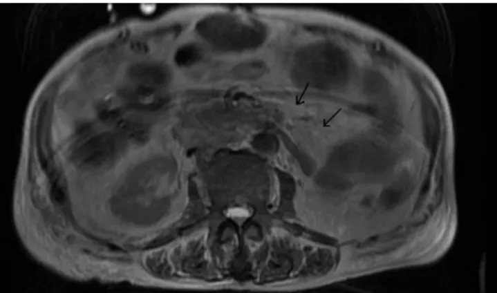
Figure 1. T2 weighted axial MR image shows full transection of the pancreas at the body with a fluid collection.
I Journal of Clinical and Analytical Medicine
112
| Journal of Clinical and Analytical Medicine Total Pancreatic Fracture Due to Blunt Trauma
4
13. Ragozzino A, Manfredi R, Scaglione M, De Ritis R, Romano S, Rotondo A: The use of MRCP in the detection of pancreatic injuries after blunt trauma. Emergency radiology 2003, 10(1):14-18.
14. Fulcher AS, Turner MA, Capps GW, Zfass AM, Baker KM: Half-Fourier RARE MR cholangiopancreatography: experience in 300 subjects. Radiology 1998, 207(1):21-32.
15. Moore EE, Cogbill TH, Malangoni MA, Jurkovich GJ, Champion HR, Gennarelli TA, McAninch JW, Pachter HL, Shackford SR, Trafton PG: Organ injury scaling, II: Pancreas, duodenum, small bowel, colon, and rectum. The Journal of trauma 1990, 30(11):1427-1429.
16. Kao LS, Bulger EM, Parks DL, Byrd GF, Jurkovich GJ: Predictors of morbidity after traumatic pancreatic injury. The Journal of trauma 2003, 55(5):898-905. 17. Cogbill TH, Moore EE, Morris JA, Jr., Hoyt DB, Jurkovich GJ, Mucha P, Jr., Ross SE, Feliciano DV, Shackford SR: Distal pancreatectomy for trauma: a multicenter experience. The Journal of trauma 1991, 31(12):1600-1606.
How to cite this article:
Gulpinar K, Ozıs SE, Konak B, Ozdemir S. Total Pancreatic Fracture Due to Blunt Trauma: Report of a Rare Case. J Clin Anal Med 2016;7(suppl 2): 110-3.
Journal of Clinical and Analytical Medicine I 113