Is There any Effect of Urogenital Cysts on Semen Parameters in
Autosomal Dominant Polycystic Kidney Disease?
Otozomal Dominant Polikistik Böbrek Hastalığında Ürogenital
Kistlerin Semen Parametreleri Üzerine Etkisi Var mı ?
Correspondence Address:
Sami UZUN
İstanbul Haseki Eğitim ve Araştırma Hastanesi, Nefroloji Bölümü, İstanbul, Turkey Phone : + 90 212 529 44 00 E-mail : [email protected] Received : 12.11.2014 Accepted : 11.12.2014 ABSTRACT
OBJECTIVE: Autosomal dominant polycystic kidney disease (ADPKD) is a systemic disease with cysts in many organs including the urogenital tract. The aim of the study was to evaluate the relationship between urogenital cysts, semen pathologies and infertility in ADPKD.
MATERIAL and METHODS: Male ADPKD patients aged 18-60 with creatinine clearance years higher than 60 ml/min were included. All patients had magnetic resonance imaging of the urinary system and pelvis, scrotal Doppler ultrasonography and sperm analysis. The results were compared with those of a healthy control group.
RESULTS: 27 patients and 17 volunteers were included. Seminal vesicle and prostate cysts were detected in four (15%) and six (22%) patients, respectively. Five of the 23 married patients (21%) had infertility and this rate was higher than in the control group (p=0.044). The ratio of sperms with normal morphology and progressive motility was lower, and the rate of hypospermia, oligozoospermia, azospermia, asthenozoospermia and teratozoospermia were higher in the patient group. There was no significant difference between patients with/without urogenital cysts regarding seminal pathologies. CONCLUSION: Seminal abnormalities and infertility are more frequent in patients with ADPKD. Defects in spermatogenesis and sperm motility may be related to urogenital cysts as well as ciliary pathologies. There is a need for further studies evaluating the role of urogenital cysts in semen pathologies.
KEY WORDS: Autosomal dominant polycystic kidney disease, Cyst, Spermiogram, Infertility ÖZ
AMAÇ: Otozomal dominant polikistik böbrek hastalığı (ODPBH) ürogenital sistemle birlikte birçok organda kist oluşumuna neden olabilen sistemik bir hastalıktır. Çalışmada, ODPBH olan bireylerde ürogenital kistler ile semen patolojileri ve infertilite ilişkisi değerlendirilmiştir.
GEREÇ ve YÖNTEMLER: Çalışmaya 18-60 yaşları arasında, kreatinin klirensi 60 ml/dakika/1.73 m2’nin üzerinde, ODPBH olan erkekler dahil edildi. Tüm hastalara üriner sistem ve pelvik manyetik rezonans(MR) inceleme, skrotal Doppler ultrasonografi ve semen analizi yapıldı. Sonuçlar sağlıklı kontrol grubu ile karşılaştırıldı.
BULGULAR: Yirmiyedi hasta ve 17 gönüllü çalışmaya dahil edildi. Seminal vezikül kisti 4(%15), prostat kisti 6 (%22) hastada tespit edildi. Kontrol grubundan daha sık olarak, evli olan 23 hastanın 5’inde (%21) infertilite mevcuttu (p=0.044). Normal morfolojili sperm oranları ve ileri motilite daha düşük, hipospermi, oligozoospermi, azospermi, asthenozoospermi ve teratozoospermi oranları hasta grubunda daha yüksekti. Ürogenital kisti olan/olmayan hastalarda semen patolojileri açısından anlamlı fark yoktu.
SONUÇ: Seminal anormallikler ve infertilite ODPBH olanlarda daha sıktır. Spermatogenez ve sperm motilite defektleri siliar patolojiler gibi urogenital kistlerle ilişkili olabilir. Ürogenital kistlerin semen patolojilerindeki rolünü değerlendirmek için ileri çalışmalara ihtiyaç vardır.
ANAHTAR SÖZCÜKLER: Otozomal dominant polikistik böbrek hastalığı, Kist, Spermiyogram, İnfertilite Sami UZUN1 Savaş ÖZTÜRK1 Meltem GÜRSU1 Mustafa DIKER2 Tolga AKMAN3 Abdullah ŞUMNU1 Serhat KARADAĞ1 Aydın ZEKI1 Egemen CEBECI1 Nadir ALPAY4 Adem KIRIŞ2 Ömer SARILAR5 Rumeyza KAZANCIOĞLU6
1 Haseki Research and Training Hospital, Department of Nephrology, İstanbul, Turkey
2 Haseki Research and Training Hospital, Department of Radiology,
İstanbul, Turkey
3 Bezmialem Vakıf Universty, Faculty of Medicine, Department of Urology, İstanbul, Turkey
4 İstanbul Medipol University, Faculty of Medicine, Department of Internal Medicine, İstanbul, Turkey
5 Haseki Research and Training Hospital, Department of Urology,
İstanbul, Turkey
6 Bezmialem Vakıf Universty, Faculty of Medicine, Department of Nephrology, İstanbul, Turkey
INTRODUCTION
Autosomal dominant polycystic kidney disease (ADPKD) is the most frequent hereditary disease of the kidney. It is regarded as a systemic disease due to its association with pathologies of cardiovascular, gastrointestinal, urogenital and central nervous systems (1,2). Autosomal dominant polycystic kidney disease is responsible for 5-10% of cases with end stage renal failure (3). The most common site of extra renal involvement is the liver. The spleen, pancreas, esophagus, lungs, thyroid gland, bladder, ovaries, uterus, testicles, epididymis, prostate gland and seminal vesicles are other sites where cysts may be detected (1-3).
Various figures have been reported for the prevalence of cysts in the seminal vesicles, prostate, testicles and epididymis in previous series. Information on the clinical significance of these cysts is limited. Torra et alreported recently that the ratio of cysts in the seminal vesicles in ADPKD was 43% (4). They demonstrated that sperm abnormalities, although frequent, were not related to these cysts. However, there is no controlled trial in which reproductive dysfunction was evaluated, whether related or unrelated to urogenital cysts.
The aims of the present study were to screen for urogenital cysts, to evaluate semen pathologies and infertility, and to investigate the relationship between these cysts and semen abnormalities in ADPKD patients.
MATERIALS and METHODS
Twenty-seven males with ADPKD followed up in nephrology clinics of two centers in our city were included. The study was approved by the local ethics committee and written consent was obtained from each participant. Among the patients from the same family, only one male was included in the study.
Patients younger than 18 or older than 60 years of age (to exclude sperm abnormalities related with age), and those with stage 4-5 chronic kidney disease (glomerular filtration ratio <30ml/min/1.73m2), diabetes mellitus or any systemic disease
other than ADPKD were excluded from the study.
Data about the history of patients, including the duration of their present marriage and fertility, were recorded as well as findings on physical examination. Biochemical parameters such as glucose, urea, creatinine, sodium, potassium, calcium, total protein, albumin, alanine transaminase-ALT, total cholesterol, HDL-cholesterol, LDL-cholesterol, triglyceride, hemoglobin, urine analysis, creatinine clearance and daily proteinuria levels were recorded. All patients underwent magnetic resonance imaging (MRI) of the urinary system and pelvis and scrotal Doppler ultrasonography (USG).
Autosomal dominant polycystic kidney disease was diagnosed according to the Ravine’s revised criteria (5). With ultrasonography, the minimum number of cysts for the diagnosis of ADPKD was three (unilateral or bilateral) between the ages of
15 and 39 years, two (bilateral) between ages of 40 and 59 years, and four (bilateral) at the age of 60 years or more.
Laboratory Analysis
Blood samples were obtained after an overnight fast for the measurement of serum glucose, urea, creatinine, uric acid, cholesterol, triglyceride, sodium, potassium, total protein, albumin, and ALT using the Siemens Advia 2400 auto-analyzer with the appropriate methods. Renal functions were evaluated measuring the creatinine clearance with 24-hour urine. Hemoglobin levels were measured using the ABX Pentra DX120 machine.
Radiological Methods
Magnetic resonance imaging: The Philips Achieva 1.5 Tesla machine was used for MRI. The sequences used for this imaging method were sagittal T2, sagittal T1-SPIR, axial T1, axial T2 and coronal T2. An abdominal coil was used while imaging the abdomen. Spatial resolution was obtained by low FOV (field of view). Hypointense cysts in T1 sequences and hyperintense cysts were regarded as simple cysts. Hyperintense cysts in T1 sequences and hypointense cysts were regarded as complicated cysts. Kidney volumes were calculated by the ellipsoid formula [volume= (π/6) x (length x width x depth)](6). Mean kidney volume was gained by calculating the average of the volumes of the two kidneys.
Doppler Ultrasonography: Doppler ultrasonographic examination of the scrotum was performed using the General Electric LOGIQ-9 machine (10 MHz linear probe). Epididymis cysts were defined as anechoic cysts with a posterior acoustic shadow and without internal echo and the minimum diameter needed for diagnosis was 5 mm.
Semen Analysis
All patients underwent sperm analysis. Semen samples were collected by masturbation in the laboratory after three days of sexual fasting. Analysis of semen was performed with the “Neubauer hemocytometer” (Isalab, International GmbH, Wertheim, Germany) after the liquefaction period in the laboratory of 13-30 minutes. Semen profile was obtained according to the criteria described by World Health Organization (WHO) (7). Hypospermia was defined as volume <2 ml, oligozoospermia as sperm concentration <20x106/ml, asthenozoospermia as <50%
of spermatozoa with forward motility, and teratozoospermia as <15% normal forms.
Infertility was defined as failure to conceive after a year of regular intercourse without using any form of contraception (7).
A control group was formed with 17 healthy male volunteers without any renal or extra renal disease to compare semen analysis with the patient group. Volunteers were chosen without any former knowledge about marital status or fertility; and those coming from families with history of ADPKD were excluded.
Liver cysts were detected in 16 patients (59%). Patients with liver cysts were older than those without liver cysts, although statistically insignificant (44±10 years vs. 39±15 years, p=0.39). With MRI, seminal vesicle cysts were detected in four patients (15%) while cysts in the prostate gland were present in six patients (22%). The age of the patients with cysts in the seminal vesicle (35±4 years vs. 43±13 years, p=0.14) and prostate gland (40±9 years vs. 43±13, p=0.62) were less than their counterparts without statistical significance.
Scrotal Doppler USG could be performed in 23 patients. Of those 23 patients, seven (30%) had epididymis cysts while none had testicle cysts. The mean age of the patients with epididymis cysts detected by Doppler USG were not different than those without epididymis cysts (39±10 years vs. 42±13 years, p=0.86).
Among the 16 patients with liver cysts, seven had epididymis cysts and four also had prostate gland cysts. The co-existence of these cysts was not statistically significant. When the patients with liver cysts were excluded, none of the remaining patients had any cysts in the urogenital tract.
Data Analysis
Statistical analysis was conducted using the Statistical Package for Social Sciences 15 package program (SPSS for Windows 15.0, standard version). Numerical data were expressed as mean ± standard deviation (SD). Intergroup comparisons were performed by Student’s t-test or the Mann-Whitney U test when necessary. For non-numerical data, 2x2 contingency tables, chi-square test and Fisher’s exact test were used when appropriate.
RESULTS
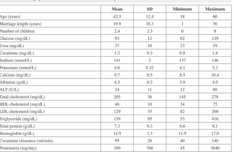
Twenty-seven patients were included in the study. The mean age was 42±12 years. Twenty patients (74%) had family history of ADPKD. Family screening of the remaining seven patients was not a part of our study. Demographics and biochemical analyses are presented in Table I. The mean creatinine clearance and daily proteinuria were 95±28 ml/min and 389±766 mg/day, respectively.
Results of the Imaging Methods
The right, left and mean kidney volumes measured by MRI were 468±377 ml, 494±360 ml and 481±357 ml, respectively.
Table I: The demographic and biochemical data of the ADPKD patients.
Mean SD Minimum Maximum
Age (years) 42.3 12.4 18 60
Marriage length (years) 19.9 10.3 1 36
Number of children 2.4 2.3 0 8 Glucose (mg/dL) 93 12 82 129 Urea (mg/dL) 37 10 23 59 Creatinine (mg/dL) 1.2 0.3 0.8 1.8 Sodium (mmol/L) 141 2 137 146 Potassium (mmol/L) 4.6 0.32 4.1 5.2 Calcium (mg/dL) 9.7 0.5 8.5 10.4 Albumin (g/dL) 4.5 0.3 3.9 4.9 ALT (U/L) 24 11 12 60 Total cholesterol (mg/dL) 205 36 145 278 HDL cholesterol (mg/dL) 46 10 34 75 LDL cholesterol (mg/dL) 129 35 82 200 Triglyceride (mg/dL) 159 95 53 416 Total protein (g/dL) 7.3 0.3 6.6 8.1 Hemoglobin (g/dL) 14.9 1.3 11.9 17.0
Creatinine clearance (ml/min) 95 28 40 140
Proteinuria (mg/day) 389 766 45 3640
II, Figure 1). Hypospermia was detected in 14 patients (52%), while it was seen in four healthy volunteers (24%). There were three patients with no sperms in the semen sample (azospermia) and therefore had infertility. Oligozoospermia was present in 37% of the patient group (10 patients) while it was not detected in the control group.
When the three patients with azospermia were excluded, 20 of the remaining 24 the patients had asthenozospermia (the ratio of sperms with forward motility was less than 50%). This ratio was statistically significantly lower than the control group. The frequency of teratozoospermia was lower in the control group as well. There was no statistically significant difference between patients with or without urogenital cysts regarding biochemical parameters and seminal pathologies.
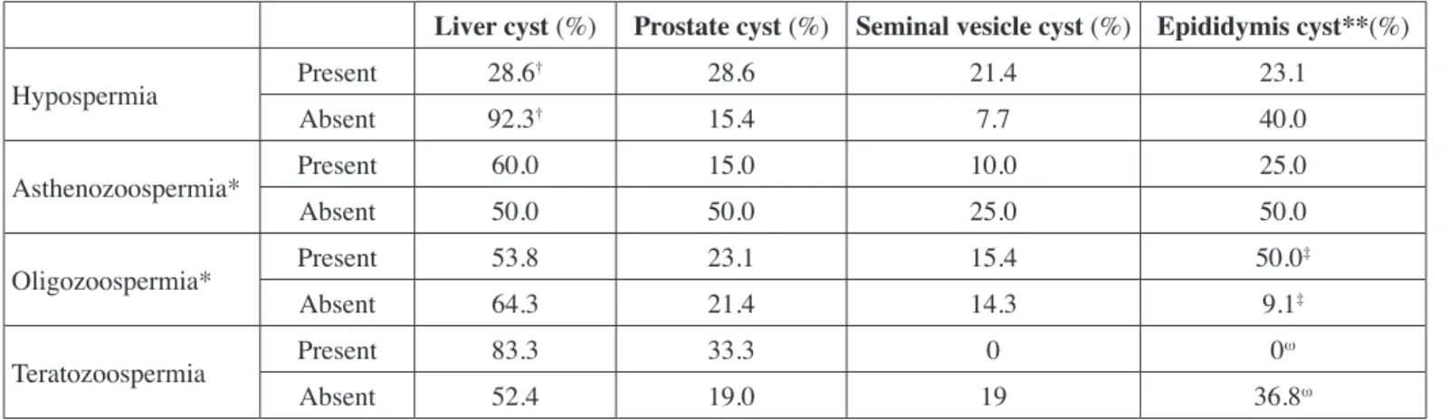
The comparisons of the results of semen analysis and imaging techniques are presented in Table III. Hypospermia was detected to be less frequent in patients with liver cysts (p=0.002). Moreover, oligozoospermia and teratozoospermia were more prevalent in patients with epididymis cysts (p=0.03 and p=0.047, respectively).
Data Related to Fertility and Semen Analysis
Four of the patients (15%) were single. The mean duration of marriage of the remaining 23 patients were 19.9±10.3 years and the mean number of children was 2.4±2.3. Five of the 23 married patients (21%) had infertility of whom three failed to conceive with standard treatment methods. The other two patients did not try any method of infertility treatment although they fulfilled the criteria for infertility proposed by WHO.
The mean age of the control group was 33±3 years. Four volunteers were single; and the remaining 13 married volunteers had at least one child without need for infertility treatment.
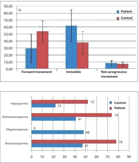
Results of the semen analysis in both patient and the control group are presented in Table II, Figures 1, 2. Semen volume and liquefaction time were similar in both groups. Number of sperms per mm3 and total sperm count were lower in the patient group.
The ratio of sperms with normal morphology was higher in the control group. Regarding sperm motility, the ratio of sperms with progressive motility was higher in the control group and the ratio of immobile sperms was higher in the patient group (Table
Figure 1: Sperm motility characterictis in both patient
and the control group.
Figure 2: Results of the semen analysis in both patient
and the control group. 90,00 80,00 70,00 60,00 50,00 40,00 30,00 20,00 10,00 0,00 % Patient Control
Forward movement Immobile Non-progressive
movement 0 10 Hypospermia Asthenozoospermia Oligozoospermia Teratozoospermia 20 30 40 50 60 52 22 41 0 74 48 78 47 70 80 90 Control Patient
Table II: Comparison of semen analysis of the patient and the control groups.
Patient group n=27 Control group n=17 P
Mean±SD Minimum Maximum Mean±SD Minimum Maximum
Semen volume (ml) 2.4±1.4 0.8 5.5 2.9±1.4 1.0 5.5 0.26
Number of sperms (X106 /mm3) 20 ±17 0 65 47±19 22 86 <0.0001
Total sperm count (X103) 55±65 0 253 155±12 56 550 <0.0001
Liquefaction time (min) 18±6 10 30 18±7 10 30 0.85
Normal morphology (%) 11±14 1 52 17±10 3 35 0.008 Abnormal morphology (%) 89±14 48 99 83±10 65 97 0.008 Progressive motility (%) 29±20 0 66 54±15 22 76 <0.0001 Immobililty (%) 62±23 26 100 38±16 15 73 0.001 Nonprogressive motility (%) 8.1±3.7 0 16 7.2±2.7 4 15 0.37 Hypospermia[n (%)] 14 (52) 4 (22) 0.063 Asthenospermia [n (%)] 20 (74) 7 (41) 0.005 Oligozoospermia [n (%)] 13 (48) 0 (0) <0.0001 Teratozoospermia [n (%)] 21 (78) 8 (47) 0.036
Table III: Analysis results comparing findings with imaging methods and spermiogram.
Liver cyst (%) Prostate cyst (%) Seminal vesicle cyst (%) Epididymis cyst**(%)
Hypospermia Present 28.6† 28.6 21.4 23.1 Absent 92.3† 15.4 7.7 40.0 Asthenozoospermia* Present 60.0 15.0 10.0 25.0 Absent 50.0 50.0 25.0 50.0 Oligozoospermia* Present 53.8 23.1 15.4 50.0‡ Absent 64.3 21.4 14.3 9.1‡ Teratozoospermia Present 83.3 33.3 0 0ω Absent 52.4 19.0 19 36.8ω
† p=0.002, ‡ p=0.03, ω p=0.047. *Three patients with azoospermia were not included. **Detection of epididymis cysts were performed by scrotal
ultrasonography in 23 patients.
DISCUSSION
Autosomal dominant polycystic kidney disease is the most frequent hereditary nephropathy. Many extra renal manifestations affecting cardiovascular, gastrointestinal, urogenital and central nervous system may co-exist with renal pathologies (1-2). We evaluated in our study the cysts in the liver, seminal vesicles and prostate gland by MRI and scrotal Doppler USG. Doppler USG was preferred for detection of epididymis cysts due to the tubular
structure of the organ. We compared these radiological results with semen analysis of which the results were also compared with those of a control group.
Various rates of cysts in the seminal vesicles, prostate gland, testicles and epididymis have been reported by different studies; although their clinical significance and relation with male fertility are not known. Torra et al investigated 30 patients with ADPKD with transrectal USG and detected cysts in seminal
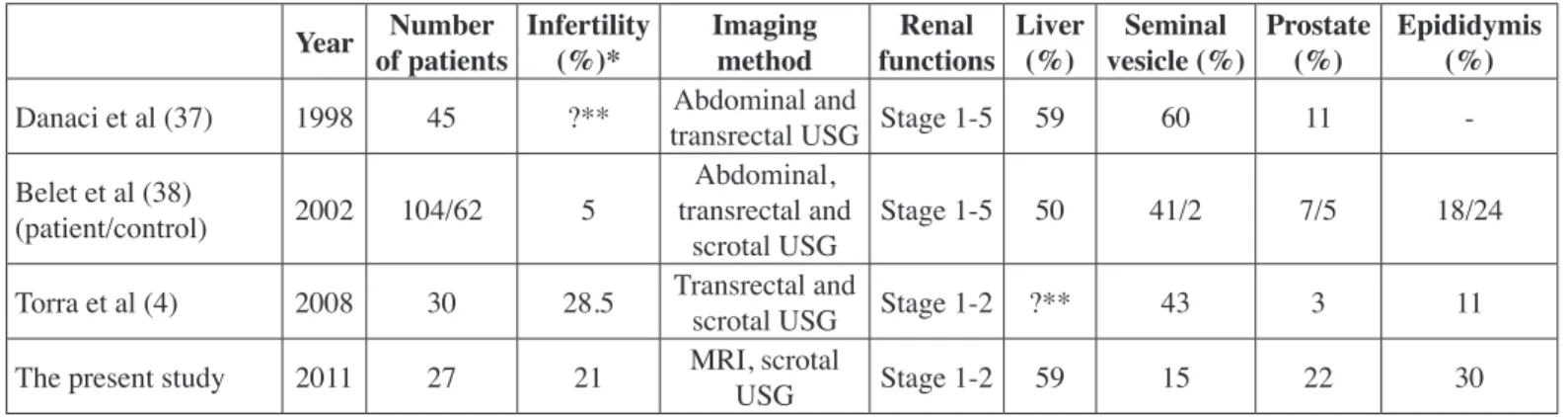
vesicles in 43%, in prostate gland in 3% and in epididymis in 11% (4). They did not report the rate of liver cysts and interestingly none of the patients had cysts in more than one site. Liver cysts were present in 59% of patients in our study. Moreover our results were different than those of Torra et al. in that cysts in the seminal vesicles were less frequent (15% vs. 43%) while those in the epididymis were more frequent (30% vs. 11%). Another study found cysts in seminal vesicles in 60%, prostate gland in 11% and in liver in 42% of patients by abdominal and transrectal USG (8). The same group reported rates of cysts in seminal vesicles, prostate gland and epididymis as 41%, 7% and 18% respectively in a larger group of patients (n=104) (9). The corresponding rates in the control group (n=62) of the same study were 2%, 5% and 24%, respectively. The primary data of these mentioned studies are presented in Table IV together with the results of the present study. Differences in rates may be due to inclusion of patients with differing renal functions. Torra et al included those with estimated glomerular filtration rate above 60 ml/min as in our study, while patients with advanced renal dysfunction were also included in the other studies.
The second aim of our study was to investigate seminal pathologies and infertility including their relation with urogenital cysts in patients with ADPKD. There are few studies and case reports showing coexistence of seminal vesicle cysts with sperm abnormalities and infertility (10,11) and we therefore need larger epidemiological studies to investigate whether a relationship between urogenital cysts and sperm abnormalities in patients with ADPKD.
Infertility was found to be more prevalent in patient group than the control group in our study. Although semen volume and liquefaction time were similar in both groups, total sperm count, ratio of sperms with normal morphology and progressive movement were lower in the patient group (Table II, Figure 1). Moreover, hypospermia, azospermia, oligospermia and teratozoospermia were more frequent in the patient group
when compared with the control group. With these findings, it can be said that seminal abnormalities and infertility are more frequent in patients with ADPKD. Hypospermia was less frequent in patients with liver cysts, while oligozoospermia and teratozoospermia was more frequent in those with epididymis cysts (Table III). However, the low number of patients and especially of those with urogenital cysts may lead to lack of statistical significance in subgroup analysis.
Advanced renal dysfunction may lead to male and female infertility due to disturbances in hypothalamus-pituitary gland-gonad axis (12). This factor was not applicable for our study group because only patients with creatinine clearance more than 40 ml/min were involved. Moreover, experimental studies reported that polycystins, which are cell membrane related proteins, play a role in sperm motility and there may be ultra structural abnormalities in the flagella of sperms causing motility disorders in ADPKD patients (13-15). In the study conducted by Torra et al, urogenital cysts were detected in 10 of 30 patients with ADPKD while seminal abnormalities were present in 20 of 22 patients who had sperm analysis with an infertility rate of 28.5% (Table IV). Nine of those with sperm abnormalities (asthenozoospermia in seven, hypospermia in two, teratozoospermia in four and oligozoospermia in one patient) had urogenital cysts. There was no relation between the presence of urogenital cysts and abnormal semen analysis findings. They compared the patient group with semen analysis of semen donors in their unit and found that abnormalities related to motility (asthenozoospermia and teratozoospermia) were more frequent in the patient group. Belet et al clinically screened (not with sperm analysis) 104 patients with ADPKD of which only five patients were found to be infertile (9) (Table IV). These five patients had a creatinine value over 177 µmol/L.
We detected that the rates of oligozoospermia and teratozoospermia were higher in patients with epididymis cysts (Table III). The epididymis plays an important role in
Table IV: Important studies conducted about genitourinary cysts in autosomal dominant polycystic kidney disease.
Year Number of patients Infertility(%)* Imaging method functionsRenal Liver (%) vesicle (%)Seminal Prostate (%) Epididymis (%) Danaci et al (37) 1998 45 ?** transrectal USG Stage 1-5 59Abdominal and 60 11 -Belet et al (38)
(patient/control) 2002 104/62 5
Abdominal, transrectal and
scrotal USG Stage 1-5 50 41/2 7/5 18/24
Torra et al (4) 2008 30 28.5 Transrectal and scrotal USG Stage 1-2 ?** 43 3 11
The present study 2011 27 21 MRI, scrotal USG Stage 1-2 59 15 22 30
USG: Ultrasonography, MRI: Magnetic resonance imaging, * Dependent on the statement of the patient without knowing whether the wife or husband is infertile, ** No data.
sperm maturation and transport. The sperm cells undergo many biochemical, molecular and metabolic changes during their transport through the epididymis. Sperm cells coming from seminiferous tubules mature to become motile sperms through these changes (16,17). Transfer to the vas deferens occurs with regular peristaltic contractions of the regional smooth muscles, not with sperm movements. Pathologies affecting this region are expected to cause oligozoospermia and/or teratozoospermia but not hypospermia because most of the semen volume is formed by seminal vesicles and prostate gland which are more distal organs. Epididymis cysts are known to cause infertility due to their mechanical obstructive effects. There are data showing increased incidence of epididymis cysts in infertile individuals (5,18). Epididymis cysts may be expected to cause abnormalities in sperm count and motility in ADPKD patients taking into account these data together with the findings in our study. There is a need for more specific advanced studies about this subject.
There are some limitations of the study. The number of the cases were low in both the ADPKD and control groups. Moreover; the study neglected the andrological hormone influence in both the ADPKD and control groups.
CONCLUSION
Urogenital cysts are frequent extra-renal findings in ADPKD and may be detected even in the early stages of the disease. The infertility prevalence is increased in males with ADPKD even in the nonuremic phase when compared with the healthy population. Infertility results from abnormalities in both the sperm count and motility. Defects in spermatogenesis and sperm motility may be related to urogenital cysts as well as ciliary pathologies. There is a need for further studies evaluating the role of urogenital cysts in semen pathologies.
Acknowledgement: No grant support was obtained for the study. None of the authors had any conflict of interest.
Competing interests: The authors declare that they have no competing interests.
REFERENCES
1. Torres VE, Harris PC, Pirson Y: Autosomal dominant polycystic kidney disease. Lancet 2007;369:1287-1301
2. Torres VE, Harris PC: Autosomal dominant polycystic kidney disease: Last three years. Kidney Int 2009;76:149-168
3. Ecder T, Fick-Brosnahan GM, Schrier RW: Polycystic kidney disease. In: Schrier RW (ed), Disease of the Kidney and Urinary Tract. 8th ed. Philadelphia, PA: Lippincott Williams & Wilkins, 2007;502-539
4. Torra R, Sarquella J, Calabia J, Martí J, Ars E, Fernández-Llama P, Ballarin J: Prevalence of cysts in seminal tract and abnormal semen parameters in patients with autosomal dominant polycystic kidney disease. Clin J Am Soc Nephrol 2008;3:790-793
5. Pierik FH, Dohle GR, Muiswinkel JM, Vreeburg JT, Weher RF: Is routine scrotal ultrasound advantageous in infertile men? J Urol 1999;162(5):1618-1620
6. Bakker J, Olree M, Kaatee R, de Lange EE, Moons KG, Beutler JJ, Beek FJ: Renal volume measurements: Accuracy and repeatability of US compared with that of MR imaging. Radiology 1999;211(3):623-628
7. World Health Organization: WHO Laboratory Manual for the Examination of Human Semen and Sperm-Cervical Mucus Interaction. 5th ed. Cambridge: Cambridge University Press, 2010 http://www.who.int/reproductivehealth/ publications/ infertility/9789241547789/en/index.html.
8. Danaci M, Akpolat T, Bastemir M, Sarikaya S, Akan P, Selcuk MB, Cengiz K: The prevalence of epididymal, seminal vesicle cysts in autosomal dominant polycystic kidney disease. Nephrol Dial Transplant 1998;13:2825-2828
9. Belet U, Danaci M, Sarikaya S, Odabas F, Utas C, Tokgoz B, Sezer T, Turgut T, Erdogan N, Akpolat T: Prevalence of epididymal, seminal vesicle, prostate and testicular cysts in autosomal dominant polycystic kidney disease. Urology 2002;60:138-141
10. Li Vecchi M, Cianfrone P, Damiano R, Fuiano G: Infertility in adults with polycystic kidney disease. Nephrol Dial Transplant 2003;18(1):190-191
11. Shefi S, Levron J, Nadu A, Raviv G: Male infertility associated with adult dominant polycystic kidney disease: A case series. Arch Gynecol Obstet 2009;280(3):457-460
12. Handelsman DJ, Dong Q: Hypothalamo-pituitary gonadal axis in chronic renal failure. Endocrinol Metab Clin North Am 1993;22(1):145-161
13. Neill AT, Moy GW, Vacquier VD: Polycystin-2 associates with the polycystin-1 homolog, suREJ3, and localizes to the acrosomal region of sea urchin spermatozoa. Mol Reprod Dev 2004;67(4):472-477
14. Fang S, Baker HW: Male infertility and adult polycystic kidney disease are associated with necrospermia. Fertil Steril 2003;79(3):643-644
15. Vora N, Perrone R, Bianchi DW: Reproductive issues for adults with Autosomal dominant polycystic kidney disease. Am J Kidney Dis 2008;51(2):307-318
16. Bedford JM: Components of sperm maturation in the human epididymis. Adv Biosci 1973;10:145-155
17. Cornwall GA: New insights into epididymal biology and function. Hum Reprod Update 2009;15(2):213-227
18. Van der Linden EF, Bartelink AK, Ike BW, van Leeuwaarden B: Polycystic kidney disease and infertility. Fertil Steril 1995;64(1):202-203