CLINICAL DENTISTRY AND RESEARCH 2017; 41(1): 10-18 Original Research Article
CLINICAL DENTISTRY AND RESEARCH 2017; 41(1): 10-18 Orijinal Araştırma
Correspondence Becen Demir, DDS, PhD
Private Practice, Egemen Sk. No:7/A Kadıköy – İstanbul, Turkey Cellular Phone: 0 533 778 7286 E-mail: [email protected] Becen Demir, DDS, PhD Private Practice, İstanbul, Turkey Nur Balcı, DDS, PhD
Assistant Professor, Department of Periodontology, Faculty of Dentistry, İstanbul Medipol University, İstanbul, Turkey
Haluk Barış Kara, DDS, PhD
Associate Professor, Department of Prosthodontics, Faculty of Dentistry, İstanbul Medipol University, İstanbul, Turkey
Umut Çakan, DDS, PhD
Associate Professor, Department of Prosthodontics, Faculty of Dentistry, İstanbul Medipol University, İstanbul, Turkey
Hilal Uslu Toygar, DDS, PhD
Associate Professor, Department of Periodontology, Faculty of Dentistry, İstanbul Medipol University, İstanbul, Turkey
SHORT-TERM RADIO FREQUENCY ANALYSIS MEASUREMENTS OF
ADIN IMPLANTS
ABSTRACT
Background and Aim: The purpose of this study was to understand and monitor the transition from primary to secondary stability of implants having sand blast large grit acid etched surface, for the timing of the prosthodontic treatment via using the Radio Frequency Analysis.
Subjects and Methods: Forty-two dental implants were placed in 19 patients and Implant Stability Quotient measurements were performed at baseline, 1st, 2nd, 3rd, 4th, 6th and 8th weeks. The lengths and diameters of the implants were also correlated with Implant Stability Quotient values. Results: The present data showed no correlation between lengths, diameters and Radio Frequency Analysis at any of the measurement times. In each group, implant stability at the baseline and 8th weeks showed significant higher Radio Frequency Analysis (RFA) values than the 2nd, 3rd and 4th weeks (p<0.05). Statistically no difference was found between the baseline and 8th week measurements (p>0.05).
Conclusion: Measuring Implant Stability Quotient values can help the clinician understand the condition of the implant without disrupting the healing implant-bone interface, allowing to decide the loading time.
Keywords: Dental Implants, Osstell, Resonance Frequency Analysis, Stability
Submitted for Publication: 07.24.2016 Accepted for Publication : 01.06.2017
Sorumlu Yazar Becen Demir
Periodontoloji Uzmanı Serbest Diş Hekimi, Egemen Sk. No:7/A Kadıköy – İstanbul, Türkiye
GSM: 0 533 778 7286 E-mail: [email protected]
Becen Demir
Periodontoloji Uzmanı, Serbest Diş Hekimi,
İstanbul, Türkiye
Nur Balcı
Yar. Doç. Dr., İstanbul Medipol Üniversitesi, Diş Hekimliği Fakültesi, Periodontoloji Anabilim Dalı, İstanbul, Türkiye
Haluk Barış Kara
Doç. Dr., İstanbul Medipol Üniversitesi, Diş Hekimliği Fakültesi, Protetik Diş Tedavisi Anabilim Dalı, İstanbul, Türkiye
Umut Çakan
Doç. Dr., İstanbul Medipol Üniversitesi, Diş Hekimliği Fakültesi, Protetik Diş Tedavisi Anabilim Dalı, İstanbul, Türkiye
Hilal Uslu Toygar
Doç. Dr., İstanbul Medipol Üniversitesi, Diş Hekimliği Fakültesi, Periodontoloji Anabilim Dalı, İstanbul, Türkiye
ADIN İMPLANTLARININ KISA DÖNEM RADYO FREKANS ANALIZ
ÖLÇÜMLERİ
ÖZ
Amaç: Bu çalışmanın amacı, kumlanmış-asitlenmiş yüzeye sahip implantların primer stabiliteden sekonder stabiliteye geçişini, Radyo Frekans Analiz yöntemi kullanarak anlamak ve gözlemlemektir.
Bireyler ve Yöntem: Ondokuz hastada 42 implant yerleştirilmiş ve başlangıç, 1, 2, 3, 4, 6 ve 8. haftalarda İmplant Stabilite ölçümleri (Implant Stability Quotient-ISQ) yapılmıştır. Bulgular: Elde edilen bilgiler, implantların boyu ve çapı ile Radyo Frekans Analiz ölçümleri arasında hiçbir zaman diliminde ilişki göstermemektedir. Tüm gruplarda implant stabilitesi, başlangıç ve 8. haftada 2, 3 ve 4. haftalara göre anlamlı derecede yüksek İmplant Stabilite ölçüm değeri göstermiştir (p<0.05). Başlangıç ve 8. hafta arasında istatistiksel fark bulunamamıştır (p>0.05). Sonuçlar: İmplant Stabilite değerlerini ölçmek, iyileşen implant-kemik bağlantısına zarar vermeden, implantın iyileşme durumu hakkında fikir vererek yükleme zamanının kararında hekime yardımcı olabilir.
Anahtar Kelimeler:Dental implant, Osstell, Radyo Frekans Analizi, Stabilite
Yayın Başvuru Tarihi : 24.07.2016 Yayına Kabul Tarihi : 06.01.2017
CLINICAL DENTISTRY AND RESEARCH
INTRODUCTION
Osseointegration is a direct and lasting connection between vital bone and titanium implants.1 To achieve a successful osseointegration, primary and secondary stability criterias must be met. While the former of these implies a mechanical contact between alveolar bone and implant, the latter is the consequence of direct bone apposition on the implant surface. The shift from primary to secondary stabilization is the result of successful wound healing, starting immediately after implant placement in the surgical site.2-4
Monitoring the osseointegration process with a non-invasive and non-destructive method is important for the clinician to decide when to load the implants. Two methods used for this purpose are use of a Periotest and Radio Frequency Analysis (RFA). A Periotest is a device basically designed to diagnose and assess the periodontopathies and occlusal load, as well as to control the treatment’s progress for natural teeth. It is also used for assessing the osseointegration of dental implants.5,6 The results of the Periotest are expressed as Periotest values (PTV), which range from −8 (lowest mobility) to +50 (highest mobility). The RFA technique is a bending test of implant-bone complex. With the help of a transducer, a small force is applied and the amount of displacement is measured.7 The results are expressed as Implant Stability Quotient (ISQ), which ranges from 1 to 100. Higher ISQ values represent less displacement and therefore more stiffness of the bone-implant interface.
Meredith et al.8 published the resonance frequency measurements of implants in maxilla and stated that this technique could help in following the changes in stability at the implant-tissue interface. The RFA technique was then commercialized as Osstell (Integration Diagnostics, Göteborg, Sweden) and studies were reported as ISQ measurements by using the Osstell machine for different implant systems.9-11 The second generation of the device, the Osstell Mentor (Integration Diagnostics AB), is wireless and Valderrama et al.12 demonstrated that both generations correlate well but cannot be compared directly in terms of ISQ measurements. Different implant systems are also not comparable although the same measuring device is used, so multiple measurements are necessary for each implant system. Therefore, the aim of this article is to present the short-term RFA measurement results of 42 implants with SLA(Sand-blasted, Large grit, Acid etched) surface.
MATERIALS AND METHODS
Nineteen subjects (aged 19-79, mean age: 51±11.07) who received 42 implants were examined. The study protocol was approved by İstanbul Medipol University Ethics Committee (no: 203). Patients with systemic diseases -such as uncontrolled diabetes mellitus, osteoporosis or autoimmune diseases- as well as immunosupressed patients and pregnant women were not included. Cases involving immediate implant placement or in need of advanced surgical techniques including sinus lifting, ridge splitting or bone grafting were not included either. Prior to surgery, panoramic radiographs were taken and the amount of available bone and anatomical relationships were evaluated. All patients underwent thorough periodontal therapy and oral hygiene instructions were given before surgical approach.
Implant Placement and Prosthetic Rehabilitation
All operations were done under infiltration anesthesia with articaine HCl containing 0.006 mg epinephrine HCl (Ultracain D-S, Sanofi Aventis, İstanbul, Turkey). Full-thickness mucoperiosteal flaps were raised and implants were placed according to the manufacturer’s protocol. Healing abutments were placed in order to make weekly measurements possible and flaps were then closed using 3-0 silk sutures. All patients received postoperative instructions and were prescribed antibiotics (amoxicillin/clavulanic acid, 2g per day for 5 days) and analgesics (diclofenac sodium, 150 mg for 5 days). Sutures were removed after 7 days. Prosthesis was delivered after the 8th week of measurement.
Implants
Implants used in this study were from a single manufacturer (ADIN Dental Implant Systems, Israel) with the SLA surface; diameters and lengths ranging between 3.5-5 mm and 8-13 mm, respectively. Twenty-two of the implants were Touareg-S implants and 20 of the implants were of the Touareg-X type. The difference between these two types is in their apex design.
RFA Measurements
RFA measurements were made with the Osstell Mentor device at baseline immediately after implant placement and then at the 1st, 2nd, 3rd, 4th, 6th and 8th weeks. The Smartpeg of the device was connected to the implant and measurements were performed and recorded from the buccal side (BL) and from the mesial side (MD).
Statistical Analysis
The statistical assessment was carried out using SPSS 15.0 for Windows. Distribution of parameters was tested with Kolmogorov-Smirnov test. Student’s t-test was used to compare the parameters between the groups. In determining the difference between the averages of different RFA periods, repeated measures analysis of variance and Bonferroni tests were used. No power analysis was used in this study.
RESULTS
All 42 implants osseointegrated successfully and could be restored after 8 weeks. Among the 42 implants, 15 (35.7 %) were placed in maxilla and 27 (64.3 %) were placed in the mandible.
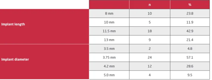
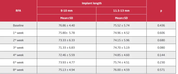
The lengths and diameters of the implants are shown in Table 1. Implant length did not have a significant effect on RFA measurements when 8-10 mm and 11.5-13 mm implants were grouped together (p>0.05) (Table 2), also implant diameters did not have an effect on RFA measurements, 3.5-3.75 mm and 4.2-5 mm implants were grouped together (p>0.05) (Table 3).
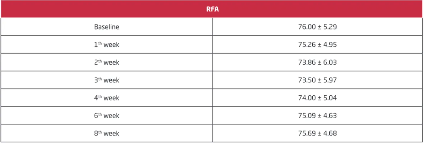
RFA measurements of all 42 implants on a weekly basis and comparisons between all measurements are shown in Table 4 and Figure 1. For all implants, there were no significant differences between the baseline (76.00 ± 5.29) and 8th week (75.69 ± 4.68) measurements (p>0.05). Third week measurements (73.50 ± 5.97) demonstrated the lowest ISQ value (p=0.038) (Figure 1). Both baseline and 8th week ISQ measurements were significantly higher from those of the
2nd, 3rd and 4th weeks, 73.86 ± 6.03, 73.50 ± 5.97 and 74.00 ± 5.04, respectively (p<0.05).
DISCUSSION
This study evaluated the short-term RFA measurements of 42 SLA surface implants to monitor the healing pattern and to decide the loading time. Two different implant types, by means of macro-design, were used. The difference between these two groups was present according to their apex design. Since statistical analysis showed no significance at any time point (data not shown) between these groups, all implants were evaluated together.
RFA, as an evaluation method, has been used in in vitro and in vivo studies for a long time.13 The technique has shown an evolution from a wired system to a wireless one and it is still evolving to be more sensitive and repeatable.8,12,14,15 Although RFA is a widely used, non-invasive technique, it is still criticized by some researchers for measuring only the general stiffness of the bone-implant interface and not the exact relationship between the bone and implant surface.16 A new method is being developed by Kim et al.17 to evaluate the stability of implants by using inductive sensors. The authors speculate that their method showed better consistency and differentiability with implant stability compared to RFA in various implantation conditions,18 but it has not yet been commercialized.
Concerning the effect of implant length on RFA measurements, there is no consensus in the literature. In the present study, the implants were grouped as short (8-10 mm) and long (11.5-13 mm). The decrease in the third week in the shorter group
Table 1. Distribution of implants by length and diameter
n % Implant length 8 mm 10 23.8 10 mm 5 11.9 11.5 mm 18 42.9 13 mm 9 21.4 Implant diameter 3.5 mm 2 4.8 3.75 mm 24 57.1 4.2 mm 12 28.6 5.0 mm 4 9.5
CLINICAL DENTISTRY AND RESEARCH
was more dramatic than the longer one but did not reach a significant level at any time point (p>0.05). Meredith et al.8 measured the resonance frequencies of implants using the first generation implant/transducer system and stated that the measurements were related to the length of the implant above the bone, which they called effective implant length. Sim and Lang19 used tissue level implants having the same diameter but different lengths. In their study, resonance frequencies were assessed by the Osstell Mentor. Although their results also showed no statistical significance, they speculated that implant length was an effective factor for
ISQ values at the baseline, but after 2 weeks short implants showed almost identical results to the long ones. Güler et al.20 and Quesada-Garcia et al.21 also published reports stating that there is no relationship between the length of the implants and ISQ measurements. In contrast to these studies, Barikani et al.22 tested the effect of implant length on primary stability in different bone types and concluded that the implant stability was affected by length especially in D3 bone. In the present study bone type was neglected. Two finite element analysis studies also showed positive correlation between the length of the implant and RFA measurements. In one of these studies,23
Table 2. Comparison of implant length and RFA measurements RFA Implant length p 8-10 mm 11.5-13 mm Mean±SD Mean±SD Baseline 76.86 ± 4.40 75.52 ± 5.74 0.436 1th week 75.80± 5.78 74.96 ± 4.52 0.606 2th week 73.33 ± 6.33 74.15 ± 5.96 0.680 3th week 71.33 ± 6.83 74.70 ± 5.19 0.080 4th week 72.46 ± 5.59 74.85 ± 4.60 0.144 6th week 73.93 ± 4.77 75.74 ± 4.51 0.230 8th week 75.13 ± 4.94 76.00 ± 4.59 0.571
Table 3. Comparison of implant diameter and RFA measurements RFA Implant diameter p 3.5-3.75 mm 4.2-5.0 mm Mean±SD Mean±SD Baseline 75.07 ± 5.17 77.50 ± 5.29 0.152 1st week 74.57 ± 5.21 76.37 ± 4.42 0.258 2nd week 75.19 ± 5.13 71.68 ± 6.88 0.067 3rd week 73.61 ± 5.71 73.31 ± 6.57 0.876 4th week 73.84 ± 4.85 74.25 ± 5.48 0.804 6th week 74.57 ± 4.49 75.94 ± 4.88 0.362 8th week 75.07 ± 4.66 76.68 ± 4.67 0.284
implants were fixed in the bone by joining corresponding nodes of implant and bone. The other study, Pattijn et al.24 also rigidly fixed the implant and bone surfaces assuming a full osseointegration. These modeling conditions may not be reflecting the exact clinical situations, which prevents a direct comparison with the present results. Another study by Östman et al.25 also reported a relationship between implant length and RFA measurements but their results are in the opposite direction of the aforementioned studies, stating lower stability for longer implants. The authors explained that lower stability may be due to reduced implant diameter in the marginal bone level to prevent friction during placement and prolonged drilling time which may cause over preparation of the implant bed. Differences between implant morphologies, experience level of the practitioners, as well as diameters of drills used in the present study also prevent a direct comparison of the results.
The diameter of the implants did not have a significant effect on ISQ measurements in the present study when 3.5-3.75 and 4.2-5mm implants were grouped together. Bischof et al.26 and Han et al.27 also reported that implant diameter was not a factor for ISQ measurements. On the contrary, there are also studies suggesting that there is a relationship between the diameter of the implant and ISQ values.21,22,28 The increase in diameter can let the implant engage more of the buccal/lingual cortical bones,25 therefore resulting in increased baseline ISQ values. Also more diameter means more surface available for osseointegration resulting in increased ISQ values in the following weeks.20
Bone healing around dental implants is a complex phenomenon. There are two different definitions depending on where osteoblasts begin to form bone: Distance osteogenesis and contact osteogenesis. In the former, new bone begins to form from native bone towards the implant, whereas in the latter osteoblasts migrate and attach at the implant surface and then start to synthesize bone matrix towards native bone.29 Other cell types, including osteoclasts, also play their parts and appear in a chronological sequence.30 In a fracture healing model, Schell et al.31 reported that osteoclasts are active from the very early phases of bone healing. Histological studies by Berglundh et al.3 and Abrahamsson et al.4,32 also reported necrosis and resorption of bone in contact with the implant, which is responsible for mechanical primary stability. This bone apposition and resorption events affect RFA measurements clinically. In our study, ISQ results started to decrease from the 1st week and reached their minimum at the 3rd week. Han et al.27 investigated the factors influencing RFA during implant tissue integration
Table 4. RFA measurements of all implants on a weekly basis
RFA Baseline 76.00 ± 5.29 1th week 75.26 ± 4.95 2th week 73.86 ± 6.03 3th week 73.50 ± 5.97 4th week 74.00 ± 5.04 6th week 75.09 ± 4.63 8th week 75.69 ± 4.68
Figure 1. RFA measurements of all implants on a weekly basis * p<0.05, compared to baseline and 8th week measurements
CLINICAL DENTISTRY AND RESEARCH
and concluded that the lowest ISQ was reached at 3 weeks. They also recommend monitoring implant stability at the 8th week, since the results were restored at that time post-surgically. Makary et al.14 also recorded a significant fall at the 3rd week, which increased at the 6th week. Another study by Shokri and Daraeighadikolaei33 monitored 15 SLA implants for 11 weeks after surgery. Their lowest recordings were reached at the 4th week within a range between 62 and 79 which was even accepted as stable.34
Although primary stability initially decreases due to osteoclastic activity and then increases as a result of osteoblastic activity, the relationship between Bone Implant Contact (BIC) and RFA measurements is still being debated. Animal32,35,36 and human37,38 histological and histomorphometrical studies fail to show a correlation between these two parameter. Degidi et al.37 explains this situation as a result of two dimensional histologic sections which represents the three dimensional BIC and the mineralized bone to implant contact may not accurately depict the strength of the connection between the implant and the bone.
Another parameter which affects osseointegration process is the biomaterial’s itself. Properties such as surface topography, surface chemistry and surface energy/wettability have influences on wound healing via cells which will eventually give rise to osseointegration.39 Since we used only SLA surface in the present study, the biomaterial influences are neglected. Ignoring the bone type evaluation is a limitation for this study. The authors suggest the placement of standardized implants into similar bone types in the forthcoming studies. CONCLUSION
In conclusion, routine RFA measurements can be easily performed without disrupting the healing implant-bone interface and allow the practitioner to decide when to load implants. Weekly measurements may help us to understand the shift from mechanical stability to biological integration, but it is not feasible to evaluate the patients every week. Studies reporting RFA measurements for different implant systems may allow clinicians to understand the loading time for each implant system only by measuring the ISQ value. ACKNOWLEDGEMENT
This paper was presented as a poster on PIEG 5th Annual International Symposium Of Advanced Protocols in Oral Implantology, April 20-23, 2013, Antalya, Turkey
REFERENCES
1. Adell R, Lekholm U, Rockler B, Branemark P-I. A 15 year study of osseointegrated implants in the treatment of the edentulous jaw. Int J Oral Surg 1981; 10: 387-416.
2. Davies JE. Understanding peri-implant endosseous healing. J Dent Educ 2003; 67: 932-949.
3. Berglundh T, Abrahamsson I, Lang NP, Lindhe J. De novo alveolar bone formation adjacent to endosseous implants. Clin Oral Implants Res 2003; 14: 251-262.
4. Abrahamsson I, Berglundh T, Linder E, Lang NP, Lindhe J. Early bone formation adjacent to rough and turned endosseous implant surfaces. Clin Oral Implants Res 2004; 15: 381-392.
5. Truhlar RS, Morris HF, Ochi S. Stability of the bone-implant complex. Results of longitudinal testing to 60 months with the periotest device on endosseous dental implants. Ann Periodontol 2000; 5: 42-55.
6. Oh JS, Kim SG. Clinical study of the relationship between implant stability measurements using periotest and osstell mentor and bone quality assessment. Oral Surg Oral Med Oral Pathol Oral Radiol 2012; 113: e35-40.
7. Sennerby L, Meredith N. Implant stability measurements using resonance frequency analysis: biological and biomechannical aspects and clinical implications. Periodontol 2000 2008; 47: 51-66.
8. Meredith N, Book K, Friberg B, Jemt T, Sennerby L. Resonance frequency measurements of implant stability in vivo. A cross-sectional and longitudinal study of resonance frequency measurements on implants in the edentulous and partially dentate maxilla. Clin Oral Implants Res 1997; 8: 226-233.
9. Nedir R, Bischof M, Szmukler-Moncler S, Bernard JP, Samson J. Predicting osseointegration by means of implant primary stability. Clin Oral Implants Res 2004; 15: 520-528.
10. Glauser R, Sennerby L, Meredith N, Rée A, Lundgren A, Gottlow J et al. Resonance frequency analysis of implants subjected to immediate or early functional occlusal loading. Successful vs. failing implants. Clin Oral Implants Res 2004; 15: 428-434.
11. Ersanli S, Karabuda C, Beck F, Leblebicioglu B. Resonance frequency analysis of one-stage dental implant stability during the osseointegration period. J Periodontol 2005; 76: 1066-1071. 12. Valderrama P, Oates TW, Jones AA, Simpson J, Schoolfield JD, Cochran DL. Evaluation of two different resonance frequency devices to detect implant stability: a clinical trial. J Periodontol 2007; 78: 262-272.
13. Meredith N, Alleyne D, Cawley P. Quantitative determination of the stability of the implant-tissue interface using resonance frequency analysis. Clin Oral Implants Res 1996; 7: 261-267. 14. Makary C, Rebaudi A, Sammartino G, Naaman N. Implant primary stability determined by resonance frequency analysis: correlation with insertion torque, histologic bone volume, and torsional stability at 6 weeks. Implant Dent 2012; 21: 474-480. 15. Tang YL, Li B, Jin W, Li DH. Torsional resonance frequency analysis: a novel method for assessment of dental implant stability. Clin Oral Implants Res 2015; 26: 615-622.
16. Veltri M, Balleri P, Ferrari M. Damping factor for monitoring the bone interface at dental implants. Clin Oral Implants Res 2007; 18: 738-742.
17. Kim DS, Lee WJ, Choi SC, Lee SS, Heo MS, Huh KH et al. A new method for the evaluation of dental implant stability using an inductive sensor. Med Eng Phys 2012; 34: 1247-1252.
18. Kim DS, Lee WJ, Choi SC, Lee SS, Heo MS, Huh KH et al. Comparison of dental implant stabilities by impact response and resonance frequencies using artificial bone. Med Eng Phys 2014; 36: 715-720.
19. Sim CP, Lang NP. Factors influencing resonance frequency analysis assessed by Osstell mentor during implant tissue integration: I. Instrument positioning, bone structure, implant length. Clin Oral Implants Res 2010; 21: 598-604.
20. Guler AU, Sumer M, Duran I, Sandikci EO, Telcioglu NT. Resonance frequency analysis of 208 Straumann dental implants during the healing period. J Oral Implantol 2013; 39: 161-167. 21. Quesada-García MP, Prados-Sánchez E, Olmedo-Gaya MV, Muñoz-Soto E, Vallecillo-Capilla M, Bravo M. Dental implant stability is influenced by implant diameter and localization and by the use of plasma rich in growth factors. J Oral Maxillofac Surg 2012; 70: 2761-2767.
22. Barikani H, Rashtak S, Akbari S, Badri S, Daneshparvar N, Rokn A. The effect of implant length and diameter on the primary stability in different bone types. J Dent (Tehran) 2013; 10: 449-455.
23. Winter W, Möhrle S, Holst S, Karl M. Parameters of implant stability measurements based on resonance frequency and damping capacity: a comparative finite element analysis. Int J Oral Maxillofac Implants 2010; 25: 532-539.
24. Pattijn V, Van Lierde C, Van der Perre G, Naert I, Vander Sloten J. The resonance frequencies and mode shapes of dental implants: Rigid body behaviour versus bending behaviour. A numerical approach. J Biomech 2006; 39: 939-947.
25. Ostman PO, Hellman M, Wendelhag I, Sennerby L. Resonance frequency analysis measurements of implants at placement surgery. Int J Prosthodont 2006; 19: 77-83.
26. Bischof M, Nedir R, Szmukler-Moncler S, Bernard JP, Samson J. Implant stability measurement of delayed and immediately loaded implants during healing. Clin Oral Implants Res 2004; 15: 529-539. 27. Han J, Lulic M, Lang NP. Factors influencing resonance frequency analysis assessed by Osstell mentor during implant tissue integration: II. Implant surface modifications and implant diameter. Clin Oral Implants Res 2010; 21: 605-611.
28. Gehrke SA, Tavares da Silva Neto U. Does the time of osseointegration in the maxilla and mandible differ? J Craniofac Surg 2014; 25: 2117-2120.
29. Davies JE. Mechanisms of endosseous integration. Int J Prosthodont 1998; 11: 391-401.
30. Terheyden H, Lang NP, Bierbaum S, Stadlinger B. Osseointegration-communication of cells. Clin Oral Implants Res 2012; 23: 1127-1135.
31. Schell H, Lienau J, Epari DR, Seebeck P, Exner C, Muchow S et al. Osteoclastic activity begins early and increases over the course of bone healing. Bone 2006; 38: 547-554.
32. Abrahamsson I, Linder E, Lang NP. Implant stability in relation to osseointegration: an experimental study in the Labrador dog. Clin Oral Implants Res 2009; 20: 313-318.
33. Shokri M, Daraeighadikolaei A. Measurement of primary and secondary stability of dental implants by resonance frequency analysis method in mandible. Int J Dent 2013; 2013: 506968. 34. Huwiler MA, Pjetursson BE, Bosshardt DD, Salvi GE, Lang NP. Resonance frequency analysis in relation to jawbone characteristics and during early healing of implant installation. Clin Oral Implants Res 2007; 18: 275-80.
35. Ito Y, Sato D, Yoneda S, Ito D, Kondo H, Kasugai S. Relevance of resonance frequency analysis to evaluate dental implant stability: simulation and histomorphometrical animal experiments. Clin Oral Implants Res 2008; 19: 9-14.
36. Rozé J, Hoornaert A, Layrolle P. Correlation between primary stability and bone healing of surface treated titanium implants in the femoral epiphyses of rabbits. J Mater Sci Mater Med 2014; 25: 1941-1951.
37. Degidi M, Perrotti V, Piattelli A, Iezzi G. Mineralized bone-implant contact and bone-implant stability quotient in 16 human implants retrieved after early healing periods: a histologic and histomorphometric evaluation. Int J Oral Maxillofac Implants 2010; 25: 45-48.
CLINICAL DENTISTRY AND RESEARCH
38. Rozé J, Babu S, Saffarzadeh A, Gayet-Delacroix M, Hoornaert A, Layrolle P. Correlating implant stability to bone structure. Clin Oral Implants Res 2009; 20: 1140-1145.
39. Feller L, Jadwat Y, Khammissa RA, Meyerov R, Schechter I, Lemmer J. Cellular responses evoked by different surface characteristics of intraosseous titanium implants. Biomed Res Int 2015; 2015: 171945.