Correspondence: Ayse Batirel, Kartal Dr. Lutfi Kirdar Education and Research Hospital, Infectious Diseases and Clinical
Microbiology, Semsi Denizer Cd. E-5 Karayolu Cevizli Mevkii 34890 Kartal, İstanbul, Turkey Email: [email protected] Received: 27.July.2014, Accepted: 09.October.2014
CASE REPORT
Primary hepatic actinomycosis mimicking a tumor (inflammatory
pseudotumor): Case report and literature review
Ayşe Batirel1, Ferhat Arslan2, Sevinç Hallaç Keser3, Hasan Fehmi Kücük4, Dilek Yavuzer3, Oğuz Karabay5,
Serdar Özer1
1 Kartal Dr. Lutfi Kirdar Education and Research Hospital, Department of Infectious Diseases and Clinical Microbiology, Istanbul, Turkey
2 Istanbul Medipol University, Department of Infectious Diseases, Istanbul, Turkey
3 Kartal Dr. Lutfi Kirdar Education and Research Hospital, Department of Pathology, Istanbul, Turkey 4 Kartal Dr. Lutfi Kirdar Education and Research Hospital, Department of General Surgery, Istanbul, Turkey 5 Sakarya University, Medical Faculty, Department of Infectious Diseases and Clinical Microbiology, Sakarya, Turkey
ABSTRACT
Actinomycosis often manifests with abscesses in the cervicofacial region. Hepatic involvement occurs usually second-ary to an intraabdominal infection. “Isolated or primsecond-ary hepatic actinomycosis (PHA) defines actinomycosis in which the source of infection cannot be demonstrated elsewhere. Herein, we aimed to highlight hepatic actinomycosis in the differential diagnosis of hepatic mass lesions, and also its occurrence even in patients without underlying risk factors. A 24-year-old man, who presented with epigastric and right-upper-quadrant abdominal pain, fever, weight loss, and had a tumor-like mass in the liver was admitted to our hospital. He had no predisposing risk factors or comorbidities. We reviewed all the cases with PHA, who had no predisposing risk factors, in English medical literature from 1993 to 2014. Actinomycotic hepatic pseudotumors should be considered in the differential diagnosis of solitary liver lesions even in patients without any predisposing factors. Multi-disciplinary approach is important in the diagnosis and management.
J Microbiol Infect Dis 2015;5(2): 79-84
Key words: Actinomycosis, Actinomyces spp, inflammatory pseudotumor, liver
Tümörü taklit eden primer hepatik aktinomikoz (Inflamatuar Psödotümör): Olgu sunumu ve literatür derlemesi
ÖZET
Aktinomikoz genellikle servikofasiyal bölgede abseler şeklinde ortaya çıkar. Karaciğer tutulumu çoğunlukla batın içi enfeksiyonlara ikincil olarak gelişir. “İzole veya Primer Hepatik Aktinomikoz (PHA)” başka bir enfeksiyon odağının gös-terilemediği karaciğer yerleşimli aktinomikozu tanımlar. Burada, hepatik aktinomikozun karaciğerde saptanan kitle lez-yonlarının ayırıcı tanısında yer alması gerektiğini ve risk faktörü olmayan hastalarda da gelişebileceğini vurgulamayı amaçladık. Epigastrik hassasiyet, sağ üst kadran ağrısı, ateş, kilo kaybı yakınmalarıyla başvuran 24 yaşında erkek hasta, görüntülemede karaciğerde tümör-benzeri kitle saptanması nedeniyle hastanemize yatırıldı. Hastanın, Aktinomikoz için herhangi bir risk faktörü veya eşlik eden hastalığı yoktu. İngilizce tıp literatüründeki 1993-2014 yılları arasındaki tüm benzer PHA olgularını derledik. Aktinomikotik karaciğer psödotümörleri, hiçbir risk faktörü veya ek hastalığı olmayan hastalarda bile soliter karaciğer lezyonlarının ayırıcı tanısında hatırlanmalıdır. Tanı ve tedavide multidisipliner yaklaşım çok önemlidir.
Anahtar kelimeler: Actinomycosis, Actinomyces spp, inflamatuvar psödotümör, karaciğer
INTRODUCTION
Actinomyces spp are Gram positive, anaerobic and
filamentous bacilli, and are commensals of the oral cavity, gastrointestinal and genital tract.
Actinomy-cosis is a chronic, slowly progressive, suppurative and granulomatous disease caused by Actinomyces israelii.1,2 It has been called “the most misdiagnosed
experienced clinicians. Although its incidence has been diminishing, it is so often confused with a tu-mor.3 Infection occurs through mucosal disruption
and invasion. Although most patients manifest with abscesses in the cervicofacial region, cases of tho-racic, abdominal, pelvic and central nervous system actinomycosis have also been reported. The ileo-cecal region is the most frequently involved region in the abdomen.4,5 Hepatic actinomycosis is a rare
disease with an overall mortality rate of 7.6%.6 The
symptoms and signs are similar to other pyogenic liver abscesses, but the course is more indolent in actinomycosis.7 Hepatic involvement which occurs
usually secondary to intraabdominal infection has been reported in 15% of abdominal disease and in 5% of all cases of actinomycosis.8 If the source of
the infection cannot be demonstrated, these cases are classified as isolated or primary hepatic actino-mycosis (PHA).7,9 Sometimes hepatic lesions may
mimic malignant lesions both clinically and radiolog-ically and those lesions are referred as “inflamma-tory pseudo-tumor”.7,8,10 In majority of the previously
reported cases with actinomycosis, there is at least one predisposing condition or comorbidity. Howev-er, our patient was a young immunocompetent adult with no underlying risk factors or diseases. There-fore, by describing this case and reviewing similar cases, we aimed to highlight hepatic actinomycosis in the differential diagnosis of hepatic mass lesions, and also its occurrence even in patients without un-derlying risk factors or predisposition, in addition, to point out the importance of a multi-disciplinary approach in the differential diagnosis and manage-ment. Written informed consent for presentation of this case has been obtained from our patient.
CASE REPORT
A 25-year-old previously healthy man presented to our hospital because of anorexia, weight loss, ab-dominal distension, right upper quadrant abab-dominal pain and fever of a year’s duration. Medical history was unremarkable. On examination, tenderness on the right upper quadrant of the abdomen of the patient and hepatomegaly (8 cm under the costal margin) were noted on palpation. He had no dental carries or signs of periodontitis. Laboratory tests re-vealed elevated erythrocyte sedimentation rate (84 mm/h, normal 0-12 mm/h), C-reactive protein level (CRP) (122 mg/L, normal 0-3 mg/L), and anemia (hemoglobin 8.9 mg/dl, hematocrit 29%, normal 12-16 mg/dl, 42-52%, respectively). Liver transaminas-es were within normal limits. Abdominal ultrasound (US) revealed a hypoechoic mass 91x71mm in
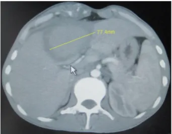
di-ameter with irregular borders in the left lobe of liver. On computed tomography (CT) scan, an isodense [denoting a tissue having a radiopacity (radioden-sity) similar to that of another or adjacent tissue] le-sion 85x75 mm in diameter with a thick and irregular capsule, and invasion of the overlying ribs which had no contrast enhancement (Figure 1) was visualized. Tumor markers [Carcinoembryonic antigen (CEA), carbohydrate antigen (CA)19-9, CA 12-5, CA 15-3, alpha fetoprotein] were normal. Serologic tests for Hepatitis B and C viruses, Human immunodeficien-cy virus (HIV), Cytomegalovirus, Herpes Simplex Virus, Echinococcus granulosus and Entamoeba
histolytica were all negative. Indirect
hemagglutina-tion test for hydatid cyst was also negative.
Figure 1. Computerized tomography image of the
abdo-men: An isodense lesion 85x75 mm in diameter with a thick and irregular capsule, and invasion of the overlying ribs which had no contrast enhancement
Intravenous ceftriaxone 2x1 g (1 gram every 12 h) and metronidazole 4x500 mg (500 milligrams ev-ery 6 h) were initiated empirically. Blood and urine cultures were negative, however fever continued for 10 days after the initiation of antibiotic treat-ment. The patient underwent exploratory laparot-omy which revealed a tumor-like mass (10x10 cm in diameter in liver segment 4, 5 and 6) infiltrating the duodenum, colon and the abdominal wall. His-topathologic examination of the core biopsy frozen section demonstrated degeneration in parenchymal cells, mild to moderate inflammatory cell infiltration in portal areas (Figure 2a, 2b, 2c).
Bacterial cultures remained sterile, possibly because of fastidious nature of growth and prior antibiotic therapy. Microscopic examination of PAS,
Giemsa and Grocott’s methenamine silver stained specimens revealed aggregates of branching fila-mentous microorganisms termed “sulfur granules” forming a granular structure in focal microabscesses (Figure 2a, 2b, 2c). These findings were compatible with actinomycosis which caused an inflammatory pseudotumor. Examination of the oral cavity, pan-oramic dental X-ray, upper and lower gastrointes-tinal endoscopy, colonoscopy, abdominal US,
tho-racic and abdominal CT scans failed to demonstrate any other likely focus of infection or site of origin. Therefore, it was considered as a PHA. Ampicillin-sulbactam 4x1.5 g (1,5 grams every 6 h) iv for 2 weeks and then amoxicillin-clavulonate 2x1 g po for the next 10 weeks were administered. A control ab-dominal USG of the patient after 12 weeks showed complete resolution of the previous hepatic lesion.
Figure 2a, 2b, 2c. Microscopic examination of PAS (2a), Giemsa (2b) and GROCOTT (2c) stained specimens revealed
aggregates of branching filamentous microorganisms termed “sulfur granules” forming a granular structure in focal microabscesses. Inflammatory reaction and fibrosis with polymorphonuclear cells, lymphoplasmocytes, and histiocytes were visualized.
DISCUSSION
Actinomyces spp cause opportunistic infections
more commonly. The anatomical distribution is 60% cervicofacial, 15% thoracic, 20% abdominal, and 5% other sites.11 Actinomyces spp produces
pro-teolytic enzymes and can spread by direct invasion and extension without regard to tissue planes.12,13
Spread to the liver occurs via direct extension from a contiguous abdominal focus or hematogenously from a distant lesion via portal vein or hepatic ar-tery. Hepatic lesions occasionally infiltrate the dia-phragm and are complicated by pleural and lung lesions, can form cutaneous fistula and sinus tracts, and extend to the abdominal and pelvic organs.11
The tumor-like lesion in our case infiltrated to the abdominal wall and was attached to the colon and duodenum.
Hepatic actinomycosis usually occurs in males (70-97% of cases) in the fourth and fifth decades.6,9,14
Risk factors include poor oral hygiene, previous dental procedures, perforated peptic ulcer, colonic diverticulitis, abdominal or pelvic surgery, abdomi-nal wall trauma, gastrointestiabdomi-nal foreign body or le-sions, gallstones, immunosupression, diabetes mel-litus, inflamatory bowel disease such as ulcerative colitis, alcoholism, intravenous drug abuse, long-standing intra-uterine device in females.6,9,11,15,16
The most common predisposing event has been reported to be appendicitis.17 Our patient had no
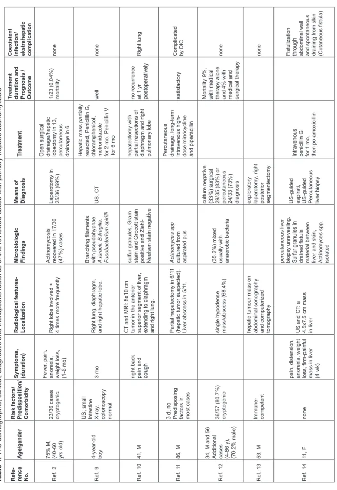
underlying risk factors. He had no history of blunt abdominal trauma or surgery. Also, no underlying cause of immunosuppression was detected. CD4, CD8 counts, serum IgG, IgA, IgM levels were nor-mal. We reviewed the English language literature (pubmed, google scholar) for similar PHA cases with no underling risk factors or predisposition from 1993 to September 2014. We excluded the cases with comorbidity or predisposing risk factors for actinomycosis. We summarized the demographic, clinical, diagnostic and therapeutic features of the reviewed similar cases in Table 1.1,2,6,9-17
In patients with hepatic actinomycosis, the pre-senting symptoms are often nonspecific. Fever, ab-dominal pain, and weight loss are common present-ing complaints, with a subacute onset and a pro-longed course.8,9,18 Symptoms are usually present
for 1-6 months prior to the patient’s presentation.9
Although most reported cases of hepatic actino-mycosis had abnormal liver function tests, trans-aminases may be within normal limits and therefore misleading.8 Laboratory examination often
demon-strates a leukocytosis (75%) with a shift to the left and elevation of alkaline phosphatase (83,3%).15
Table 1.
The demographic, clinical, diagnostic and therapeutic features of the reviewed cases with primary hepatic actinomycosis
Refe- rence No.
Age/gender
Risk factors/ Predisposition/ Comorbidity Symptoms, (duration) Radiological features- Localization Microbiologic Findings Means of Diagnosis
Treatment
Treatment duration and Prognosis / Outcome Coexistent infection/ ekstrahepatic complication
Ref. 2
75% M, (40-60 yrs old) 23/36 cases cryptogenic
Fever
, pain,
anorexia, weight loss, (1-6 mo) Right lobe involved > 4 times more frequently Actinomycosis recovered in 17/36 (47%) cases
Laparotomy in 25/36 (69%) Open surgical drainage/hepatic lobectomy in 13, percutaneous drainage in 6
1/23 (0,04%) mortality
none
Ref. 9
4-year-old boy US, small Intestine X-ray
,
colonoscopy normal
3 mo
Right lung, diaphragm, and right hepatic lobe. Branching filaments with pseudohyphae A.israeli, B.fragilis, Fusobacterium spirilli
US, CT
Hepatic mass partially resected, Penicillin G, chloramphenicol, metronidazole for 2 mo, Penicillin V for 6 mo
well
none
Ref. 10
41, M
right back pain and cough
CT
and MRI: 5x10 cm
tumor in the anterior superior segment of liver
,
extending to diaphragm and right lung. sulfur granules, Gram stain and Grocott stain positive and Ziehl- Neelsen stain negative hepatectomy with partial resections of diaphragm and right pulmonary lobe no recurrence at 1 yr postoperatively
Right lung
Ref. 1
1
86, M
3 d, no Predisposing factors in most cases
Partial hepatectomy in 6/1
1
(hepatic tumor suspected). Liver abscess in 5/1
1.
Actinomyces spp cultured from aspirated pus
Percutaneous drainage, long-term intravenous high- dose minocycline and piperacillin
satisfactory
Complicated by DIC
Ref. 12
34, M and 56 Additional cases (4-86 y), (70.2% male) 36/57 (80.7%) cryptogenic single hypodense mass/abscess (68.4%) (35.2%) mixed usually with anaerobic bacteria culture negative (33%) surgical 29/35 (83%) or percutaneous 24/33 (73%) diagnosis Mortality 9%, with medical therapy alone and 4% with medical and surgical therapy
none
Ref. 13
53, M
Immune- competent hepatic tumour mass on abdominal sonography and computerized tomography exploratory laparotomy , right posterior segmentectomy none Ref. 14 11, F none
pain, distension, anorexia, weight loss, firm-painful mass in liver (4 wk)
US and CT
: a
4.5x7.5 cm mass in liver percutaneous liver biopsy unrevealing. Sulfur granules in drained fistula material between liver and skin, Actinomyces spp. isolated
US-guided aspirati, US-guided Percutaneous liver biopsy Intravenous penicillin G for 1 mo, then po amoxicillin Fistulization through abdominal wall and spontaneous draining from skin (Cutaneous fistula)
Refe- rence No.
Age/gender
Risk factors/ Predisposition/ Comorbidity Symptoms, (duration) Radiological features- Localization Microbiologic Findings Means of Diagnosis
Treatment
Treatment duration and Prognosis / Outcome Coexistent infection/ ekstrahepatic complication
Ref. 6
40, F
none
8 mo
US: 2 subcapsular hypoechoic heterogeneous lesions in right liver lobe. CT: 2 hypodense subcapsular lesions with thick walls enhanced in postcontrast series in right liver lobe chronic inflammation, abscess formation containing actinomyces colonies
Laparotomy and biopsy 6-wk parenteral penicillin G and, following 6-mo oral amoxicillin.
good
Massive pleural effusion, atelectasia. Right pulmonary empyema
Ref. 15
50, F
Immune- competent dyspnea, significant weight loss and intermittent fever (2 mo)
Abdominal US and CT
:
metastatic liver disease? Pleural and pericardial ef
fusion
histological examination Prolonged treatment with high dose penicillin Complete resolution, control US normal
none
Ref. 1
67 cases, Predominantly M (50/67) 75% cryptogenic right lobe in half, mimicked a liver tumor in 45%, single in 2/3 coexisted with infections by common bacteria in 32% examination of surgical (84%) or percutaneous (79%) specimens Antibiotic therapy alone in 50% combined antibiotic treatment with surgical or percutaneous drainage in 50%
Mortality rate: 7.6%.
none
Ref. 16
40, M
none
abdominal pain, weight loss (1 mo)
CT
: hypodense cystic
lesions in right and left lobe. US and CT
:
cystic lesions with necrotic debris in right and left lobe actinomycotic colonies with a surrounding suppurative granulomatous reaction
Laparotomy and biopsy Surgical debridement and drainage surgery iv penicillin G, po penicillin V
4 wk iv + 2 mo po
None
Ref. 17
37, M
Immune- competent intermittent fever and upper left abdominal pain (2 mo) US: heterogeneous abscess with a hypoechoic center and honeycomb enlarged spleen. CT
: ill-de
fined
hypodense lesion in spleen. MRI: multiloculated splenic lesion, laparotomy showed multiple liver nodular lesions and a splenic abscess. filamentous branching bacilli, sulfur granules within H&E stained necrotic foci. Grocott methenamine silver nitrate staining splenic and hepatic actinomycosis
Explaratory laparotomy
,
histopatholo- gically splenectomy and ceftriaxone for 6 days + penicillin for 1 mo iv and oral for 3 mo Control Abdominal MRI after 4 mo: complete resolution, patient asymptomatic for 2 years
Splenic involvement M: male, F: female, CT : computerized tomography , MRI: magnetic resonance imaging, US: ultrasound, DIC: disseminated intravascular coagulation, mo: month/-s, wk: week/-s, H&E: hematoxylin and
eosin, iv: intravenously
A single hypodense mass, as in our case, is the most common radiological finding (observed in 2/3 of the cases), but it may also cause multiple lesions.16,18 Lesions were most often described as
single or multiple abscesses and soft central locu-lations containing white or yellow pus. As the dif-ferential diagnosis from malignant lesions could not be made with radiologic studies7, actinomycosis is
often misdiagnosed as primary or metastatic liver tumor.6,15 Hepatic lesions mimicking malignant
le-sions both radiologically and clinically are called “in-flammatory pseudo-tumor”. Right liver lobe is four times more frequently affected then the left lobe.13
Infectious diseases in the differential diagnosis in-clude pyogenic liver abscesses, amebiasis and echinococcosis.
Because of its fastidious nature, it is difficult to cultivate the microorganism and it may take up to two weeks to grow in culture media. Blood cultures grew the bacteria in 15.4% of the cases. In 82.9% of the patients, diagnostic specimen was obtained by surgical intervention.18 Gram staining and
micros-copy can reveal yellow clusters of filaments sur-rounded by neutrophils, known as “sulfur granules”. Definitive diagnosis is based upon microscopic vi-sualization of typical ‘sulfur granules’ (microcolonies of the organism) or recovery of Actinomyces organ-isms in anaerobic culture. The diagnosis also may be suggested by visualization of organisms with the characteristic branched, gram-positive, filamentous, and often beaded morphology.9,13 Nocardia species
are indistinguishable from Actinomyces organisms by Gram stain but can be excluded by staining specimens with a modified acid-fast protocol.9
Be-sides histopathological appearance, positive Gram staining and presence of sulfur granules were other findings in diagnosis of actinomycosis.
The treatment of isolated hepatic actinomyco-sis is managed by antibiotic treatment only in one half of the cases, surgical excision or percutaneous drainage in addition to antibiotic therapy may be necessary in the other half.6,9 Actinomycosis species
are generally susceptible to penicillin G and amoxi-cillin but resistant to ciprofloxacin.11 Tetracycline,
erythromycin, doxycycline, clindamycin, rifampicin, chloramphenicol, ceftriaxone and imipenem are al-ternative agents for antimicrobial therapy.6,18
Com-bined surgical excision /drainage and 12 weeks of antimicrobial therapy resulted in the recovery of the patient.
CONCLUSION
PHA is a rare disease among solitary liver masses and can mimick malignant tumors clinically and ra-diologically. Therefore, it should be considered in the differential diagnosis of space-occupying soli-tary liver lesions even in patients with no predispos-ing risk factor. Multi-disciplinary collaboration is im-portant in the diagnosis and management.
REFERENCES
1. Burden P. Actinomycosis. J Infect 1989;19:95-99.
2. Cope Z. Actinomycosis involving the colon and the rectum. J Int Coll Surg 1949;12:401-404.
3. Weese WC, Smith IM. A study of 57 cases of actinomycosis over a 36-year period. A diagnostic ‘failure’ with good prog-nosis after treatment. Arch Intern Med 1975;135:1562-1568. 4. Cintron JR, Del Pino A, Duarte B, Wood D. Abdominal
actino-mycosis. Dis Colon Rectum 1996;39:105-108.
5. Piper MH, Schaberg DR, Ross JM, et al. Endoscopic detection and therapy of colonic actinomycosis. Am J Gastroenterol 1992;87:1040-1042.
6. Kanellopoulou T, Alexopoulou A, Tanouli MI, et al. Primary he-patic actinomycosis. Am J Med Sci 2010;339:362-365. 7. Tamsel S, Demirpolat G, Killi R, Elmas N. [Primary hepatic
actinomycosis: a case of inflammatory pseudotumor (case report)]. Tani Girisim Radyol 2004;10:154-157.
8. Wong JJ, Kinney TB, Miller FJ, Rivera-Sanfeliz G. Hepatic actinomycotic abscesses: diagnosis and management. AJR Am J Roentgenol 2006;186:174-176.
9. Miyamoto MI, Fang FC. Pyogenic liver abscess involv-ing Actinomyces: case report and review. Clin Infect Dis 1993;16:303-309.
10. Lai AT, Lam CM, Ng KK, et al. Hepatic actinomycosis pre-senting as a liver tumour: case report and literature review. Asian J Surg 2004;27:345-347.
11. Uehara Y, Takahashi T, Yagoshi M, et al. Liver abscess of Actinomyces israelii in a hemodialysis patient: case report and review of the literature. Intern Med 2010;49:2017-2020. 12. Sheth S, Fishman EK, Sanders R. Actinomycosis involving
the liver. Computed tomography/ultrasound correlation. J Ul-trasound Med 1987;6:329-331.
13. Kocabay G, Cagatay A, Eraksoy H, et al. A case of isolated hepatic actinomycosis causing right pulmonary empyema. Chin Med J (Engl) 2006;119:1133-1135.
14. Putman HC, Jr., Dockerty MB, Waugh JM. Abdominal acti-nomycosis; an analysis of 122 cases. Surgery 1950;28:781-800.
15. Lall T, Shehab TM, Valenstein P. Isolated hepatic actinomy-cosis: a case report. J Med Case Rep 2010;4:45.
16. Islam T, Athar MN, Athar MK, et al. Hepatic actinomycosis with infiltration of the diaphragm and right lung: a case report. Can Respir J 2005;12:336-337.
17. Deshmukh N, Heaney SJ. Actinomycosis at multiple colonic sites. Am J Gastroenterol 1986;81:1212-1214.
18. Sharma M, Briski LE, Khatib R. Hepatic actinomycosis: an overview of salient features and outcome of therapy. Scand J Infect Dis 2002;34:386-391.