KADIR HAS UNIVERSITY
GRADUATE SCHOOL OF SCIENCE AND ENGINEERING
IN SILICO DESIGN OF SELECTIVE NEURONAL NITRIC OXIDE
SYNTHASE INHIBITORS IN ORDER TO PREVENT
NEURODEGENERAT˙IVE DISEASES
NURDAN KAYRAK
IN SILICO DESIGN OF SELECTIVE NEURONAL NITRIC OXIDE
SYNTHASE INHIBITORS IN ORDER TO PREVENT
NEURODEGENERAT˙IVE DISEASES
NURDAN KAYRAK
B.S., Molecular Biology and Genetics Weighted Biology Ege University, 2010
M.S., Computational Biology and Bioinformatics Kadir Has University, 2013
Submitted to the Graduate School of Science and Engineering in partial fulfillment of the requirements for the degree of
Master of Science in
Computational Biology and Bioinformatics
KADIR HAS UNIVERSITY May, 2013 APPENDIX B Nur da n K ayr ak M.S . The sis 2013 S tudent’ s F ull Na me P h.D. (or M.S . or M.A .) The sis 20 11
KADIR HAS UNIVERSITY GRADUATE SCHOOL OF SCIENCE AND ENGINEERING
IN SILICO DESIGN OF SELECTIVE NEURONAL NITRIC OXIDE SYNTHASE INHIBITORS IN ORDER TO PREVENT NEURODEGENERAT˙IVE DISEASES
NURDAN KAYRAK
APPROVED BY:
Prof. Dr. KEMAL YELEKCI Thesis Supervisor
Kadir Has University Prof. Dr. KUTLU ÜLGEN Boğaziçi University
Asst. Prof. Dr. EBRU DEMET AKTEN AKDOĞAN Kadir Has University
APPROVAL DATE: 15/05/2013 APPENDIX B
1
Table of Contents
1 INTRODUCTION ... 13
1.1 Background of Nitric Oxide Synthase ... 16
1.2 eNOS ... 17
1.3 iNOS ... 19
1.4 nNOS ... 21
1.5 Nitric-oxide ... 23
1.6 Mechanism of NO Production ... 25
1.7 The Substrate L-ARG of NOS ... 26
1.8 Active site and CO-Factors of NOS ... 27
1.8.1 H4B ... 27
1.8.2 Zn ... 29
1.8.3 The Heme Iron ... 29
2 Computational Methodology ... 30
2.1 General Approach to the Molecular Docking ... 30
2.2 Docking Tool : AutoDock Version 4 ... 34
2.2.1 Genetic Algorithm ... 34
2.3 Accelrys Discovery Studio ... 36
2.3.1 Docking Tool: Ligand Docking with Libdock ... 37
2.3.2 Docking Tool: Ligand Docking with C-Dock ... 40
2.4 DE-NOVO ... 41
2.4.1 Fragment Docking Using De-Novo Receptor ... 42
2
3 Molecular Docking Setups ... 43
3.1 Dockıng setup of Autodock ... 43
3.2 Dockıng setup of Libdock ... 44
3.3 Dockıng setup of C-Docker ... 46
3.4 Setup of De Novo ... 47
4 RESULT and DISCUSSION ... 49
4.1 Preparation ... 49
4.2 DOCKING ... 54
4.3 Selection of Nnos Enzymes ... 57
4.4 Selection of eNOS and iNOS ... 65
4.5 Comparison of Experimental and Computational Data ... 74
4.6 nNos Selective Inhibitor Design ... 74
4.7 Scanning the inhibitor in the ZINC library ... 75
4.8 Docking Studies of Selected Inhibitors Candidates for nNOS, eNOS and iNOS Enzymes... 80
3 Table of Figures
Figure 1 Original three-dimensional appearance of the eNOS enzyme ... 18
Figure 2 Original three-dimensional view of the eNOS enzyme has been minimized. (single domain)... 19
Figure 3 Original three-dimensional appearance of the iNOS enzyme ... 20
Figure 4Original three-dimensional view of the iNOS enzyme has been minimized.(single domain) ... 21
Figure 5 Original three-dimensional appearance of the nNOS enzyme ... 22
Figure 6 Original three-dimensional view of the nNOS enzyme has been minimized.(single domain) ... 23
Figure 7 Nitric Oxide Production from L-Arginine by NOS enzym ... 25
Figure 8: Chemical structure of L-Arginine ... 26
Figure 9 H4B layout in nos enzymes in stick representation ... 28
Figure 10 Chemical structure of tetrahidrobiopterin ... 28
Figure 11 Shows that iron of the heme and Fe-S bound in stick representation Computational Methodology ... 30
Figure 12 : :Inhibition behavior of inhibition constant of 20 compound against the nNOS enzyme ... 63
Figure 13: Inhibition behavior of inhibition constant of 20 compound against the nNOS enzymes.(10,12,13 compunds are extracted ) ... 64
Figure 14 : Inhibition behavior of logarithmic constant of 17 compound against the eNOS ... 69
Figure 15 Inhibition behavior of logarithmic constant of 17 compound against the iNOS ... 73
4
Figure 17 2D and 3D structures of KN5 Inhibitor in the eNOS enzyme ... 87 Figure 18 3D and 2D Structures of KN5 Inhibitor in the iNOS enzyme ... 89 Figure 19 2D and 3D structure of KN5 Inhibitor in the nNOS enzyme ... 90
5 List of Table
Table 1 : Parameters of Dpf files ... 44
Table 2 : Parameters of Libdock ... 45
Table 3: : Parameters of CDocker ... 46
Table 4: : Referanced 20 Compound that are compared with computational values ... 55
Table 5:Computational and Experimental inhibition constant of 20 compound and 10 nNOS enzymes from referances.(All values are in nanomolar scale) ... 59
Table 6: Logaritmic values of Cmputational and Experimental inhibition constant of 20 compound and 10 nNOS enzymes from referances.(All values are in nanomolar scale) 61 Table 7: :Experimental and Computational values of 17 compound and 3 eNOS enzymes that are selected referances (All values are in nanomolar scale) ... 66
Table 8: :Logaritmic values of experimental and Computational values of 17 compound and 3 eNOS enzymes that are selected referances (All values are in nanomolar scale) .. 68
Table 9:: Experimental and Computational values of 17 compound and iNOS enzymes that are selected referances (All values are in nanomolar scale) ... 71
Table 10: Logaritmic values of Experimental and Computational values of 17 compound and iNOS enzymes that are selected referances (All values are in nanomolar scale) ... 72
Table 11 The Leads used in this study ... 76
Table 12 Selected inhibitor candidates ... 79
Table 13 Autodock resultsof obtained 19 inhibitors for eNOS,iNOS and nNOS enzymes (All values are in nanomolar scales, Ki is inhibition constant) ... 82
Table 14 Logaritmic values of autodock results of obtained 19 inhibitors for eNOS, iNOS and nNOS enzymes (All values are in nanomolar scales, Ki is inhibition constant) ... 84
7 ABSTRACT
Nitric Oxide syntheses (NOS) are the family of enzymes which catalyzes the oxidation L-Arginine amino acid to nitric oxide molecule (NO) L-citrulline. Mammals contain three different NOS isozymes: Neuronal NOS (nNOS, in the brain), inducible NOS (iNOS, in macrophage cells), endothelial NOS (eNOS, the inner walls of blood vessels). Nitric Oxide (NO) is an important messenger molecule, which regulates several
physiological functions in cardiovascular system and neuronal cells in the brain.
Indeed, NO is a free radical gaseous molecule under normal conditions and highly toxic substance to our cells. In our body, it is produced locally at proper concentration at proper time. In endothelial cells, it relaxes smooth muscle causing to decrease blood pressure. Macrophage cells generate NO as an immune defense system to destroy microorganisms and pathogens.
In our brain under certain pathological conditions after a certain ages produced excessive NO, causes tissue damage and oxidative stress. This leads are a variety of neurodegenerative diseases, such as Alzheimer's disease, rheumatoid arthritis and Parkinson's diseases. For this reason, it is important to inhibit selectively neuronal isozymes of NOS, nNOS in the brain. Three isozymes show extraordinarily structure similarities hindering the selective inhibitor design. In the literature there are many outstanding studies, however there has not being developed any drug which
8
accomplished the required affinity and selectivity. Neurodegenerative diseases were very common death cause after cardiovascular diseases and cancer in the developed
countries. We plan to use computer modeling based on the known crystal structure of three NO isozymes. In this project computationally, we developed, highly selective nNOS inhibitors via in silico screening. The inhibitors whose experimental inhibition values reported up to now were tested within our prepared model NOS isozymes. The obtained computational binding constants were compared with literature experimental values. The enzymes whose experimental values agreed with computational values were chosen for further studies. First, several suitable scaffolds (leads) were determined from lead library of ZINC database. These leads were optimized using fragment library of ZINC and Accelrys in the nNOS active site. The new selective and potent inhibitors were determined as a result of in silico screening. The inhibitors binding energy and inhibition constants toward nNOS, eNOS and iNOS enzymes are reported. In the future project, a collaborative work is going to be searched for the synthesis and enzymatic work of these determined inhibitors.
Key words: Nitric oxide synthase, nNOS, eNOS, iNOS, docking, scoring, molecular modeling, in silico screening
9 Özet
Nitrik oksit sentazlar (NOS) L-Arginin aminoasidini L-sitrüline oksitleyerek Nitrik oksit molekülü üreten bir enzim ailesidir. Memelilerde NOS enziminin nöronal NOS (nNOS, beyinde ve omurilikte), indüklenebilir NOS (iNOS, makrofajlarda), endotelyal NOS (eNOS, kan damarlarımızın iç çeperlerinde) olmak üzere üç izoenzimi bulunur. Bu üç değişik izoenzimden üretilen Nitrik oksit (NO) kardiovasküler sistemden, bağışıklık sistemindeki fizyolojik fonksiyonları da içeren bir dizi görevi üstlenen bir sinyal molekülüdür. Aslında NO normal
şartlarda gaz halinde, radikal ve çok toksik bir moleküldür. Bünyemizde uygun zamanda uygun derişimde üretilir ve lokal olarak kullanılır. Beyinde
nörotransmitter görevi üslenen NO ileri yaşlarda kontrolsüz üretildiğinde doku hasarına ve oksidatif strese sebep olur. Bu durum da Alzheimer, romatoid artrit ve Parkinson gibi nörodejeneratif hastalıklarının oluşumuna katkıda bulunur. Damar iç çeperlerinde endoteliyel tabakada üretilen NO damara esneklik vererek kan basıncının düşmesine neden olur. Vücudumuza giren mikroplara karşı bizi koruyan makrofajlarda NO molekülünü silah olarak kullanır. Dolayısı ile sadece beyinde üretilen NO molekülünün azaltılması için nNOS enziminin
10
seçimli olarak inhibe edilmesi çok önemlidir. Üç izoenzimin yapısı olağanüstü bir şekilde birbirlerine benzemektedir ve bu benzerlik nNOS seçimli inhibitör tasarımını zora sokmaktadır. Birçok çalışma olmasına rağmen henüz bunu başaracak bir ilaç geliştirilememiştir.Nörodejeneratif hastalıklar, gelişmiş ülkelerde kalp damar hastalıkları ve kanserden sonraki en sık ölüm nedenidir. Bu projede amacımıza uygun olarak nNOS'a seçimli bir dizi inhibitör
tasarlanmıştır. Bunun için hedeflenen izoenzimlerin yapılarını temel alan bilgisayar destekli ilaç tasarımından yararlanılmıştır. İlk olarak bugüne kadar çalışılmış önemli NOS hedefli inhibitörler sistemimizde her 3 izoenzim için test edilip benzerlik ve farklılıklar bir model oluşturulmak amacıyla incelenmiştır. Deneysel ve hesapsal sonuçları en çok destekleyen 3 izoenzimin ( nNOS, eNOS ve iNOS) kristal yapıları in silico tarama yapılmak için seçilmiştir. Tespit edilen bu enzimlere karşı ZINC veri bankası taranarak uygun ana yapılar
(leads), daha sonra da de novo programı kullanılarak ana iskeletlere fragmentler (ZINC ve Accelrys fragmant kütüphanesi) eklenerek nNOS seçimli olan
inhibitörler tespit edilmiştir. Çalışmanın sonucunda nNOS enziminin aktif
bağlanma bölgesine seçimli olarak yüksek ilgiyle bağlanan inhibitörlerin yapıları, bağlanma enerjileri ve inhibisyon katsayıları verilmiştir.Bu sonuç raporu
kapsamında ve ümit vaat edenler ileriki projelerde bu konuda çok ileri düzeyde araştırmalar yapan laboratuvarlar ile işbirliğine gidilerek denemeleri
11
Anahtar Kelimeler: Nitrik Oksit sentaz, nNOS, eNOS, iNOS, docking, skorlama, moleküler modelleme, in silico tarama.
12
ACKNOWLEDGMENTS
First of all I would like to thank my advisor Prof. Dr. Kemal Yelekçi who always supported and helped me during the thesis period.Thank you for his useful advices and encouragement.And also I thank to Mr. Bora Büyüktürk. For overcoming through the difficulties I encounted with computer systems.
Last but not least, I would like to thank to my family for their support and patience and encouragement as well as to my friends.
13
1 INTRODUCTION
Considering the examples of national economies in the world, pharmaceutical industry and the R&D activities are becoming important increasingly. In addition to the high cost of research and development activities, laborious research-based pharmaceutical
companies have increased their risks. At the end of the R&D processes enduring 12-14 years on average. (Silverman R. B., 2009)Thousand of molecules and time consuming development process are needed for a drug to place into a shelf on the pharmacy. Intelligent drug design is method to accelerate the finding procedure of new molecules. Determination of chemical drug candidates by screening and searching in silico
environment gives direction to experimental studies.
Due to the potential benefit of treatment of neurodegenerative diseases, many
pharmaceutical companies started programs to identify the selective nNOS compounds by the early 1990’s . (Silverman R. B., 2009) NOS enzyme has three isoenzymic forms. Mammals contain three different NOS isozymes: Neuronal NOS (nNOS, in the brain), inducible NOS (iNOS, in macrophage cells), endothelial NOS (eNOS, the inner walls of blood vessels). ENOS in endothelial cells produce NO to regulate blood pressure. NNOS in neuronal cells produces NO for neurotransmission. INOS in macrophage cells
produces nitric oxide to combat infection and microorganisms stimulated by pathogens. (Silverman R. B., 2009) Under the presence of cofactors nicotinamidadenine
14
mononucleotide (FMN) and tetrahidrobiyopterin(BH4) NOS enzyme produces NO by oxidation of L-arginin terminal guadino group. (Silverman R. B., 2009) Nitric Oxide syntheses (NOS) are the family of enzymes which catalyzes the oxidation L-Arginine amino acid to nitric oxide molecule (NO) L-citrulline. Nitric Oxide (NO) is an important messenger molecule, which regulates several physiological function in cardiovascular system and neuronal cells in the brain. (Silverman R. B., 2009)The overproduction of NO by nNOS or iNOS and the underproduction by eNOS have been shown to lead to pathophysiological conditions such as neurodegenerative diseases Alzheimer, and Parkinson diseases, stroke, rheumatoid arthritis, hypertension and atherosclerosis. Inhibition of nNOS can be therapeutic benefit. Five cofactors are required for the catalyses by NOS izoenzymes. (Silverman R. B., 2009) NADPH is in reductase domain and transfers two electrons to FAD, FMN, transfers one electron to heme in oxygenase domain. BH4 in oxygenase domain facilitates catalysis from L-arginin to L-sitruline (Abu-Soud, Presta, Mayer, & Stuehr, 1997).
The expected lacking effects of Nitric Oxide enzymes are observed on the transgenic mice for all NOS isoenzymes (knock-out). (Silverman R. B., 2009) As a result of these experiments, without the hypertensive effect of inhibition of eNOS or without causing immune problem for iNOS inhibition, nNOS inhibitor will have protective effect on neuro-degenerative diseases. Due to the potential benefit of treatment of
neurodegenerative diseases, many pharmaceutical companies identify compounds by the selective nNOS programs initiated in the early 1990s. Since the crystal structure could
15
not obtaine previously , the general approach is using of L-arginine as a substrate for the lead and applying structural changes on it with the hope of selective binding analogues to nNOS ,Enos and iNOS. (Silverman et al.2009). As a result of the lack of selectivity studies, these three enzyme substrate, an active region and the reaction are very similar. Any modifications on L-arginine in the active sites will have similar effects. A few designed compounds bind like an anchor to active site, try to reach out second cavity of aminoacids (Silverman et al. 2003).
In this thesis we aimed to determine the potential inhibitors based on the eNOS, iNOS, nNOS enzymes structures but only which is selective for nNOS enzymes. An inhibitor that prevents neuronal degenerative formations which is caused by accumulation of excess nitric oxide in brain cells in elderly ages contributing to Alzheimer treatment research will be designed. Autodock protocols, Accelrys Discovery Studio, Lipdock protocols that have computer aided drug design methods are utilized based on the crystal structures of NOS isozymes. The most important reported inhibitors up to now are tested with our prepared NOS isozymes and their obtained computational binding constants are compared with literature experimental values. From these lead scaffolds new , more selective and potent inhibitors are designed. The results of this study, tens of thousands derivatives scanned with experimental high-throughput screening method (HTS) and identified for potential drug molecules ,so time and money will be saved.
16
1.1 Background of Nitric Oxide Synthase
Nitric Oxide syntheses (NOS) are the family of enzymes which catalyzes the oxidation L-Arginine amino acid to nitric oxide molecule (NO) L-citrulline. (Silverman, Roman, Martasek, Gomez-Vidal, & Ji, 2006) Mammals contain three different NOS isozymes: Neuronal NOS (nNOS, in the brain), Inducible NOS (iNOS, in macrophage cells), Endothelial NOS (eNOS, the inner walls of blood vessels). Nitric Oxide (NO) is an important messenger molecule, which regulates several physiological function in cardiovascular system and neuronal cells in the brain. (Silverman, Roman, Martasek, Gomez-Vidal, & Ji, 2006) Different isoforms of NOS have different cell and tissue structures. Catalytic center of nitric-oxide synthease is composed of two domains. The C-terminal domain is reductase domain that binds FMN, FAD and NADPH. The N-terminal oxygenase domain contains heme,H4B and the substrate L-Arginine. (Abu-Soud, Presta, Mayer, & Stuehr, 1997) The oxygenase domain of the three NOS isoforms is almost identical. (Abu-Soud, Presta, Mayer, & Stuehr, 1997) Five cofactors are necessary for catalyzes by NOS. Reductase domain can transfer the electron to the oxygenase domain for enzymatic activity. Reductase domain and oxygenase domain are linked by binding of a calcium-calmodulin motif. (After & Feldman, 1993)Electrons flow from NADPH through FMN and FAD in one subunit to the other subunit. (Silverman R. B., 2009). There are three known NOS isoforms. Two are constitutive form and the third one is inducible form. The gene coding for iNOS is located on
17
chromosome 17 and for nNOS is located on chromosome 12. Gene coding for eNOS is located on chromosome 7. Three isozymes show extraordinarily structure similarities for preventing the selective inhibitor design. For this reason, it is important to inhibit
neuronal isozymes of NOS in the brain selectively. (Silverman,, et al., 2008)
1.2 eNOS
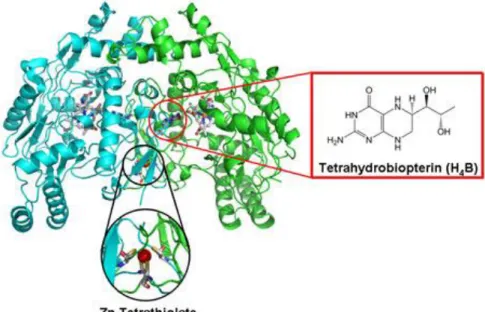
ENOS controls the smooth muscle tone primarily. It relaxes smooth muscle so blood pressure is decreased in endothelial cells. (Hah, Martasek, Roman, & Silverman, 2003)The crystal structure for the heme domain of eNOS has been solved. Zinc ion is coordinated tetrahedral by two pairs of cystine residues and is located at the eNOS dimer interface. Enos is a dimer that contains two similar monomers and it should have dimeric form to be functional. The gene that codes monomers is on the 7q35-36 chromosome and contains 26 ekson. Firstly heme molecule links the enzymes and then enzymes take dimeric form. The absence of the heme group, enzymes remains monomer. When the enzyme goes to be dimeric form, BH4 come up to link and the dimer becomes stable. Then Zn ions are linked to the stable form of enzymes. (Erdal, Litzhger, Seo, Zuhu, Ji, & Silverman, 2005) (Poulos, Silverman, Jamal, Li, Xue, & Delker, 2010) Functional activity of the dimer depends on the number of connected BH4. Not connection of the BH4 can produce the dimer form of O2 and connection of the one BH4 molecule; both O2 and NO are produced by eNOS dimer. If the presence of high level of BH4 creates
18
dimer enzymes, NO are created. The location of the enzyme in the cell is un clear. Enos is located depending on the cell membrane in the cell. (Arnet, McMillan, Dinerman, Ballermann, & Lowenstein, 1996)
19
Figure 2 Original three-dimensional view of the eNOS enzyme has been minimized. (Single domain)
1.3 iNOS
NO is generated by macrophage cells as an immune defense system to demolish
microorganisms and pathogens. Large amounts of NO in the immune defense system are produced by the inducible isoforms iNOS. (Silverman R. B., 2009)
20
21
Figure 4Original three-dimensional view of the iNOS enzyme has been minimized. (Single domain)
1.4 nNOS
The neuronal nitric oxide synthesis that is involved in the development of nervous system products such as NO in neuronal cells. It regulates the release of
neurotransmitters so synaptic plasticity, memory function and neuroendocrine secretion are controlled by NO. It is significant for memory and learning. (Erdal, Litzhger, Seo, Zuhu, Ji, & Silverman, 2005) Neuronal nos is related plasma membranes so nNOS carry out a role in cell communication. NOS1 gene encodes nNOS in chromosomal region 12q24 and this gene contains large amount polymorphic repeats of eNOS and nNOS.
22
NNOS are located in cytosol. (Ji, Li, Flinspach, Poulos, & Silverman, 2003)
23
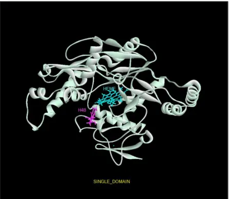
Figure 6 Original three-dimensional view of the nNOS enzyme has been minimized. (Single domain)
1.5 Nitric-oxide
Nitric-oxide is a free radical molecule in both intracellular and extracellular structure form. Half-life of NO is 2-5 seconds. Many cellular processes are regulated by the release of NO from NO-producing cells. In addition NO is a neurotransmitter that pass freely through the cell membrane. (Silverman R. B., 2009) NO is a free radical gaseous molecule under normal conditions and highly toxic substance to our cells. In our body, it
24
is produced locally at proper concentration at proper time. If its production and
consumption go wrong, then serious pathological and physiological diseases will arise. In neuronal cells, NO regulates the release of neurotransmitters and is involved in synaptic plasticity, memory function and neuroendocrine secretion. In endothelial cells, it relaxes smooth muscle causing to decrease blood pressure.
Macrophage cells generate NO as an immune defense system to destroy microorganisms and pathogens. In our brain, under certain pathological conditions after certain ages produced excessive NO, causes tissue damage and oxidative stress. (Silverman, Roman, Martasek, Gomez-Vidal, & Ji, 2006)
The overproduction of NO by nNOS or iNOS and the underproduction by eNOS have been shown to lead to pathophysiological conditions such as neurodegenerative diseases Alzheimer, and Parkinson diseases, stroke, rheumatoid arthritis, hypertension and atherosclerosis. Inhibition of nNOS can thus be of considerable therapeutic benefit. However inhibition must be isoforms selective so that only NO formation by the disease-associated NOS will be inhibited by the treatment, while the physiological function of the other isoforms often eNOS is unaffected. Therefore, it is important to inhibit selectively neuronal isozymes of NOS, nNOS in the brain. (Silverman,, et al., 2008)
25
1.6 Mechanism of NO Production
No production are took place in two identical steps by all three NOS isoforms. In the first step, L-Arg is transformed Ng-hydroxy-L-Arg (NHA) at the heme active site of NOS enzymes. In the second step, NHA is oxidized to NO and citrulline. (Silverman R. B., 2009) For catalysis reducing enquired are transferred from NADPH through FAD to FMN. Finally they transfer to the heme group. NADPH is in reductase domain and transfers two electrons to FAD, FMN, and transfers one electron to hem in oxygenase domain. BH4 in oxygenase domain facilitates catalysis from L-margining to L-citrulline (Silverman,, et al., 2008)
26
1.7 The Substrate L-ARG of NOS
The original substrate L-Arg of Nos binds directly above the heme iron (Fe) atom. Another cofactor H4B binds along the side of the heme. Moreover, the substrate L-Arg and H4B are joined together through an extended H-bonding mediated network by one of the two propionate groups of the heme. (Zhang, Fast, Marletta, Martasek, &
Silverman, 1997)
27
1.8 Active site and CO-Factors of NOS
1.8.1 H
4B
H4B is a cofactor of all three nos enzymes isoforms that is essential for the catalytic .Biochemical studies showed that H4B play a role in protein stability, monomer-dimer equilibrium, proteolytic susceptibility, heme-ligand binding and substrate binding properties. (Shearer, et al., 1998) A cellular level is more important for structure of NOS enzymes. H4B binding make happen to increases L-arginine binding and to shift heme iron of NOS to high-spin state so in NOS enzymes stabilizes the active form of enzymes. Generation of NO is occurred by suboptimal concentration of H4B. (Silverman R. B., 2009)Another possible role of H4B is to perform a one electron donor to the heme iron for cleavage of the o-o bond.
28
Figure 9 H4B layout in nos enzymes in stick representation
29
1.8.2 Zn
Zn plays a role such as cofactor in the catalytic activity. In the dimer interface of NOS enzymes Zn-tetrathiolate center helps dimer stabilization. It creates formation of the pterin binding site. (Silverman R. B., 2009)
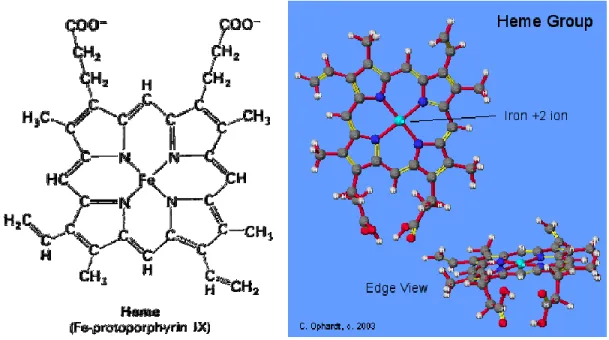
1.8.3 The Heme Iron
The iron of heme takes a place in the porphyrin ring. It is coordinated by pyrole nitrogen atoms of the porphyrin ring. A study shows that Fe-S bond is important for the catalytic activity of all NOS. Only Fe+2-diatomic ligand complex cause to be stable the NO complex. On the heme iron the decrease of positive charge in being reduced from Fe+3 to Fe+2 allows to move closer to the heme iron for more desirable no bounded contacts. (Li, Raman, Martasek, Masters, & Poulos, 2001)
30
Figure 11 Shows that iron of the heme and Fe-S bound in stick representation
Computational Methodology
1.9 General Approach to the Molecular Docking
Molecular docking is the most important and useful key tool in computational biology. Molecular docking studies are used to define the interaction of two molecular structures. These molecules are small molecules known as ligand. In this context large molecules known as a protein. It finds the best orientation of ligand in receptor active site. When
31
ligand come in contact any residue of receptor molecules, this protein cavity become receptor active site so a protein cavity where binding interaction come true, is described a receptor binding site by using the position of known ligands. The receptor binding site is most important for all docking. Molecular docking form is a complex form with minimum energy.
Aim of molecular docking is predicting molecular identification, conformation and determining optimal binding energy between two molecular structures.
For my thesis, molecular docking has main application in Drug discovery and designing. The result of molecular docking is analyzed by scoring function. Scoring function converts interacting energy that called as the docking score. Scoring function is
mathematical methods for the predicting non_covalent interaction. It is called as binding affinity between ligand and target protein after they have been docked. Scoring functions have been improved for predicting intermolecular interactions,
Binding Energy:
∆G bind = ∆Gvdw + ∆Ghbond + ∆Gelect + ∆Gconform + ∆G tor + ∆G sol
32
Docking score is numerical values so interaction energy is calculated. Then the most important state is analyzing the 3D pose of the bound ligand. It can be visualizing tools like Accelrys Discovery Studio. Modes of protein ligand interaction help to protein and ligand annotation. (DISCOVERY STUDIO 3.0,protocols)
There are majorly two type of docking. Flexible docking and rigid docking. For drug discovery and designing performed rigid docking. In rigid docking the target receptor protein and ligand is kept fixed during the molecular docking. This docking algorithm is based on geometric, volumetric and atomic coordinates of the target receptor protein. But sometimes some problems may occur. For example rigid body molecule as a target receptor cannot be a model all receptor molecules and in reality proteins are not stable in cells. Rigid-body docking occurs more quickly than the others. The flexible docking approach is harder than performing of rigid-body docking. (DISCOVERY STUDIO 3.0,protocols)
There are four major steps in molecular docking:
1. Preparing the Target Receptor: Firstly 3D structure of target protein should be downloaded from protein-data bank. This structure includes water molecules, non stabilize charge, missing residue. They should be completed according to parameters.
33
2. Determining of the Active Site: Active site where binding interaction come true should be identified. The target receptor protein has many active sites but of interest should be chosen.
3. Ligand Preparation: Ligands are in the various databases like ZINK, and literature. The Lipinski’s Rule of 5 should be applied when the ligands are selected. These rules are very important for drug development. Lipinski' rule for the selecting ligand
•Not more than 5 –H bond donors.
•Molecular Weight not more than 500 Da.
•Log P not over 5 for octanol water partition coefficient.
•Not more than 10 H bond acceptors. (LIPINSKI@PFIZER.COM)
4. Docking: The last step, the ligand is docked in the target protein. Then interactions are checked. The ligand the best fit is selected.
34
1.10
Docking Tool : Auto Dock Version 4
AutoDock 4.0 is a docking tool that is open source to simulate the interactions between flexible ligands and target macromolecules. It was used genetic algorithm and grid-based method for energy evaluation. Standard procedure for docking produces per ten
independent results for each ligand for docking flexible ligands and twist angles. AutoDock is contemplated to guess how ligands bind to a target protein of known 3D structures. Therefore, autodock is a crucial tool for drug design. (AutoDock Protocol)
1.10.1 Genetic Algorithm
Energy calculations in AutoDock are managed by producing grid maps before starting the docking. Grid maps are calculated by Auto grid and then created by placing the receptor protein inside that it is defined 3D grid box by using. Probe atom is located on each grid point by Auto grid. It divided grid spacing so grid maps are created for each type of created atom in docking. Grid maps are collected in separate grid files.
Recalculation of the distances included in scoring function at each energy evaluation.
35
energy conformation can be found (Autodock Tutorial).
AutoDock use Lamarckian genetic algorithm. The ligand is explained as a chromosome that has seen standard genes accounting for the ligand's cartesian coordinates. Torsional angles of ligands can also be identified. After that, genetic algorithm creates a population of random individuals with in grid box that also contain the target protein. Three translation(x, y, and z), four orientation and torsional angle genes are appointed randomly for each individual. These random individuals are converted into similar phenotype. (Autodock Tutorial)
AutoDock docking environment uses a semi-empirical force field based on the AMBER force fields. AutoDock uses a molecular mechanics model for enthalpic contributions. Van der Waals and hydrogen bonding is an empirical model for entropic changes. Each of these components is multiplied by obtained empirical weights from the calibration against a set of knowed experimental binding constants. The Lamarckian genetic algorithm of Autodock was used to research the conformational space. They are randomly assigned torsion angles to rotatable bonds and an overall rotation. 50
independent runs were carried out for each docking with each run that consist maximum 100 million energy evaluation. 300 distinct ligand conformers are initially generated and positioned randomly in the binding cavity. For all docking Lamarckian genetic algorithm was applied as well as all other docking parameters. Size of grid box was 70 A0 in all dimensions. The distance between two grid points is set to 0.375 A0 . (GRID, 2002 )It is centered at the contained Fe atom in the center of Heme group. It covers the entire
36
binding site and its neighbour residues. Every docking consisted out of 100 GA runs and 10 independent runs. They are performed for each molecule. Maximum 50 million energy evaluations are allowed for each docking. All inhibition constants derived from binding energies that are calculated at the temperature 298.15 K.
Semi empirical free energy force field is used by Autodock 4.2 for estimation of ligand conformation. (AutoDock Protocol) Force field regards two parts. Ligand and target macromolecule are in the unbounded conformation.
At the beginning, intermolecular energy are calculated from conformation of ligand and target protein in unbounded state to bounded state. Secondly, intramolecular energetic of ligand-target receptor pair is estimated in their bound conformation. Free energy of binding equation is shown as below. The molecular energy calculated from total energy of hydrogen bond, desolvation, electrostatic energies, vander Waals, internal energy and unbound system energy.
1.11
Accelrys Discovery Studio
Accelrys Discovery Studio is a soft ware program. It provides molecular design solution for especially drug discovery area. Computational chemist and computational biologist
37
also use this program. It includes many modern methods for molecular docking and design new ligands. In my thesis Libdock, Cdock and Denovo protocols are used. For docking, enzymes are handled as a rigid. Ligands are handled as flexible. (DISCOVERY STUDIO 3.0,protocols)
1.11.1 Docking Tool: Ligand Docking with Libdock
Libdock is a docking tool that is hotspot matching for docking ligand molecules into an active receptor site. In other words it is fast, accurate docking method. At the beginning, a hotspot map is computed for the enzyme active site. There are two hot spot types: polar and a polar. Hotspot map is used to align the ligand conformation to form the best interactions in the enzymes. It uses enzyme site features to refer as Hotspots (K). After from favorable interactions, a final energy-minimization is calculated. Then the best scoring ligand poses are saved. First of all, before the docking, it is inevitable to bring out a receptor binding site so multiple conformations of ligands are into it. A binding sphere is necessary for libdock docking operation. (LibDock Protocol)
We used to cofactor's coordinate for creating the binding sphere and the default value of 100 for the number of hotspots. So the conformational time is increased. For binding site
38
image, a polar ligand atom such as a hydrogen bond donor or acceptor selects a polar hotspots and a polar atom such as a carbon atom selects an a polar hotspots. We also used the default value of 0.25 for the docking tolerance. RMSD tolerance (A0) contributes the matching algorithm to accept or reject any given match.
In Libdock Protocol, there are three catalyst algorithms for producing diverse ligand conformations. These conformational search procedures are Fast, Best and Caesar. Fast mode supplies low energy conformations. Best mode makes curtain more rigid energy minimization in cartesian space by using poling. Caesar is a very fast mode. This approach is also combined with consideration of local rotational symmetry.
Conformation duplicates due to topological symmetry in a systematic search can be eliminated. (LibDock Protocol)
Lipdock achieve docking in four different conformation methods
-High Quality
-High Search
-High Search for SASA (Solvent Accessible solvent Area)
39
We also prefer fast search so we have most appropriate docking result and very large data set. (LibDock Protocol)
Libdock matching procedure, for example: A triplet of atoms A1, A2, A3 is considered a match to a triplet of hotspots, H1, H2, H3 so multiple conformations of the ligand generated. LibDock workflow starts conformational search like fast, caesar and then hotspots are generated related to target receptor protein. The next step is triplet match. Final optimization is the last procedure. A higher Libdock-score value indicates more favorable binding with small molecule and target protein. The ligands are sorted by the best LibDockScore for the best pose. (LibDock Protocol).
Libdock was preferred because of the following features:
-Doing features-based docking
-Fast speed
-It can be run in parallel
40
1.11.2 Docking Tool: Ligand Docking with C-Dock
C-dock is a grid-based molecular docking method that applies CHARMm force fields. It uses molecular dynamics grid-based docking algorithm and find the random
conformation in the target protein. While the small molecules are permitted to flex, the enzyme is held rigid. Small molecules like ligands are assured to approximately docked into the protein binding site. At high-temperature molecular dynamics are used to find possible ligand position in target rigid protein. Then ligand poses are evaluated with a grid representation in c-docker result. (Brooks, Bruccoleri, Olafson, States,
Swaminathan, & Karplus,, 1983)
Firstly C-docker protocol improves pre-docked ligands using charmm. The binding receptor site sphere parameter can be bring out, if ligand is not pre-docked. It must be pointed out that before docking. Ligands must be into the binding receptor side. Several random ligand conformations have generated in the protein active site and procedure goes on heating and cooling. And then consisting of various heating and cooling stages of MD-based makes energy-minimize. C-docker uses soft-core potential to explore a variety of conformational fields of small organics and macromolecules. (Brooks, Bruccoleri, Olafson, States, Swaminathan, & Karplus,, 1983)
41
A higher Cdocker energy value indicates more favorable binding with small molecule and target protein. Receptor-ligand energy and internal ligand energy are in the Cdocker energy. This method gives more accurate results than others. (Brooks, Bruccoleri, Olafson, States, Swaminathan, & Karplus,, 1983)
1.12
DE-NOVO
Denovo is most important ligand binding occurrences. The initial aim of new drug discovery is attempt to design new novel ligand compounds that are important target proteins therapeutically. New ligands are modified by changing the known inhibitors to increase the binding affinity of target proteins and ligands. Computational approaches to settling the inhibitor binding mode in target protein active site so called binding site, have been improved ((Friesner, et al., 2004)
De-novo drug design needs a three-dimensional structure of the target receptor protein. These methods used to design new ligand structures by serially adding new molecules to growing ligand structure. So functionality and molecular weight are changed
comparatively. These methods can be used for creating different molecules structures. New ligands which are generated from de-novo producer are designed by adding fragments so this is crucial in ligand binding. New ligands are designed for increase the binding affinity and improve specificity of the ligands. (Denovo Protocol)
42
1.12.1 Fragment Docking Using De-Novo Receptor
Structure- based design is founded method for using the receptor structure to discover new ligand. These protocols use the LUDI algorithm and prioritize lead candidates via the exploration of library of ligand scaffolds. (Denovo Protocol)
1.12.2 Fragment Docking Using De-Novo Fragment
This method improves the ligand to the target receptor by finding appropriate fragments so it creates new ligands from the beginning ligand scaffold by Ludi Algorithm. (Denovo Protocol)
43
2 Molecular Docking Setups
2.1 Dockıng setup of Autodock
In my thesis, firstly our study was asked to test the correct function of the system in order to determine the potential eNOS, iNOS and nNOS inhibitors based on structure. Selective inhibitors that only connected nNOS design are very difficult. For this purpose, the result of our system is tried to be a suitable by way of the various articles in the literature crystal structures of tested inhibitors in the available experimental data. So we use different docking software tools to determine the effective potential inhibitor and enzyme based on their binding affinity. AutoDock which is open source program, Libdock and C-Docker protocols in Accerlys software are used.
Four input parameter files are required for autodock so autodock tools creating pdbqt files which stores the atomic coordinates, partial charges and solvation parameters for all the atoms in the target proteins.,gpf files that is grid parameter files to set up Autogrid calculations and dpf files that creates docking parameter files for AutoDock calculations. Hydrogen atoms and gasteiger charges are added in pdbqt files. Grid files (gpf) include points of grid size, grid center points so grid files are defined as these parameters. In my thesis we use grid files as much as a number of enzymes. Grid center points of each
44
target enzymes on the x y z coordinates. Docking settings are designed for best docking. 70 70 70 grid points are chosen for three-dimension. Docking space is kept default 0.375 A0. Dpf files contain docking algorithm. Number of energy evaluation of dockings, number of generation and number of runs of docking are defined in dpf files. These parameters of each docking that ı chosen are in the Table 1.
Table 1 : Parameters of Dpf files
2.2 Docking setup of Libdock
Libdock docking tools requires two input files. These files are target enzyme file and and ligand file that must be dsv format so at the beginning the pdb format for each enzymes are converted to the dsv format by using Accelrys. All NOS enzymes are accrosed this process. The binding site of target enzymes can use as volume of distance for all NOS enzymes. Therefore before the docking process binding site of each
enzymes are defined. Radiuses of binding site of each enzyme are chosen 16 by using the position of known ligands so binding site volume is defined as spheres. Doing current selection is more important. Before the docking process, parameters are defined.
Parameters of dpf file of Autodock
Population s ize
150
Num be r of e valuation5 000000
Num be r of ge ne ration27000
Num be r of run50-100
45
Libdock parameter is showed in Table 2. After the libdocking, libdock scores are ranked.
Table 2 : Parameters of Libdock
Parameters of Libdock
Num be r of hot s pots
100
Dock ing tole rance
0,25
Dock ing pre fe re nce s
high quality
Conform ation m e thod
fast
46
2.3 Docking setup of C-Docker
In CDOCKER, 10 replicas for each ligand are generated in the active site of the target receptor protein. The receptor binding site where ligand interact the receptor is created spherical shape with a diameter of 12A0 and centered on the FAD molecule. Flexible ligand and rigid receptor are used for performing of simulated annealing. The ligand-protein interactions in the binding site are computed from grid extension 8.0. Random conformations are generated using 1000 molecular dynamic steps. In the simulated annealing, the number of heating step is set to 2000 steps, whereas cooling steps to 5000, and temperature of the heating is 700 K and temperature of the cooling is 300 K. The final refinement step of minimization is performed with full potential. Final
minimized docking poses are clustered, based on a heavy atom. RMSD approach use 0.5 tolerance but the Discovery Studio 3.0 is analyzed the 10 top hit conformation.
Table 3: Parameters of CDocker
Parameters of CDocker
Top Hits
10
Random Conform ations
10
Sim ulated Annealling
True
47
2.4 Setup of De Novo
De novo receptor protocol was applied by using developed University of California ZINC library. 4 lead (scaffold) molecules that molecule for the enzyme produced from over 5000 molecules and that have high binding energy were selected. These scaffolds have the highest Ludi Score in the denovo protocols. Then these lead molecules are made fragment base de novo for modifying. Libdock and Autodock perform for each molecule.
1OM4, 3DQS and 1NSI enzymes are selected enzymes as a result of the Autodock and Libdock protocols. Leads that have high ludi score are selected for each enzyme. Potential inhibitory candidates are created using fragment-base de-novo. All ligands are docked to choose the best inhibitor using AutoDock and libdock. Results are discussed in result and discussion part.
When denovo protocols are used, we used an in stock subset of the following
information. Zinc library compose of the third cluster in the database these are Standard, clean and in stock.
Standard subsets, numbers are 1-10. These are our approximations of popular subsets that appear commonly in the literature. Clean subsets numbers are 11-20. Immediate availability subsets, numbers are 21-30.
49
3 RESULT and DISCUSSION
3.1 Preparation
In our study, we tested our system to determine the accuracy and precision of it by aiming to identify potential inhibitors based on the structures of nNOS (neural, in brain), eNOS (endothelial, inside veins) and iNOS (induced, in macrophage) enzymes but only nNOS selective ones.
Because all three enzymes have enormous similarities for the structure and shapes, it is very hard problem to design an inhibitor only binding to nNOS.
Therefore, the obtained results are fixed to the system by testing crystal structures of inhibitors whose experimental results are published on variety of related articles.
Initially, 3PNE, 3PNF, 3PNG, 3SVP, 3SVQ, 1OM4, 1P6I, 1P6J, 1QWC, 1RS7, 3B3N, 3B3O, 3DQR, 3N2R, 3SVP, 3B3M for nNOS, 3PNH, 1FOI, 3DQS, 3DQT for eNOS and 1NSI for iNOS are selected and retrieved from protein databank at the end of literature search.
50
Since our study will be carried on a subunit of 3D structure which is crystallized together with Arginine complex, proteins are converted to single domain chain.
Not only inhibitors but also water molecules and irrelevant molecules placed in the crystal complex obtained from protein databank are removed from the pdb files.
The charge of Fe atom which is placed in heme cofactor is changed to “+3” from “+2”. The bond in between CYS amino acid which is generally located behind the Heme cofactor and Fe atom of Heme molecule is intentionally broken in Accelrys DS
environment. Then, the hydrogen atoms are inserted. Distortions on the protein chain are corrected by the same program. Under the influence of a short minimization, the bond angles and distances are adjusted in an optimized manner. Hence, this optimized pro-structures became suitable to our docking (the insertion of ligands to the active sides of proteins) studies. (Figure 1 Original three-dimensional appearance of the eNOS
51
52
53
54
3.2 DOCKING
Compounds which are know their inhibition coefficient (Ki) are selected from different sources to enzymes. (Table 1). Structures have been minimized by drawing and computational binding energies were calculated using the Autodoc 4.2 . Also Structure drawing program module and protocols in Accelrys
Discovery Studio used for minimization.
Grid box dimensions 60 x 60 x 60 Angstrom, grid box center X, Y, Z coordinates and heme Fe atom that acted as a cofactor for each enzyme are chosen for inhibitors of the enzyme reach to the active site in Autodock study. For each of docking 10 independent runs is activated according to set 50 million for each of the evolution of energy.
Ki values that are obtained from Autodock4.2 and computational values are given in Table 2. Table 3all values are converted logarithmic values. All results are nanomolar.
55
Table 4: Referanced 20 Compound that are compared with computational values
1 Ref. 1 11 Ref. 10 2 Ref. 2 12 Ref. 11 3 Ref. 2 13 Ref. 12 4 Ref. 3 14 Ref. 13 5 Ref. 4 15 Ref. 14
56 6 Ref. 5 16 Ref.15 7 Ref. 6 17 Ref.16 8 Ref. 7 18 Ref. 17 9 Ref. 8 19 Ref. 18 10 Ref. 9 20 Ref. 19
57
3.3 Selection of Nnos Enzymes
Computational values of 20 compounds that are known their experimental values (Table 4) were found by docking to nNOS enzymes. Table 5 shows the 1OM4 nNOS enzyme that is the best fit with experimental data was chosen for modeling. Also graph plotted again as removed 3 computational our conclusions (10, 12 and 13 outliers) that are very difference according to experimental data. (Figure 5)
As can be seen in the figure a 1OM4 nNOS enzyme has become more compatible with assay.
58
Compound 3B3M 1OM4 1P6I IP6J 1QWC 3B3N 3B3O 3DQR 3N2R 3SVP Experimental
1 421,78 370,16 42,66 84,2 62,59 162,22 192,87 152,86 60,87 97,22 760 2 1710 59,57 1650 509,1 8,99 905,57 964,7 1030 265,79 8,53 120 3 12,97 14,58 6,36 44,96 0,1 5,97 16,86 145,3 10,61 1,06 100 4 368,53 217,86 702,81 959,27 2,29 439,24 1000 406 249,02 36,19 130 5 308,52 416,67 218,16 393,3 301,79 327,46 213,9 420,61 99,25 124,22 38 6 1960 64,54 1630 395,99 0,63 611,71 1370 210,79 603,38 14,39 120 7 194,02 0,05 13,9 33,44 0,11 222,36 53,19 136,84 37,99 8,79 100 8 157,5 47,49 99,08 276,64 0,7 269,51 573,45 855,42 134,67 22,74 50 9 1560 111,62 2680 3340 0,53 13310 3220 932,65 2810 37,02 600 10 652,11 30,54 361,54 990,85 1,49 1250 2630 2690 882,73 11,26 20000 11 431,56 17,22 248,48 947,27 1,33 1440 1810 2150 765,08 29,09 57 12 599,28 13570 561,42 1030 2940 366,09 263,8 4640 823,77 254,35 26 13 39240 60720 42370 70790 67590 49310 45800 27700 43720 31980 320 14 10590 6450 14780 10650 17030 8080 11510 22710 8310 6450 40 15 585,84 22,77 430,22 40,51 0,3 472,66 369,78 619,01 171,04 7,49 120 16 349,86 27,91 335,72 397,25 1,43 703,23 612,95 1010 171,97 4,94 170
59
17 6640 49,9 1340 941,75 4,22 1040 374,36 1530 652,15 2,05 130
18 3980 188,43 1070 630,45 1270 1880 1470 2410 477,17 566,21 15
19 1580 886,24 655,46 795,41 880,57 1260 338,52 324,3 807,45 249,89 25
20 960,71 863,54 171,28 286,39 2200 345,22 127,52 8660 874,6 1150 52
Table 5: Computational and Experimental inhibition constant of 20 compound and 10 nNOS enzymes from references. (All values are in nanomolar scale)
60
Compounds 3B3M 1OM4 1P6I IP6J 1QWC 3B3N 3B3O 3DQR 3N2R 3SVP Experimental
1 2,63 2,57 1,63 1,93 1,80 2,21 2,29 2,18 1,78 1,99 2,88 2 3,23 1,78 3,22 2,71 0,95 2,96 2,98 3,01 2,42 0,93 2,08 3 1,11 1,16 0,80 1,65 -1,00 0,78 1,23 2,16 1,03 0,03 2,00 4 2,57 2,34 2,85 2,98 0,36 2,64 3,00 2,61 2,40 1,56 2,11 5 2,49 2,62 2,34 2,59 2,48 2,52 2,33 2,62 2,00 2,09 1,58 6 3,29 1,81 3,21 2,60 -0,20 2,79 3,14 2,32 2,78 1,16 2,08 7 2,29 -1,30 1,14 1,52 -0,96 2,35 1,73 2,14 1,58 0,94 2,00 8 2,20 1,68 2,00 2,44 -0,15 2,43 2,76 2,93 2,13 1,36 1,70 9 3,19 2,05 3,43 3,52 -0,28 4,12 3,51 2,97 3,45 1,57 2,78 10 2,81 1,48 2,56 3,00 0,17 3,10 3,42 3,43 2,95 1,05 4,30 11 2,64 1,24 2,40 2,98 0,12 3,16 3,26 3,33 2,88 1,46 1,76 12 2,78 4,13 2,75 3,01 3,47 2,56 2,42 3,67 2,92 2,41 1,41 13 4,59 4,78 4,63 4,85 4,83 4,69 4,66 4,44 4,64 4,50 2,51 14 4,02 3,81 4,17 4,03 4,23 3,91 4,06 4,36 3,92 3,81 1,60 15 2,77 1,36 2,63 1,61 -0,52 2,67 2,57 2,79 2,23 0,87 2,08
61 16 2,54 1,45 2,53 2,60 0,16 2,85 2,79 3,00 2,24 0,69 2,23 17 3,82 1,70 3,13 2,97 0,63 3,02 2,57 3,18 2,81 0,31 2,11 18 3,60 2,28 3,03 2,80 3,10 3,27 3,17 3,38 2,68 2,75 1,18 19 3,20 2,95 2,82 2,90 2,94 3,10 2,53 2,51 2,91 2,40 1,40 20 2,98 2,94 2,23 2,46 3,34 2,54 2,11 3,94 2,94 3,06 1,72
Table 6: Logarithmic values of Computational and Experimental inhibition constant of 20 compound and 10 nNOS enzymes from references. (All values are in nanomolar scale)
63
Figure 12 : Inhibition behavior of inhibition constant of 20 compound against the nNOS enzyme -2 -1 0 1 2 3 4 5 1 2 3 4 5 6 7 8 9 10 11 12 13 14 15 16 17 18 19 20 Coumpounds 3B3M 1OM4 1P6I IP6J 1QWC 3B3N 3B3O 3DQR 3N2R 3SVP Deneysel
64
Figure 13: Inhibition behavior of inhibition constant of 20 compound against the nNOS enzymes. (10, 12, 13 compounds are extracted)
-2 -1 0 1 2 3 4 5 1 2 3 4 5 6 7 8 9 11 14 15 16 17 18 19 20 Bileşikler 3B3M 1OM4 1P6I IP6J 1QWC 3B3N 3B3O 3DQR
65
3.4 Selection of eNOS and iNOS
A 3DQS eNOS enzyme that is the best fit with experimental data was chosen according to the figure 6. INOS enzyme is the only one used in our studies so Inos minimized. Computational and experimental results show differences but it is very normal. In this study experimental compounds differ in the different studies. Enzymes results in different conformations can be changed again. In order to minimize error, making calculations with the average structure is the most accurate way.
Coumpounds Experimentals 1FOI(eNOS) 3DQS(eNOS) 3DQT(eNOS)
1 13300 822,72 661,69 579,00 2 314000 119,45 736,55 1,32 3 128000 32,75 11,48 1,93 4 200000 718,31 701,88 2,49 5 4200 360,21 291,57 1030,00 6 73000 180,07 4600,00 1,94
66 7 128000 26,76 174,50 0,17 8 105000 34,44 170,85 0,73 9 1200 54,07 1930,00 0,10 10 8500 26,72 2240,00 0,28 11 19000 1470,00 551,51 1940,00 12 9400 45790,00 78250,00 61780,00 13 30 7980,00 5490,00 7640,00 14 314000 16,72 624,81 1,97 15 191000 82,56 3070,00 0,55 16 2000000 173,48 1730,00 1,21 17 31000 4340,00 848,41 727,16
Table 7: Experimental and Computational values of 17 compound and 3 eNOS enzymes that are selected references (All values are in nanomolar scale)
67
Coumpounds Experimentals 1FOI(eNOS) 3DQS(eNOS) 3DQT(eNOS)
1 4,12 2,92 2,82 2,76 2 5,50 2,08 2,87 0,12 3 5,11 1,52 1,06 0,29 4 5,30 2,86 2,85 0,40 5 3,62 2,56 2,46 3,01 6 4,86 2,26 3,66 0,29 7 5,11 1,43 2,24 -0,77 8 5,02 1,54 2,23 -0,14 9 3,08 1,73 3,29 -1,02 10 3,93 1,43 3,35 -0,55 11 4,28 3,17 2,74 3,29 12 3,97 4,66 4,89 4,79
68 13 1,48 3,90 3,74 3,88 14 5,50 1,22 2,80 0,29 15 5,28 1,92 3,49 -0,26 16 6,30 2,24 3,24 0,08 17 4,49 3,64 2,93 2,86
Table 8: Logarithmic values of experimental and Computational values of 17 compound and 3 eNOS enzymes that are selected references (All values are in nanomolar scale)
69
Figure 14 : Inhibition behavior of logarithmic constant of 17 compounds against the eNOS
Compound Experimental 1NSI(iNOS)
1 50000 197,57 2 39000 0,04 3 29000 0,01 4 25000 0,09 5 2730 116,10 -2 -1 0 1 2 3 4 5 6 7 1 2 3 4 5 6 7 8 9 10 11 12 13 14 15 16 17 Compounds Deneysel 1FOI(eNOS) 3DQS(eNOS )
70 6 16000 0,30 7 29000 0,00 8 3510 0,11 9 14000 0,03 10 180000 0,07 11 26000 1900,00 12 37000 20430,00 13 670 55220,00 14 39000 0,14 15 18000 0,03 16 25000 0,07 17 9500 1890,00
71
Table 9: Experimental and Computational values of 17 compound and iNOS enzymes that are selected references (All values are in nanomolar scale)
Coumpounds Experimental 1NSI(iNOS)
1 4,70 2,30 2 4,59 -1,37 3 4,46 -2,11 4 4,40 -1,05 5 3,44 2,06 6 4,20 -0,52 7 4,46 -2,44 8 3,55 -0,95 9 4,15 -1,56
72 10 5,26 -1,13 11 4,41 3,28 12 4,57 4,31 13 2,83 4,74 14 4,59 -0,84 15 4,26 -1,53 16 4,40 -1,14 17 3,98 3,28
Table 10: Logarithmic values of Experimental and Computational values of 17
compound and iNOS enzymes that are selected references (All values are in nanomolar scale)
73
Figure 15 Inhibition behavior of logarithmic constant of 17 compounds against the iNOS -3 -2 -1 0 1 2 3 4 5 6 1 2 3 4 5 6 7 8 9 10 11 12 13 14 15 16 17 Compounds Deneysel
74
3.5 Comparison of Experimental and Computational Data
When the Table 5, Figure 13 are examined, experimental values of compound 9,12,13 and 14 higher than the experimental values of nNOS enzymes. Experimental values of compound 10 are significantly higher than the computational values. However the harmony was found for 1OM4 nNOS enzyme. As already mentioned graphic again draw by removing the three compounds (10, 12 and 13) that are very deviations at the figure 5. Experimental and computational data of 1OM4 shows that it is the most compliable enzymes. Consequently, 1OM4 neuronal enzymes will be used for computational studies.
3.6 nNOS Selective Inhibitor Design
As a result of our work, scaffolds were developed in the 1OM4 nNOS enzyme that is the most compliance with the experimental result.
75
De Novo receptor protocol was applied to 1OM4 nNOS enzyme by using Zinc library of California University.
4 lead scaffold molecules that have high binding energy with1OM4 nNOS enzyme and have the highest score have been select. Computational Later these scaffold are modified to produce 10 ligands by applying Denovo Fragment base library.Autodock program made for each of the docking in order to examine the layout of the three-dimensional, two-dimensional structure and select the scaffold in the produced ligands.
3.7 Scanning the inhibitor in the ZINC library
100 leads that are suitable with active site of nNOS enzymes produced using ZINCV12 library which includes one million molecules. Considering the De Novo Score and synthesizability 4 leads are selected for use fragment base de novo Table. From the chosen 4 potantion lead, 200 potential inhibitor candidates were produced by using ZINCv12 library and library of Accelrys 3.1.fragment-based module. The candidates are selected from the 19 compounds considering the molecular weight, molecular structure and Denovo score Table 12
76 Lead1
Lead2
Lead3
Lead4
Table 11 The Leads used in this study
77
KN3 KN4
KN5 KN6
KN7 KN8
78 KN11 KN12 KN13 KN14 KN15 KN16 KN 17 KN18
79 KN19
80
3.8 Docking Studies of Selected Inhibitors Candidates for nNOS,
eNOS and iNOS Enzymes
Molecule No nNOS Autodock eNOS Autodock iNOS Autodock Molecule Weight Inhibition Constant (nM)
nNos eNOS iNOS
KN1 704.77 6310 5160 501
KN2 534.79 1370 3560 539
KN3 185.88 2920 1280 573
KN4 93.28 200.88 97.08 497
81 KN6 196.87 749.69 227.65 530 KN7 25.21 598.82 88.48 546 KN8 89.43 2420 505.29 436 KN9 734.32 3090 1550 341 KN10 20.22 131.72 129.91 441 KN11 19.97 145.77 129.56 439 KN12 20.18 145.01 133.51 553 KN13 20.11 134.45 129.73 443 KN14 20.23 145.07 130.61 441 KN15 19.9 135.43 128.52 414 KN16 20.35 146.33 132.5 555 KN17 20.4 129.75 137.59 416 KN18 20.28 146.85 131.64 427
82
KN19 20.11 137.29 133.21 425
Table 13 Autodock results of obtained 19 inhibitors for eNOS, iNOS and nNOS enzymes (All values are in nanomolar scales, Ki is inhibition constant)
Inhibition Constant (nM)
nNos eNOS iNOS
KN1 2.848047409 3.800029359 3.712649702 KN2 2.728183278 3.136720567 3.551449998 KN3 2.269232664 3.465382851 3.10720997 KN4 1.969788537 2.3029367 1.987129768 KN5 1.380030248 2.408426536 2.590886398 KN6 2.294179541 2.874881718 2.357267655
83 KN7 1.401572846 2.777296297 1.946845114 KN8 1.951483231 3.383815366 2.703540703 KN9 2.865885357 3.489958479 3.190331698 KN10 1.305781151 2.119651722 2.113642583 KN11 1.300378065 2.163668154 2.112470939 KN12 1.304921162 2.161397953 2.125513796 KN13 1.303412071 2.128560807 2.113040418 KN14 1.305995883 2.161577611 2.115976429 KN15 1.298853076 2.131714878 2.108970717 KN16 1.308564414 2.165333373 2.122215878 KN17 1.309630167 2.113107367 2.138586871 KN18 1.307067951 2.166873951 2.119387874 KN19 1.303412071 2.137638905 2.124536828
84
Table 14 Logarithmic values of AutoDock results of obtained 19 inhibitors for eNOS, iNOS and nNOS enzymes (All values are in nanomolar scales, Ki is inhibition constant)
85
Figure 16 Graphical representation of logarithmic values in table 14 0 2 4 6 8 10 12 KN1 KN2 KN3 KN4 KN5 KN6 KN7 KN8 KN9 KN10 KN11 KN12 KN13 KN14 KN15 KN16 KN17 KN18 KN19 iNOS eNOS nNos
87
Figure 17 2D and 3D structures of KN5 Inhibitor in the eNOS enzyme
(Upper site) position of 3-D structure of KN5 inhibitor is shown in active site of eNOS enzymes. Amino acid side chains are shown in stick form and inhibitor in stick and circle form and also cofactor H4B (tetrahidrobiopterin) is shown in stick form and fuchsia color. Heme group is yellow.
(Lower site) Position of 2-D structure of eNOS site in the active site is shown. Hydrogen bonding or polar initiatives are shown fuchsia (magenta) colored circles and also
hydrophobic and vdW interactions are shown as a green circle.
Blue circles that are around the crescent-shaped are located on the solvent accessible area.
89
90
91
3.9 Result
For molecular modeling studies, we selected published ligands in the literature against the experimental values of the enzymes that are known. In our study, 1OM4 for nNOS, 1NSI for eNOS and 3DQS for iNOS isoenzymes that is compatible with the
computational and experimental results have been identified. These enzymes have been used to improve new inhibitors for future studies.
Leads (scaffolds) that have highest affinity in the active sites of 1OM4 nNOS enzymes have been improved using De Novo receptor protocol of Accelrys. These four lead molecules were selected considering their molecular weights, de novo scores and synthesizability.
Utilizing Zinc and Accelrys de novo fragment libraries, binding affinities of lead were optimized in the 1OM4 active site by adding fragments. Obtained molecules were identified selectively nNOS by docking each three enzymes. Molecular structures and inhibition constant of the best 19 compounds are shown in Table 10 and Table 11.
As shown in figure 8, among the selected 19 inhibitors coded KN5 inhibitor show the best inhibition. İmages of these compound binding posses in the actives site of 3DQS, 1NSI and 1OM4 enzymes were given figure 9, figure 10 and figure 11 respectively as 2D and 3D.