Rev
iew
ABSTRACT
Tumor-to-tumor metastasis is a well recognized phenomenon. Although any tumor may be potential recipient of metastasis, renal cell carcinoma and meningioma are the most common malignant and benign recipients, respectively, whereas the lung and breast are the most common metastatic donors respectively, in both settings. Patients with hereditary cancer syndromes may be at higher risk for the development of tumor-to-tumor metastases. The most common pattern of tumor-to-tumor metastasis for intracranial neoplasms is the type in which an aggressive high-grade malignancy serves as the source of tumor and a more indolent neoplasm serves as the recipient tumor. The development of tumor metastasis from a second primary malignancy is uncommon and remains biologically puzzling. Its low incidence has made its full biological characterization evasive. Although rare, neurosurgeons should be aware of the entity of tumor-to-tumor metastasis. KeywoRds: Tumor, Metastasis, Central nervous system
ÖZ
Tümörden tümöre metastaz, iyi bilinen bir fenomendir. Herhangi bir tümör, metastazlar için potansiyel hedef olsa da en sık yayıldıkları malign ve benign tümörler sırasıyla renal hücreli karsinom ve menenjiyomlardır, en sık kaynaklandıkları bölgeler ise akciğer ve memedir. Herediter kanser sendromlarına sahip olan hastalar tümörden tümöre metastaz gelişimi açısından daha fazla risk taşıyabilirler. İntrakranial neoplazilerde tümörden tümöre metastazların en sık görülen şekli, saldırgan ve yüksek evreli bir tümörün daha iyi huylu bir tümöre metastazı ile olandır. Tümör metastazının ikinci bir primer malign tümörden gelişmesi az görülür ve biyolojik olarak halen şaşırtıcıdır. Bu durumun insidansının düşük olması biyolojik tanımlamanın tam olarak yapılmasını da zorlaştırmaktadır. Nadir olsa bile nöroşirürjiyenler, bir antite olarak tümörden tümöre metastaz hakkında bilgi sahibi olmalıdırlar.
ANAHTAR sÖZCÜKLeR: Tümör, Metastaz, Santral sinir sistemi
Corresponding Author: Hakan ERDOGAn / E-mail: [email protected]
Hakan ERDOGAN1, Mehmet Volkan AYDIN2, Erol TASDEMIROGLU2 1Maltepe University, Faculty of Medicine, Department of Neurosurgery, Istanbul, Turkey 2Gelisim University, Institute of Neurological Sciences, Istanbul, Turkey
Tumor-to-Tumor Metastasis of the Central nervous
System
Santral Sinir Sisteminde Tümörden Tümöre Metastaz
InTRoduCTIon
Tumor-to-tumor metastasis is a well-documented phenom-enon since the first documented case of bronchogenic car-cinoma metastatic to a meningioma (50). However, this phe-nomenon remains fairly uncommon and less than 100 cases have been published so far in the medical literature (94). We mean different entities with the terms “tumor-to-tumor metastasis” and “collision tumor”. However, they may be confused with one another. Two neighboring neoplasms must invade one another to be called “collision tumors”. Extending this definition, collision lesions refer to histologically different pathological conditions found in combination and may include neoplastic, vascular, congenital, or infectious/ inflammatory lesions (75). However, according to Campbell et al, tumor-to-tumor metastasis should include certain strict criteria. These are as follows: 1) More than one primary tumor exists; 2) The recipient tumor must be a true neoplasm; 3) The donor tumor must be the source of a true metastasis. Direct contagious spread is not acceptable as metastasis; and 4) Tumors that have metastasized to the lymphatic system
where lymphoreticular malignant tumors already exist are excluded (25). Interestingly, in the majority of the cases, the metastatic lesions are occult and only discovered during autopsy (14). Although any tumor may be the potential recipient of metastasis, renal cell carcinoma is by far the most common recipient among the malignant tumors (121), and lung cancer metastasis to renal cell carcinoma represents the most common combination (116).
The most common pattern of tumor-to-tumor metastasis for intracranial neoplasms is the type in which an aggressive high-grade malignancy serves as the source of tumor and a more indolent neoplasm serves as the recipient tumor. How-ever, a unique case characterized with secondary lymphoma involving metastatic follicular thyroid carcinoma of the skull has been reported (85). This case represents an unusual pre-sentation of a patient with metastatic follicular thyroid carci-noma to the skull (recipient tumor), who then developed a diffuse large B-cell lymphoma (donor tumor) that secondarily infiltrated the skull metastasis (85).
Literature Review Methods
In this review, we intended to direct the attention of neurosurgeons to this well-recognized but rarely encountered entity by performing a PubMed search to identify all cases published up to now. Articles that were referenced by reviewed articles were also evaluated. Data were extracted from articles after a search for the words “tumor”, “metastasis” and “central nervous system”. Exclusion criteria included collision tumors reported as tumor-to-tumor metastasis, and doubtful cases reported with insufficient surgical or post-mortem evidence.
Metastasis to Meningiomas
In the central nervous system, meningiomas have been found to be the most common intracranial benign tumors to host a metastatic cancer (59), the majority of which arise from lung or breast carcinomas (81). Certain similarities between meningioma and breast cancer exist. They each occur more frequently in women in the fifth and sixth decade, and pregnancy accelerates the symptoms of both entities. Since metastases to leptomeninges commonly occurs, a leptomeningeal metastatic focus can grow adjacent to the meningioma followed by successive fusion of both tumors (collision tumors). These cases should be eliminated from tumor-to-tumor metastasis, and for this purpose, Pamphlett proposed basic criteria for the diagnosis of true tumor-to-meningioma metastasis: 1) The metastatic focus must at least be partially enclosed by a rim of histologically distinct host tumor tissue; and 2) The existence of metastasizing primary carcinoma must be proven and compatible with the metastasis (105). Furthermore an increase in the estrogen and progesterone receptor proteins has been noted.
According to Barz, 88 meningiomas (0.9%), were found among 8371 autopsies. 29 meningiomas (0.3%) were associated with extraneural primary malignancies. In 4 cases (0.04%), a carcinoma metastasized into a meningioma (8). Less common primary sites of genitourinary system yielding to such metastasis have been reported, including the kidneys and prostate (96). A higher predisposition to metastasize to meningiomas has been reported for breast carcinoma (82). A review of 20 cases of metastatic carcinoma disclosed several interesting facts; 1) metastasis to an intracranial meningioma or neurilemmoma was twice as frequent in autopsy material as in surgical material, 2) this phenomenon was twice as common in females, 3) adenocarcinoma of the lung in males and of the breast in females were the most commonly encountered donor tumors, 4)the metastasizing tumor was always found to be widely disseminated in autopsy studies, 5) despite the presence of widespread metastases, the existence of the primary carcinoma was known prior to autopsy in only about 60% of cases, 6) CNS symptoms were present in 2/3 of the patients, however, additional metastases to organs outside the CNS were more common in those without symptoms (30).
The predisposing factors are predisposing factors:
- The loss of a tumor-suppressor gene associated with syndromes of multiple primary tumors (often including meningiomas), increasing the likelihood of two or more tumors coexisting in the same patient, a hormonal relationship between meningioma and breast cancer, also increasing the likelihood of both tumors, E-cadherin expression by both meningiomas and breast cancer (3). - The low metabolic rate and indolent growth of these
tumors (55).
- The rich vascular supply that may act as a vascular filter or the low flow rate within the cranial venous system that may increase the likelihood of tumor cell adherence (118). - Absence of the host immune response within the
meningiomas rendering these tumors as immunological havens for metastases.
- The high collagen and lipid content of meningiomas that can provide a “fertile soil” for the seeding of malignant cells (47).
Surely, disruption of the blood-brain barrier should be considered as a possible route of invasion since the metastatic spread is almost certainly hematogeneous (111). Most of the cases with metastases to meningioma were intracranially located except three cases that were spinal meningiomas (3, 63, 102).
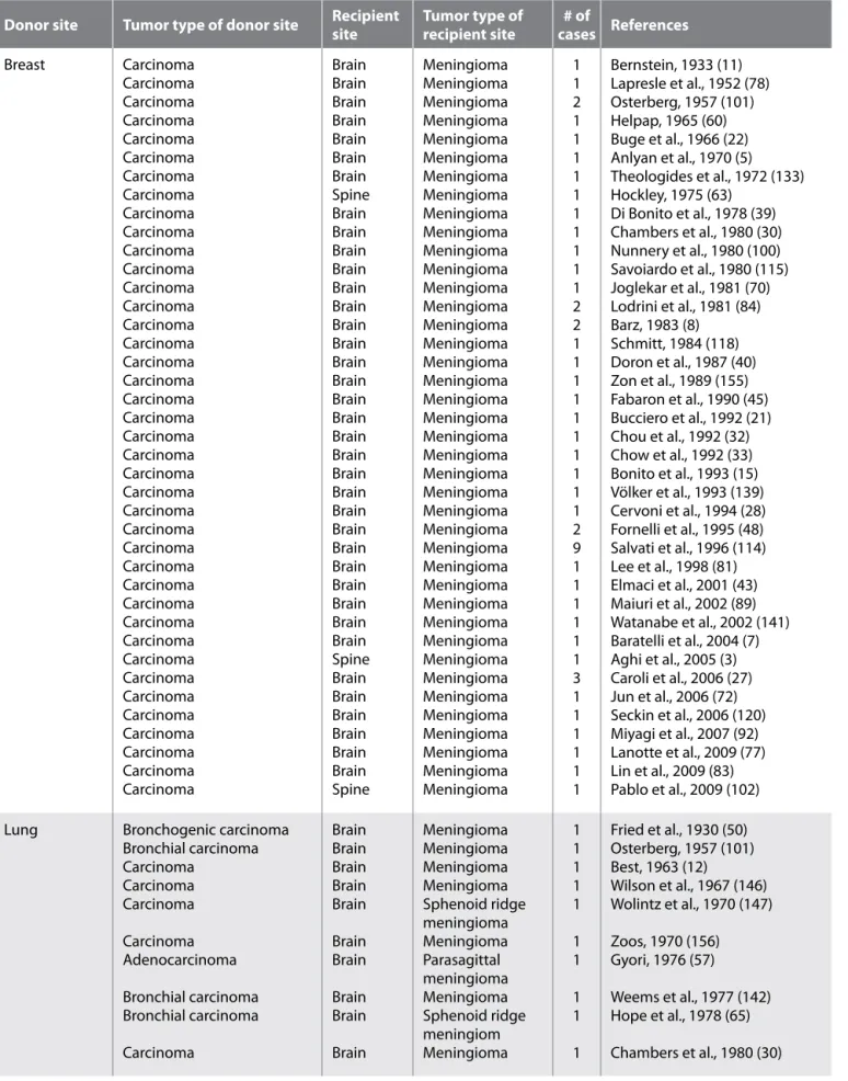
There is no reliable radiological imaging technique to detect the presence of metastasis within meningioma at present. It is supposed that computed tomography may show a hyperdense area or, when associated with necrotic component, a hypodense area (71, 115). A case of metastatic malignant melanoma within meningioma with intratumoral infarct that caused hypodensity was reported (104). There are some other cases of focal hypodensity within meningioma (30, 81). However, this appearance usually reflects the presence of calcification, necrotic foci, cystic degeneration or haemorrhage. Also, atypical signal characteristics were reported after intravenous administration of gadolinium on MRI in a few cases in the literature (27, 43, 81) without particular features. Since the physiological features of the tumor change after metastasis, neuroimaging methods such as perfusion MR and MR spectroscopy may be useful in differentiating tumor histology (23, 72). Even so, the correct diagnosis might be made by histological examination. However, the clinical picture may provide a clue about metastasis. Acute deterioration in the consciousness level and rapid onset of neurological symptoms should keep the surgeon aware of changes due to perilesional edema and increased mass effect. Analysis of Table I discloses the biggest numbers in the literature for metastasis to meningioma. There are 114 cases of tumor-to-meningioma metastasis that have been reported. Similar to previous reviews, most primary tumors were of the breast (54 cases), lung (23 cases) and kidney (10 cases). In contrast, breast and prostate metastases (8 cases) were much more common than previous studies. This increase may be explained by routine screening and early diagnosis.
Table I: Summary of Meningiomas Containing Metastases Reported in the Literature
donor site Tumor type of donor site Recipient site Tumor type of recipient site cases References # of Breast Carcinoma Carcinoma Carcinoma Carcinoma Carcinoma Carcinoma Carcinoma Carcinoma Carcinoma Carcinoma Carcinoma Carcinoma Carcinoma Carcinoma Carcinoma Carcinoma Carcinoma Carcinoma Carcinoma Carcinoma Carcinoma Carcinoma Carcinoma Carcinoma Carcinoma Carcinoma Carcinoma Carcinoma Carcinoma Carcinoma Carcinoma Carcinoma Carcinoma Carcinoma Carcinoma Carcinoma Carcinoma Carcinoma Carcinoma Carcinoma Brain Brain Brain Brain Brain Brain Brain Spine Brain Brain Brain Brain Brain Brain Brain Brain Brain Brain Brain Brain Brain Brain Brain Brain Brain Brain Brain Brain Brain Brain Brain Brain Spine Brain Brain Brain Brain Brain Brain Spine Meningioma Meningioma Meningioma Meningioma Meningioma Meningioma Meningioma Meningioma Meningioma Meningioma Meningioma Meningioma Meningioma Meningioma Meningioma Meningioma Meningioma Meningioma Meningioma Meningioma Meningioma Meningioma Meningioma Meningioma Meningioma Meningioma Meningioma Meningioma Meningioma Meningioma Meningioma Meningioma Meningioma Meningioma Meningioma Meningioma Meningioma Meningioma Meningioma Meningioma 1 1 2 1 1 1 1 1 1 1 1 1 1 2 2 1 1 1 1 1 1 1 1 1 1 2 9 1 1 1 1 1 1 3 1 1 1 1 1 1 Bernstein, 1933 (11) Lapresle et al., 1952 (78) Osterberg, 1957 (101) Helpap, 1965 (60) Buge et al., 1966 (22) Anlyan et al., 1970 (5) Theologides et al., 1972 (133) Hockley, 1975 (63) Di Bonito et al., 1978 (39) Chambers et al., 1980 (30) Nunnery et al., 1980 (100) Savoiardo et al., 1980 (115) Joglekar et al., 1981 (70) Lodrini et al., 1981 (84) Barz, 1983 (8) Schmitt, 1984 (118) Doron et al., 1987 (40) Zon et al., 1989 (155) Fabaron et al., 1990 (45) Bucciero et al., 1992 (21) Chou et al., 1992 (32) Chow et al., 1992 (33) Bonito et al., 1993 (15) Völker et al., 1993 (139) Cervoni et al., 1994 (28) Fornelli et al., 1995 (48) Salvati et al., 1996 (114) Lee et al., 1998 (81) Elmaci et al., 2001 (43) Maiuri et al., 2002 (89) Watanabe et al., 2002 (141) Baratelli et al., 2004 (7) Aghi et al., 2005 (3) Caroli et al., 2006 (27) Jun et al., 2006 (72) Seckin et al., 2006 (120) Miyagi et al., 2007 (92) Lanotte et al., 2009 (77) Lin et al., 2009 (83) Pablo et al., 2009 (102) Lung Bronchogenic carcinoma
Bronchial carcinoma Carcinoma Carcinoma Carcinoma Carcinoma Adenocarcinoma Bronchial carcinoma Bronchial carcinoma Carcinoma Brain Brain Brain Brain Brain Brain Brain Brain Brain Brain Meningioma Meningioma Meningioma Meningioma Sphenoid ridge meningioma Meningioma Parasagittal meningioma Meningioma Sphenoid ridge meningiom Meningioma 1 1 1 1 1 1 1 1 1 1 Fried et al., 1930 (50) Osterberg, 1957 (101) Best, 1963 (12) Wilson et al., 1967 (146) Wolintz et al., 1970 (147) Zoos, 1970 (156) Gyori, 1976 (57) Weems et al., 1977 (142) Hope et al., 1978 (65) Chambers et al., 1980 (30)
Lung Carcinoid tumor Carcinoma Carcinoma Carcinoma Carcinoma Adenocarcinoma Adenocarcinoma Bronchogenic carcinoma Carcinoma Carcinoma Pulmonary adenocarcinoma Carcinoma Adenocarcinoma Brain Brain Brain Brain Brain Brain Brain Brain Brain Brain Brain Brain Brain Fibroblastic meningioma Meningioma Meningioma Meningioma Meningioma Meningioma Optic nerve sheath meningioma Meningioma Meningioma Meningioma Secretory meningioma Meningioma Microcycstic meningioma 1 1 1 1 1 1 1 1 1 1 1 1 1 Smith et al., 1981 (124) Jomin et al., 1982 (71) Barz, 1983 (8) Pamphlett, 1984 (105) Schmitt, 1984 (118) Conzen et al., 1986 (36) Arnold et al., 1995 (6) Gardiman et al., 1996 (53) Bhargava et al., 1999 (13) Bori et al., 2002 (16) Cserni et al., 2002 (38) Duprez et al., 2009 (42) Takei et al., 2009 (130) Renal Adenocarcinoma Renal cell ca
Renal cell ca (with doubt) Carcinoma Renal cell ca Renal cell ca Renal cell ca Renal cell ca Renal cell ca Renal cell ca Brain Brain Brain Brain Brain Brain Brain Brain Brain Brain Meningioma Meningioma Meningioma Meningioma Meningioma Meningioma Meningioma Meningioma Meningioma Meningioma 1 1 1 1 1 1 1 1 1 1 Stortebecker, 1951 (126)
Gutierrez Morales et al., 1957 (56) Osterberg, 1957 (101) Barz, 1983, (8) Breadmore et al., 1994 (17) Han et al., 2000 (59) Kimiwada et al., 2004 (74) Chahlavi et al., 2005 (29) Lanotte et al., 2009 (77) Tsunoo et al., 2010 (135) Prostate Carcinoma Carsinoma Carcinoma Carcinoma Adenocarcinoma Adenocarcinoma Carcinoma Brain Brain Brain Brain Brain Brain Brain Meningioma Meningioma Meningioma Meningioma Meningioma Meningioma Meningioma 1 1 1 1 1 1 2 Döring, 1975 (41) Chambers et al., 1980 (30) Bernstein et al., 1983 (10) Cluroe, 2006 (34) Pugsley et al., 2009 (111) Mitchell et al., 2011 (90) Moody et al., 2012 (94) Skin (MM) Malignant melanoma
Malignant melanoma Malignant melanoma of vulva Malignant melanoma Brain Brain Brain Brain Meningioma Meningioma Fibroblastic meningioma Sphenoid wing meningioma 1 1 1 1 Wong et al., 1999 (148) Shariff et al., 2009 (122) Takei et al., 2009 (130) Pal et al., 2010 (104) Colorectal Carcinoma
Carcinoma BrainBrain MeningiomaMeningioma 11 Benedetto et al., 2007 (9)Moody et al., 2012 (94) Stomach Gastric carcinoma Brain Meningioma 1 Honma et al., 1989 (64) Esophagus Carcinoma Brain Meningioma 1 Kepes et al., 1982 (73) Gallbladder Adenocarcinoma Brain Suprasellar
meningioma 1 Peison et al., 1961 (106) Cervix Carcinoma brain Meningioma 1 Wu et al., 1977 (150) Unknown Carcinoma Brain Meningioma 1 Cappabianca et al., 1985 (26) Salivary gl. Parotid adenocarcinoma Brain Meningioma 1 Van Zandijcke et al., 1996 (138) Table I: Cont.
histological origin although the location was the skin for both (31, 54).
Metastasis to Gliomas
As can be seen from Table III, metastasis to gliomas has been reported 16 times previously. Similar to meningiomas, gliomas were metastasized by lung and breast carcinomas. The mechanisms of metastasis to gliomas remains obscure due to the fact that there are few cases and a remarkable number are from necropsy studies. Mörk et al. have suggested that no casual relationship can be invoked in these cases and the concomitance of a primary glioma and metastatic carcinoma should be regarded as coincidental (96). It is important to mention the studies of gliomas mimicking metastatic carcinomas (52, 73) as they can give an idea about the process in metastasis or differentiation. However, it can be said that the behavioral characteristics of tumor cells are the determining factors for tumor-to-tumor metastasis. Aggressive and malignant tumors usually act as donors, not recipients. Slow-growing characteristic of the host provides a longer period for metastases. This might explain the limited number of gliomas acting as the host lesions.
dISCuSSIon
Although rare, neurosurgeons should be aware of the entity of tumor-to-tumor metastasis. Meningiomas are the third and pituitary adenomas are the fifth most frequent tumor types in large series, probably reflecting their rich vascularity (144). Metastasis to a meningioma usually results in rapid enlargement of the long-standing meningioma, and leads to the first clinical manifestation of the donor malignancy. The donor tumor is usually found to be widely disseminated (30). These predisposing factors of meningiomas may render these tumors particularly susceptible to receiving seeds from extracranial malignancies. The low metabolic rate acts as a noncompeting metabolic environment conductive to cell growth. The high lipid and collagen content of meningiomas provides a fertile soil not only for the seeding of malignant cells, but also for hematogenous spread of bacterial infections (123). This is consistent with Paget’s “seed and soil theory”, which postulates that tumor development is a consequence of the supply of fertile environment (the soil) in which tumor cells (the seed) can proliferate (103). Two theories have been developed to explain the relationship between primary
Metastasis to Other Benign CNS Tumors
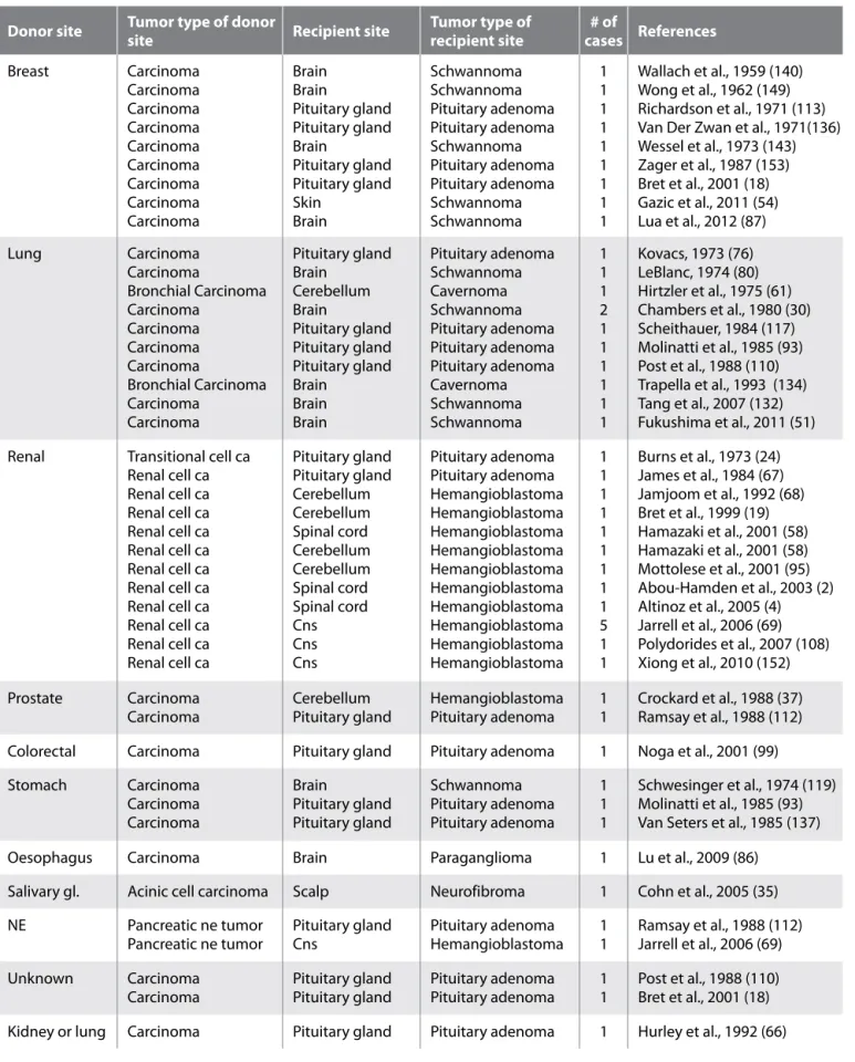
Patients with hereditary cancer syndromes may be at increased risk for the development of tumor-to-tumor metastases. Among the von Hippel-Lindau (VHL) disease cases with CNS hemangioblastomas, Jarrel et al. found six cases characterized with metastasis to CNS hemangioblastoma (69). The primary site of metastatic disease was the kidney in five patients (renal cell carcinoma) and the pancreas in one (a pancreatic neuroendocrine tumor). Two patients (including one who was also in the surgical group) were found at autopsy to have CNS metastases exclusively to spinal hemangioblastomas. Hemangioblastomas are usually an early and preferred site for metastasis in VHL cases (69). Altinoz et al. revealed that renal cell carcinoma-to-CNS hemangioblastoma is the most common donor-recipient tumor association among tumor-to-tumor metastasis cases (4) (Table II).
Similar to meningiomas, schwannomas have also been mostly metastasized by carcinomas of the breast and lung. Although there are only 11 reported cases of metastasis to schwannoma, similar mechanisms with meningiomas can be suggested except for hormonal tendency. Fukushima et al. have suggested that [(18)F]-fluorodeoxyglucose positron emission tomography might play an effective role in the preoperative diagnosis of tumor-to-tumor metastasis in schwannomas preoperatively (51) (Table II). This could be another difference with meningiomas.
Hypotheses regarding a high collagen or lipid content that have been proposed for meningiomas seem unlikely in pituitary tumors (18). On the contrary, it was suggested that the organized trombi in cavernomas may play the same role with lipid aggregation and provide metastasis a fertile soil for seeding and growth (31). Alterations of vasculature have been thought to be another reason for development of metastases in pituitary adenomas. Direct arterial supply from the carotid and meningeal systems may give rise to metastases by bypassing portal vessels. Likewise, the architecture of cavernomas may increase the chances of hematogenous metastases by slowing the flow within the lesion and increasing the likelihood of adherence to vessel wall by the metastatic emboli (31). According to our review, there are two exceptional cases among all others of benign CNS tumours. They were included in our study due to their Table I: Cont.
NE Pituitary carcinoma Brain Meningioma 1 Zhou et al., 2012 (154) Leukemia Chronic myeloid leukemia Brain Meningioma 1 Sonet et al., 2001 (125) Genitourinary Endometrial carcinoma Brain Meningioma 1 Ho, 1980 (62)
Hematopoietic Testicular lymphoma Lymphoma Lymphoma Multiple myeloma Brain Brain Brain Brain Meningioma Meningioma Meningioma Meningioma 1 1 1 1 Kepes et al., 1982 (73) Takakura et al., 1982 (129) Widdel et al., 2010 (144) Widdel et al., 2010 (144)
Table II: Summary of Other Benign CNS Tumors Containing Metastases Reported in the Literature
donor site Tumor type of donor site Recipient site Tumor type of recipient site cases References # of Breast Carcinoma Carcinoma Carcinoma Carcinoma Carcinoma Carcinoma Carcinoma Carcinoma Carcinoma Brain Brain Pituitary gland Pituitary gland Brain Pituitary gland Pituitary gland Skin Brain Schwannoma Schwannoma Pituitary adenoma Pituitary adenoma Schwannoma Pituitary adenoma Pituitary adenoma Schwannoma Schwannoma 1 1 1 1 1 1 1 1 1 Wallach et al., 1959 (140) Wong et al., 1962 (149) Richardson et al., 1971 (113) Van Der Zwan et al., 1971(136) Wessel et al., 1973 (143) Zager et al., 1987 (153) Bret et al., 2001 (18) Gazic et al., 2011 (54) Lua et al., 2012 (87) Lung Carcinoma Carcinoma Bronchial Carcinoma Carcinoma Carcinoma Carcinoma Carcinoma Bronchial Carcinoma Carcinoma Carcinoma Pituitary gland Brain Cerebellum Brain Pituitary gland Pituitary gland Pituitary gland Brain Brain Brain Pituitary adenoma Schwannoma Cavernoma Schwannoma Pituitary adenoma Pituitary adenoma Pituitary adenoma Cavernoma Schwannoma Schwannoma 1 1 1 2 1 1 1 1 1 1 Kovacs, 1973 (76) LeBlanc, 1974 (80) Hirtzler et al., 1975 (61) Chambers et al., 1980 (30) Scheithauer, 1984 (117) Molinatti et al., 1985 (93) Post et al., 1988 (110) Trapella et al., 1993 (134) Tang et al., 2007 (132) Fukushima et al., 2011 (51) Renal Transitional cell ca
Renal cell ca Renal cell ca Renal cell ca Renal cell ca Renal cell ca Renal cell ca Renal cell ca Renal cell ca Renal cell ca Renal cell ca Renal cell ca Pituitary gland Pituitary gland Cerebellum Cerebellum Spinal cord Cerebellum Cerebellum Spinal cord Spinal cord Cns Cns Cns Pituitary adenoma Pituitary adenoma Hemangioblastoma Hemangioblastoma Hemangioblastoma Hemangioblastoma Hemangioblastoma Hemangioblastoma Hemangioblastoma Hemangioblastoma Hemangioblastoma Hemangioblastoma 1 1 1 1 1 1 1 1 1 5 1 1 Burns et al., 1973 (24) James et al., 1984 (67) Jamjoom et al., 1992 (68) Bret et al., 1999 (19) Hamazaki et al., 2001 (58) Hamazaki et al., 2001 (58) Mottolese et al., 2001 (95) Abou-Hamden et al., 2003 (2) Altinoz et al., 2005 (4) Jarrell et al., 2006 (69) Polydorides et al., 2007 (108) Xiong et al., 2010 (152) Prostate Carcinoma
Carcinoma Cerebellum Pituitary gland Hemangioblastoma Pituitary adenoma 11 Crockard et al., 1988 (37)Ramsay et al., 1988 (112) Colorectal Carcinoma Pituitary gland Pituitary adenoma 1 Noga et al., 2001 (99) Stomach Carcinoma Carcinoma Carcinoma Brain Pituitary gland Pituitary gland Schwannoma Pituitary adenoma Pituitary adenoma 1 1 1 Schwesinger et al., 1974 (119) Molinatti et al., 1985 (93) Van Seters et al., 1985 (137) Oesophagus Carcinoma Brain Paraganglioma 1 Lu et al., 2009 (86)
Salivary gl. Acinic cell carcinoma Scalp Neurofibroma 1 Cohn et al., 2005 (35) NE Pancreatic ne tumor
Pancreatic ne tumor Pituitary gland Cns Pituitary adenoma Hemangioblastoma 11 Ramsay et al., 1988 (112)Jarrell et al., 2006 (69) Unknown Carcinoma
Carcinoma Pituitary glandPituitary gland Pituitary adenomaPituitary adenoma 11 Post et al., 1988 (110)Bret et al., 2001 (18) Kidney or lung Carcinoma Pituitary gland Pituitary adenoma 1 Hurley et al., 1992 (66)
tissues (extravasation) where they settle, proliferate, and induce angiogenesis, creating metastases. The activation of proteolytic enzymes that are capable of degrading the extracellular matrix (ECM) surrounding the endothelium or creating the basement membrane of epithelial tissue in different organs is indispensable in the process of intra- and extravasation (145). In this stage the activation of proteolytic enzymes, such as proteinases of the plasmin system, serineproteinases, and matrix metalloproteinases (MMPs), is necessary. Simultaneously, changes occur in the expression of many superficial glycoproteins and factors responsible for cell adhesion (integrins) and intercellular communication (cadherins). Moreover, Muller et al. showed that the chemokines and their receptors have critical roles in determining the metastatic destination of tumor cells (97). Cross talk between the primary CNS tumor and the malignant tumor usually occurs by means of expressions. For example, E-cadherin expression by both meningiomas and breast cancer may affect the choice of “seed” (cancer cells) to find the “soil” (recipient CNS tumor) (3). Neoangiogenesis is connected with the expression of many markers of this process, among them vascular endothelial growth factor (VEGF), endoglin (CD105), a transmembranous glycoprotein which is a tumor and metastasis. These are “The Mechanical Theory” of
Ewing and “The Soil and Seed Theory” of Paget (44, 103). It is not surprising that nearly all CNS tumor-to-tumor metastasis cases have eluded preoperative radiological diagnosis and were only discovered incidentally during postoperative pathological examination (72).
Cancer metastasis results from a multi-step cascading process that includes; 1) vascularization of the primary tumor; 2) detachment and invasion of cancer cells; 3) intravasation into lymphatic and blood vessels; 4) survival and arrest in the circulation; 5) extravasation into distant organs; and 6) colonization and growth of metastatic tumors. MicroRNAs (miRNAs) play critical roles in this multi-step process, both promoting and suppressing metastasis (79). Specific miRNA can cause cancer cells to invade and metastasize (88).
In the first step, vascularization of the primary tumor takes place. In the second and third stages the cancer cells released from the primary tumor have to penetrate to the blood or lymphatic vessels (intravasation), the road that dissemination follows. In the fourth stage, cancer cells should survive and in the fifth stage circulating cells can migrate through the walls of vessels to surrounding
Hematopoietic Lymphoma Infundibulum İnfundibular
choristoma 1 Takakura et al., 1982 (129) Mediastinum Carcinoid tumor Pituitary gland Pituitary adenoma 1 Abe, 1997 (1)
Skin (MM) Melanoma Brain Cavernoma 1 Chan et al., 2006 (31) Table II: Cont.
Table III: Summary of Gliomas Containing Metastases Reported in the Literature
donor site Tumor type of donor site Recipient site Tumor type of recipient site cases# of references
Breast Carcinoma Carcinoma Carcinoma Carcinoma Brain Brain Brain Brain Oligodendroglioma Oligodendroglioma Oligodendroglioma Pilocytic Astrocytoma 1 1 1 1 Strang, 1965 (127) Tally et al., 1988 (131) Wurm et al., 1994 (151) Muller et al., 1999 (98) Lung Bronchial carcinoma Bronchial carcinoma Bronchial carcinoma Brain Brain Brain Astrocytoma Fourth ventricle ependymoma Astrocytoma 2 1 1 Mörk et al., 1988 (96) Mörk et al., 1988 (96) Tajika et al., 1990 (128) Skin (MM) Melanoma Melanoma Melanoma Melanoma Brain Brain Brain Brain Glioma Oligodendroglioma Glioma Neurocytoma 1 1 1 1 Zulch, 1965 (157) Farnsworth, 1972 (46) Takakura et al., 1982 (129) Brown et al., 2003 (20) Renal Carcinoma Brain Glioblastoma 1 Franke et al., 1990 (49) Thyroid Carcinoma Brain Glioblastoma 1 Posnikoff et al., 1960 (109) Colorectal Carcinoma brain Oligodendroglioma 1 Mizutani et al., 1987 (91) Unknown Carcinoma Brain Glioma 1 Joglekar et al., 1981 (70)
3. Aghi M, Kiehl TR, Brisman JL: Breast adenocarcinoma metastatic to epidural cervical spine meningioma: Case report and review of the literature. J Neurooncol 75(2): 149-155, 2005. Review
4. Altinoz MA, Santaguida C, Guiot MC, Del Maestro RF: Spinal hemangioblastoma containing metastatic renal cell carcinoma in von Hippel-Lindau disease. Case report and review of the literature. J Neurosurg Spine 3:495-500, 2005 5. Anlyan FH, Heinzen BR, Carras R: Metastasis of tumor to
second different tumor: Collision tumors. JAMA 212(12): 2124, 1970
6. Arnold AC, Hepler RS, Badr MA, Lufkin RB, Anzai Y, Konrad PN, Vinters HV: Metastasis of adenocarcinoma of the lung to optic nevre sheath meningioma. Arch Ophthalmol 113:346-351, 1995
7. Baratelli GM, Ciccaglioni B, Dainese E, Arnaboldi L: Metastasis of breast carcinoma to intracranial meningioma. J Neurosurg Sci 48(2):71-73, 2004
8. Barz H: The incidence of metastatic carcinomas in meningiomas. A report of 4 cases. Zentralbl Allg Pathol 127:367-374, 1983
9. Benedetto N, Perrini P, Scollato A, Buccoliero AM, Di Lorenzo N: Intracranial meningioma containing metastatic colon carcinoma. Acta Neurochir (Wien) 149(8):799-803; discussion 803, 2007
10. Bernstein RA, Grumet KA, Wetzel N: Metastasis of prostatic carcinoma to intracranial meningioma. Case report. J Neurosurg 58:774-777, 1983
11. Bernstein SA: Ueber Karsinommetastase in einem Duraendo-thelom. Centralblatt für Allgemenie Pathologie und Patholo-gische Anatomie 58:163-166, 1933
12. Best PV: Metastatic carcinoma in a meningioma: Report of a case. J Neurosurg 20:892-894, 1963
13. Bhargava P, McGrail KM, Manz HJ, Baidas S: Lung carcinoma presenting as metastasis to intracranial meningioma: Case report and review of the literature. Am J Clin Oncol 22: 199-202, 1999
14. Bohn OL, De las Casas LE, Leon ME: Tumor-to-tumor metastasis: Renal cell carcinoma metastatic to papillary carcinoma of thyroid-report of a case and review of the literature. Head and Neck Pathol 3:327-330, 2009
15. Bonito D, Giarelli L, Falconieri G, Bonifacio-Gori D, Tomasic G, Vielh P: Association of breast cancer and meningioma. Report of 12 new cases and review of the literature. Pathol Res Pract 189(4):399-404, 1993
16. Bori R, Kiss CA, Huszka E, Szucs M, Tusa M, Cserni G: A rare case of tumor-to-tumor metastasis: Secondary deposits of pulmonary adenocarcinoma in a secretory meningioma. Magy Onkol 46: 261-264, 2002
17. Breadmore R, House R, Gonzales M: Metastasis of renal cell carcinoma to a meningioma. Australas Radiol 38:143-144, 1994
18. Bret P, Jouvet A, Madarassy G, Guyotat J, Trouillas J. Visceral cancer metastasis to pituitary adenoma: Report of two cases. Surg Neurol 55: 284-290, 2001
component of the receptor for transforming growth factor beta (TGFbeta), as well as neuropilin (NRP), the co-receptor for VEGF. These molecular and cellular markers play key roles in tumor progression.
As stated above, metastasis to the brain and primary brain tumors is augmented by the activation of homologous molecules between the systemic cancer and primary CNS neoplasm tissue. These molecules are not only expressed by the primary cancers, but are also echoed in the brain parenchyma and especially in primary CNS neoplasms. These molecules may act as a “fertilizer” for cancer cells and selectively reside in the primary CNS neoplasms. These findings are also consistent with Paget’s “seed and soil theory”. It is conceivable that cancer cells will metastasize to organs from similar embryological origins. Hence, malignant melanoma has a greater likelihood of forming leptomeningeal metastasis since they are both derived from neural crest cells (97).
Although these results suggest that integrins, cathedrins and chemokines play a prominent role in the dissemination of tumor cells and their subsequent invasion to a second malignancy in tumor-to-tumor metastasis cases, the development of tumor metastasis from a second primary malignancy is uncommon and remains biologically puzzling. The collected data depends mostly on post mortem studies. This suggests a higher incidence for tumor-to-tumor metastasis. Benign tumor-to-tumors should undergo periodic radiologial and physical examinations just like malignant tumors. Likewise, in patients with carcinoma, lesions of the central nervous system should not be labeled as metastasis. Whenever possible, the whole tumor should be examined pathologically.
ConCluSIon
We conclude that neurosurgeons should be aware of the phenomenon of tumor-to-tumor metastasis. Knowing this potential association is important in appropriate patient management. One can say that the preoperative diagnosis of metastasis to a tumor would not change the indication for surgical removal of the lesion. However, it will surely change the technical approach and imply an en bloc removal of the lesion to avoid any intraoperative seeding of metastatic cells.
REFEREnCES
1. Abe T, Matsumoto K, Iida M, Hayashi M, Sanno N, Osamura RY: Malignant carcinoid tumor of the anterior mediastinum metastasis to a prolactin-secreting pituitary adenoma: A case report. Surg Neurol 48(4):389- 394, 1997
2. Abou-Hamden A, Koszyca B, Carney PG, Sandhu N, Blumbergs PC: Metastasis of renal cell carcinoma to hemangioblastoma of the spinal cord in von Hippel-Lindau disease: Case report and review of the literature. Pathology 35:224-227, 2003
36. Conzen M, Sollman H, Schnabel R: Metastasis of lung carcinoma to intracranial meningioma. Case report and review of literature. Neurochirrurgia (Stuttg) 29:206-209, 1986 37. Crockard HA, Barnard RO, Isaacson PG: Metastasis of
carcinoma to haemangioblastoma cerebelli: Case report. Neurosurg 23(3):382- 384, 1988
38. Cserni G, Bori R, Huszka E, Kiss AC: Metastasis of pulmonary adenocarcinoma in right sylvian secretory meningioma. Br J Neurosurg 16:66-68, 2002
39. Di Bonito L, Bianchi C: Metastasis of breast cancer to a meningioma. Arch Anat Cytol Pathol 26:175-176, 1978 40. Doron Y, Gruszkiewicz J: Metastasis of invasive carcinoma of
the breast to an extradural meningioma of the cranial wault. Cancer 60:1081-1084, 1987
41. Döring L: Metastasis of carcinoma of prostate to meningioma. Virchows Arch A Pathol Anat Histol 366:87-91, 1975
42. Duprez R, Thimon S, Kerdraon R, Bonneau C, Metois D, Michenet P: Tumor-to-tumor metastasis: Report of three cases. Ann Pathol 29:507-511, 2009
43. Elmaci L, Ekinci G, Kurtkaya O, Sav A, Pamir MN: Tumor in tumor: Metastasis of breast carcinoma to intracranial meningioma. Tumor 87(6):423-427, 2001
44. Ewing J: Neoplastic Diseases. Philadelphia: Saunders,1928 45. Fabaron F, Bainier L, Vende B, Babin P, Morin M: Metastasis
of breast cancer in frontal meningioma. Ann Radiol (Paris) 33:48-50, 1990
46. Farnsworth J: Regressing melanoma metastasizing to an oligodendroglioma. Pathology 4:253-257, 1972
47. Fidler IJ: The pathogenesis of cancer metastasis: The ‘seed and soil’ hypothesis revisited. Nat Rev Cancer 3: 453-458, 2003 48. Fornelli A, Bacci A, Collina G, Eusebi V: Breast carcinoma
metastatic to meningioma: Review of the literature and description of 2 new cases. Pathologica 87:506-512, 1995 49. Franke FE, Altmannsberger M, Schachenmayr W: Metastasis
of renal carcinoma colliding with glioblastoma. Acta Neuropathol 80:448 – 552, 1990
50. Fried BM: Metastatic inoculation of a meningioma by cancer cells from a bronchogenic carcinoma. Am J Pathol 6:47-52, 1930
51. Fukushima Y, Ota T, Mukasa A, Uozaki H, Kawai K, Saito N: Tumor to tumor metastasis: Lung adenocarcinoma metasta-sizing to vestibular schwannoma suspected on preoperative [(18) F]-Fluorodeoxyglucose Positron Emission Tomography Imaging. World Neurosurg 78(5):553.e9-553.e13, 2011 52. Galloway PG, Roessmann U: Anaplastic astrocytoma
mimick-ing metastatic carcinoma. Am J Surg Pathol 10: 728-732, 1986 53. Gardiman M, Altavilla G, Marchioro L, Alessio L, Parenti A,
Piazza M: Metastasis to intracranial meningioma as first clinical manifestation of occult primary lung carcinoma. Tumori 82:256-258, 1996
54. Gazic B, Pizem J: Lobular breast carcinoma metastasis to a superficial plexiform schwannoma as the first evidence of an occult breast cancer. Am J Dermatopathol 33:845-849, 2011 19. Bret P, Streichenberger N, Guyotat J: Metastasis of renal
carcinoma to a cerebellar hemangioblastoma in a patient with von Hippel-Lindau disease: Case report. Br J Neurosurg 13:413-416, 1999
20. Brown HM, McCutcheon TE, Leeds NE, Schomer DS, Luna MA, Fuller GN: Melanoma metastatic to central neurocytoma: A novel case of tumor-to-tumor metastasis. J Neurooncol 61:209-214, 2003
21. Bucciero A, del Basso de Caro M, Vizioli L, Carraturo S, Cerillo A, Tedeschi G: Metastasis of breast carcinoma to intracranial meningioma. Case report and review of the literature. J Neurosurg Sci 36(3):169-172, 1992
22. Buge A, Escourolle R, Martin M, Poirier J, Devoise C: Cerebromeningeal metastases of an epithelioma of the breast. Multiple meningiomatosis. Intrication of the 2 processes. Rev Neurol (Paris) 114(4):308-312, 1966
23. Bulakbasi N, Kocaoglu M, Ors F, Tayfun C, Ucoz T: Combination of single-voxel proton MR spectroscopy and apparent diffusion coefficient calculation in the evaluation of common brain tumors. AJNR Am J Neuroradiol 24(2):225-233, 2003 24. Burns WA, Kadar AT: Unusual metastases from a transitional
cell carcinoma of the renal pelvis and ureter. Med Ann D C 42:65 – 66, 1973
25. Campbell LV, Gilbert E, Chamberlein CR, Watne AL: Metastases of cancer to cancer. Cancer 22:635-643, 1968
26. Cappabianca P, De Chiara A, Spaziante R, Del Basso De Caro M, Cirillo S, Stella L: Metastasis of carcinoma to an intracranial meningioma. Case report and revision of the literature. Acta Neurol (Napoli) 7:482-493, 1985
27. Caroli E, Salvati M, Giangaspero F, Ferrante L, Santoro A: Intrameningioma metastasis as first clinical manifestation of occult primary breast carcinoma. Neurosurg Rev 29(1):49-54, 2006
28. Cervoni L, Salvati M, Gagliardi D, Delfini R: Metastasis of breast carcinoma to intracranial meningioma. Case report. Neurosurg Rev 17(3):233-236, 1994
29. Chahlavi A, Staugaitis SM, Yahya R, Vogelbaum MA: Intracranial collision tumor mimicking an octreotide-SPECT positive and FDG-PET negative meningioma. J Clin Neurosci 12: 720-723, 2005
30. Chambers PW, Davis RL, Blanding JD, Buck FS: Metastases to primary intracranial meningiomas and neurilemmomas. Arch Pathol Lab med 104:350-354, 1980
31. Chan CH, Fabinyi GC, Kalnins RM: An unusual case of tumor-to-cavernoma metastasis. A case report and literature review. Surg Neurol 65(4):402-408; discussion 409, 2006
32. Chou LW, Ho KH, Fong CM: Intracranial meningioma with metastatic breast carcinoma. Ann Oncol 5:409-410, 1992 33. Chow LWC, Ho KH, Fong CM: Intracranial meningioma with
metastatic breast carcinoma. Ann Oncol 3: 409–410, 1992 34. Cluroe AD: Metastasis to meningioma: Clues and investigation.
Pathology 38:76-78, 2006
35. Cohn ML, Elliott DD, El-Naggar AK: Metastatic acinic cell carcinoma in a neurofibroma mistaken for carcinosarcoma. Head and Neck 27:76-80, 2005
73. Kepes JJ, Fulling KH, Garcia JH: The clinical significance of “adenoid” formations of neoplastic astrocytes, imitating metastatic carcinoma, in gliosarcomas. A review of five cases. Clinical Neuropathol 4:139-150, 1982
74. Kimiwada T, Motohashi O, Kumabe T, Watanabe M, Tominaga T: Lipomatous meningioma of the brain harboring metastatic renal-cell carcinoma: A case report. Brain Tumor Pathol 21(1):47-52, 2004
75. Koutourousiou M, Kontogeorgos G, Wesseling P, Grotenhuis AJ, Seretis A: Collision sellar lesions: Experience with eight cases and review of the literature. Pituitary 13:8-17, 2010 76. Kovacs K: Metastatic cancer of the pituitary gland. Oncology
27:533– 542, 1973
77. Lanotte M, Bench F, Panciani PP, Cassoni P, Ducati A: Systemic cancer metastasis in a meningioma: Report of two cases and review of the literature. Clin neurol Neurosurg 111:87-93, 2009
78. Lapresle J, Netsky MG, Zimmerman HM: The pathology of meningiomas. A study of 121 cases. Am J Pathol 28:757-791, 1952
79. Le XF, Merchant O, Bast RC, Calin GA: The role of MicroRNAs in the cancer invasion-metastasis cascade. Cancer Microenviron 3:137-147, 2010
80. LeBlanc RA: Metastasis of bronchogenic carcinoma to acoustic neurinoma. Case report. J Neurosurg 41(5):614-617, 1974
81. Lee A, Wallace C, Rewcastle B, Sutherland G: Metastases to meningioma. AJNR 19:1120-1122, 1998
82. Lieu AS, Hwang SL, Howng SL: Intracranial meningioma and breast cancer. J Clin Neurosci 10:553-556, 2003
83. Lin JW, Su FW, Wang HC, Lee TC, Ho JT, Lin CH, Lin YJ: Breast carcinoma metastasis to intracranial meningioma. J Clin Neurosci 16(12):1636-1639, 2009
84. Lodrini S, Savoiardo M: Metastases of carcinoma to intracranial meningioma: Report of two cases and review of the literature. Cancer 48:2668-2673, 1981
85. Lu J, Frater JL, Kreisel FH, Marcus JN, Hassan A: Secondary lymphoma involving metastatic follicular thyroid carcinoma to the skull: A unique example of tumor-to-tumor metastasis. Head and Neck Pathol 2:209-212, 2008
86. Lu JQ, Khalil M, Hu W, Sutherland GR, Clark AW: Tumor-to-tumor metastasis: Oesophageal carcinoma metastatic to an intracranial paraganglioma. J Neurosurg 110:744-748, 2009 87. Lua BK, Lieu AS, Hwang SL: Breast carcinoma metastasized
to vestibular schwannoma: A rare case of tumor-to-tumor metastasis and literature review. Kaohsiung J Med Sci 28: 397-399, 2012
88. Ma L, Weinberg RA: MicroRNAs in malignant progression. Cell Cycle 7:570-572, 2008
89. Maiuri F, Cappabianca P, Iaconetta G, D’Acunzi G: Menin-giomas associated with metastases. Zentralb Neurochir 63: 111-115, 2002
90. Mitchell RA, Dimou J, Tsui A, Kavar B: Metastatic prostate adenocarcinoma invading an atypical meningioma. J Clin Neurosci 18:1723-1725, 2011
55. Gore I, Barr R: Metastasis of cancer to cancer. Arch Pathol 66:293-298,1958
56. Gutierrez Morales JC, Gutierrez Morales SE, Astudillo Gonzales A: 72 year old man with new seizure while dancing. Brain Pathol 19:347-348,2009
57. Gyori E: Metastatic carcinoma in meningioma. South Med J 69:514-517, 1976
58. Hamazaki S, Nakashima H, Matsumoto K, Taguchi K, Okada S: Metastasis of renal cell carcinoma to central nervous system hemangioblastoma in two patients with von Hippel-Lindau disease. Pathol Int 51:948-953, 2001
59. Han HS, Kim EY, Han JY, Kim YB, Hwang TS, Chu YC: Metastatic renal cell carcinoma in a meningioma: A case report. J Korean Med Sci 15:593-597, 2000
60. Helpap B: Mammakarzinom metastase in einem meningiom. Zentralbl Allg Pathol 108: 242–245, 1965
61. Hirtzler R, Pavelić Z, Kubović M, Salić M: Metastases of lung cancer within benign tumours (author’s transl). Zentralbl Allg Pathol 119(4):272-275, 1975
62. Ho KL: Metastasis of carcinoma to meningioma. Arch Pathol Lasb Med 104: 394-395, 1980
63. Hockley AD: Metastatic carcinoma in a spinal meningioma. J Neurol Neurosurg Psychiatry 38:695-697, 1975
64. Honma K, Hara K, Sawai T: Tumour-to-tumour metastasis. A report of two unusual autopsy cases. Virchows Arch A Pathol Anat Histopathol 416:153-157, 1989
65. Hope DT, Symon L: Metastasis of carcinoma to meningioma. Acta neurochir (Wien) 40:307-313, 1978
66. Hurley TR, D’Angelo CM, Clasen RA, DiGianfilippo A, Ryan GW: Adenocarcinoma metastatic to a growth-hormone-secreting pituitary adenoma: Case report. Surg Neurol 37:361-365, 1992
67. James RL Jr, Arsenis G, Stoler M, Nelson C, Baran D: Hypophyseal metastatic renal cell carcinoma and pituitary adenoma. Case report and review of the literature. Am J Med 76:337-340, 1984
68. Jamjoom A, Kane N, Nicoll J: Metastasis of a renal carcinoma to a cerebellar hemangioblastoma in a case of von Hippel-Lindau disease. Neurosurg Rev 15:231-234, 1992
69. Jarrell ST, Vortmeyer AO, Linehan WM, Oldfield EH, Lonser RR: Metastases to hemangioblastomas in von Hippel-Lindau disease. J Neurosurg 105:256-263, 2006
70. Joglekar VM, Davis CH, Blakeney CG: Metastasis of carcinoma to meningioma and glioma. Acta Neurochir (Wien) 58:67-74, 1981
71. Jomin M, Dupont A, Wemeau J, Krivosic I, Montagne B, Lesoin F, Adenis L: Metastasis of visceral tumors in intracranial tumors. Apropos of a metastasis of a lung cancer in an intracranial meningioma. Neurochirurgie 28:343-347, 1982 72. Jun P, Garcia J, Tihan T, McDermott MW, Cha S: Perfusion
MR imaging of an intracranial collision tumor confirmed by image-guided biopsy. AJNR 27:94-97, 2006
107. Polajeva J, Sjösten AM, Lager N, Kastemar M, Waern I, Alufuzoff I, Smits A, Westermark B, Pejler G, Uhrbom L, Tchougounova E: Mast cell accumulation in glioblastoma with potential role for stem cell factor and chemokine CXCL12. PLoS One 6(9): e252222, 2011
108. Polydorides AD, Rosenblum MK, Edgar MA: Metastatic renal cell carcinomato hemangioblastoma in von Hippel-Lindau disease. Arch Pathol Lab Med 131:641-645, 2007
109. Posnikoff J, Stratford J: Carcinoma metastasis to malignant glioma. Arch Neurol 5:559-563, 1960
110. Post KD, McCormick PC, Hays AP, Kandji AG: Metastatic carcinoma to pituitary adenoma. Report of two cases. Surg Neurol 30:286-292, 1988
111. Pugsley D, Bailly G, Gupta R, Wilke D, Wood L: A case of metastatic adenocarcinoma of the prostate arising in a meningioma. Can Urol Assoc J 3:E4-E6, 2009
112. Ramsay JA, Kovacs K, Scheithauer BW, Ezrin C, Weiss MH: Metastatic carcinoma to pituitary adenomas: A report of two cases. Exp Clin Endocrinol 92:69-76, 1988
113. Richardson JF, Katayama I: Neoplasm to neoplasm metastasis. An acidophilic adenoma harbouring metastatic carcinoma: A case report. Arch Pathol 91: 135-139, 1971 114. Salvati M, Cervoni L: Association of breast carcinoma and
meningioma: Report of nine new cases and review of the literature. Tumori 82(5):491-493, 1996
115. Savoiardo M, Lodrini S: Hypodense area within a menin-gioma: Metastasis from breast cancer. Neuroradiology 20(2):107-110, 1980
116. Sawada T, Takahashi H, Hasatani K, Yoshida I, Oyama O, Inoue R, Miwa K, Kawashiri M, Misawa K, Kyoi M, Miyamoto S: Tumor-tumor metastasis: Report of an autopsy case of lung adenocarcinoma metastasizing to renal cell carcinoma. Inter Med 48:1525-1529, 2009
117. Scheithauer BW: Surgical pathology of the pituitary: The adenomas. Pathol Annu 19(2):269- 329, 1984
118. Schmitt HP: Metastases of malignant neoplasms to intra-cranial tumors: The “tumour-in-a-tumour” phenomenon. Virchows Arch A pathol Anat Histopathol 405:155-160, 1984 119. Schwesinger G, Pusinelli WD: Acoustic neurinoma with
metastases of a carcinoma of the stomach. Zentralbl Allg Pathol 118(5):473 – 475, 1974
120. Seckin H, Yigitkanli K, Ilhan O, Han U, Bavbek M: Breast carcinoma metastasis and meningioma. A case report. Surg Neurol 66(3):324-327; discussion 327, 2006
121. Sella A, Ro JY: Renal cell cancer: Best recipient of tumor-to-tumor metastasis. Urology 30:35-38, 1987
122. Shariff Z, Lim P, Wright A, Al-Ghazal S: Tumor to tumor metastasis of malignant melanoma to intracranial tumor. J Clin Med Res 1:300-301, 2009
123. Shimomura T, Hori S, Kasai N, Tsuruta K, Okada H: Menin-gioma associated with intratumoral abscess formation-case report. Neurol Med Chir (Tokyo) 34:440-443, 1994
124. Smith TW, Wanf SY, Schoene WC: Malignant carcinoid tumor metastatic to a meningioma. Cancer 47:1872-1877, 1981 91. Mizutani H, Suzuki H, Wakabayashi S, Mitake A, Mizutani M,
Banno T, Oba S, Tada T, Kuhara H: A case of metastasis from a colon carcinoma to an intracranial oligodendroglioma. Gan No Rinsho 33:1733-1739, 1987
92. Miyagi N, Hara S, Terasaki M, Orito K, Yamashita S, Hirohata M, Tokutomi T, Shigemori M: A rare case of intracranial meningioma with intratumoral metastatic breast cancers. No Shinkei Geka 35(9):901-905, 2007
93. Molinatti PA, Scheithauer BW, Randall RV, Laws ER Jr: Metastasis to pituitary adenoma. Arch Pathol Lab Med 109:287-289, 1985
94. Moody P, Murtagh K, Piduru S, Brem S, Murtagh R, Rojani AM: Tumor-to-tumor metastasis: Pathology and neuroimaging considerations. Int J Clin Exp Pathol 5:367-373, 2012
95. Mottolese C, Stan H, Giordano F, Frappaz D, Alexei D, Sreichenberger N: Metastasis of clear-cell renal carcinoma to cerebellar hemangioblastoma in von Hippel-Lindau disease: Rare or not investigated. Acta Neurochir (Wien) 143: 1059-1063, 2001
96. Mörk SJ, Rubinstein LJ: Metastatic carcinoma to glioma: A report of three cases with a critical review of the literature. J Neurol Neurosurg and Psychiatry 51:256-259, 1988
97. Muller A, Homey B, Soto H, Ge N, Catron D, Buchanan ME, McClanahan T, Murphy E, Yuan W, Wagner SN, Barrera JL, Mohar A, Verastegui E, Zlotnik A: Involvement of chemokine receptors in breast cancer metastasis. Nature 410:50-56, 2001 98. Muller W, Schroder R: Spreading of metastases into cranial
tumors: Metastasis of a breast carcinoma to a pilocytic astrocytoma. Clin Neuropathol 18(3):109–112, 1999
99. Noga C, Prayson RA, Kowalski R, Sweeney PJ, Mayberg M: Metastatic adenocarcinoma to a pituitary adenoma. Ann Diagn Pathol 5:354-360, 2001
100. Nunnery E Jr, Kahn LB, Rudnick SA: Breast carcinoma metastatic to meningioma. Arch Pathol Lab Med 104: 392-393, 1980
101. Osterberg DH: Metastases of carcinoma to meningioma. J Neurosurg 14:337-343, 1957
102. Pablo LJ, Patricia C, Julian I, Ariel Z, Roberto S, Jorge C, Juan L, Carlos V, Bernardo L: Metastasis of breast carcinoma to spinal meningioma. Breast J 15:539-540, 2009
103. Paget S: The distribution secondary growths in cancer of the breast. Lancet 1: 571-573, 1889
104. Pal D, Bhargava D, Bucur SD, Shivane A, Chakrabarty A, Van Hille P: Metastatic malignant melanoma within meningio-ma with intratumoral infarct: Report of an unusal case and literature review. Clin Neuropathol 29:105-108, 2010 105. Pamphlett R: Carcinoma metastasis to meningioma. J
Neurol Neurosurg Psychiatry 47:561-563, 1984
106. Peison WB, Feigin I: Suprasellar meningioma containing metastatic carcinoma. Report of case. J Neurosurg 18: 688-689, 1961
142. Weems TD, Garcia JH: Intracranial meningioma containing metastatic foci. South Med J 70:503-505, 1977
143. Wessel H, Gerlach H: Metastases of a breast cancer to an acoustic neurinoma (author’s transl). Zentralbl Allg Pathol 117(2):146-151, 1973
144. Widdel L, Kleinschmidt-DeMasters BK, Kindt G: Tumor-to-tumor metastasis from hemopoietic neoplasms to meningiomas: Report of two patients with significant cerebral edema. World Neurosurg 74:165-171, 2010
145. Widel MS, Widel M: Mechanisms of metastasis and molecular markers of malignant tumor progression. l. Colorectal cancer. Postepy Hig Med Dosw (Online) 60:453-470, 2006 146. Wilson CB, Jenevein EP, Bryant LR: Carcinoma of the lung
metastatic to falx meningioma: Case report. J Neurosurg 27: 161–165, 1967
147. Wolintz AH, Mastri A: Metastasis of a carcinoma of lung to sphenoid ridge meningioma. NY State J Med 70:2592-2598, 1970
148. Wong A, Koszyca B, Blumbergs PC, Sandhu N, Halcrow S: Malignant melanoma metastatic to meningioma. Pathology 31:162-165, 1999
149. Wong TW, Bennington JL: Metastasis of a mammary carcinoma to an acoustic neurinoma. J Neurosurg 19: 1088-1093, 1962
150. Wu WQ, Hiszczynskyj R: Metastasis of carcinoma or cervix uteri to convexity meningioma. Surg Neurol 8:327-329, 1977 151. Wurm G1, Huber A, Hiertz H, Fischer J, Cervos-Navarro J:
Composite cerebral metastasis and oligodendroglioma: An exceptional form of mixed neoplasia. Neurosurg Rev 17(4): 291- 294, 1994
152. Xiong J, Chu SG, Wang Y, Zhu JJ, Li C, Mao Y: Metastasis of renal cell carcinoma to a hemangioblastoma of the medulla oblongata in von Hippel-Lindau syndrome. J Clin Neurosci 17:1213-1215, 2010
153. Zager EL, Hedley-Whyte ET: Metastasis within a pituitary adenoma presenting with bilateral abducens palsies: Case report and review of the literature. Neurosurgery 21(3): 383-386, 1987
154. Zhou Q, Chang H, Gao Y, Cui L: Tumor-to-tumor metastasis from pituitary carcinoma to radiation-induced meningioma. Neuropathology 33(2):209-212, 2013
155. Zon LI, Johns WD, Stomper PC, Kaplan WD, Connolly JL, Morris JH, Harris JR, Henderson IC, Skarin AT: Breast carcinoma metastatic to a meningioma. Case report and review of the literature. Arch Intern Med 149(4):959-962, 1989
156. Zoos BA: Neoplastic metastasis into a tumor. Russ Arch Pathol 32: 68–70, 1970
157. Zulch KJ: Brain tumours: Their biology and pathology. 2nd ed. London: Heinemann, 1965:255
125. Sonet A, Hustin J, De Coene B, Gilliard C, Gustin T, Doyen C, Vandenbossche L, Jost E, Robin V, Chatelain B, Bosly A: Unusual growth within a meningioma (Leukemic infiltrate). Am J Surg Pathol 25:127-130, 2001
126. Stortebecker TP: Metastatic hypernephroma of the brain from a neurosurgical point of view; a report of 19 cases. J Neurosurg 8(2):185-197, 1951
127. Strang RR: Metastasis of breast carcinoma to an intracere-bral oligodendroglioma. Zentbl Neurochir 25:206-209, 1965 128. Tajika Y, Reifenberger G, Kiwit JC, Wechsler W: Metastatic ad-enocarcinoma in cerebral astrocytoma: Clinicopathological and immunohistochemical study with review of the litera-ture. Acta Neurochir (Wien) 105:50-55, 1990
129. Takakura K, Sano K: Clinical features of intracranial metastatic tumors. In: Takakura K, Sano K, Hojo S, Hirano A (eds), Metastatic Tumors of the Central Nervous System. Tokyo-New York: Igaku-Shoin, 1982:112-377
130. Takei H, Powell SZ: Tumor-to-tumor metastasis to the central nervous system. Neuropathology 29(3):303-308, 2009 131. Tally PW, Laws ER Jr, Scheithauer BW: Metastases of central
nervous system neoplasms. Case report. J Neurosurg 68:811-816, 1988
132. Tang GC, Piao YS, Zhao L, Lu DH: Lung adenocarcinoma metastasizing to cerebellopontine angle schwannoma (collision tumor). Acta Neurochir (Wien) 149:87-90, 2007 133. Theologides A, Lee JC: Tumor to tumor metastasis. JAMA
219(3):384, 1972
134. Trapella G, Migliore MM, Rocella P, Cavazzini L: Metastatic carcinoma to arterio-venous haemangioma. Case report. J Neurosurg Sci 37(1):35 – 38, 1993
135. Tsunoo M, Meoded A, Bonetti M, Gasparotti R: MR imaging of metastatic renal cell carcinoma to a meningioma. J Neuroradiol 37:248-250, 2010
136. van der Zwan A, Luyendijk W, Bots GT: Metastasis of mammary carcinima in a chromophobe adenoma of the hypophysis. Psychiatr Neurol Neurochir 74(5):369-377, 1971 137. van Seters AP, Bots GT, van Dulken H, Luyendijk W, Vielvoye GJ: Metastasis of an occult gastric carcinoma suggesting growth of a prolactinoma during bromocriptine therapy: A case report with a review of the literature. Neurosurgery 16(6):813-817, 1985
138. Van Zandijcke M, Casselman J: MR imaging of a metastasis in a meningioma. Acta Neurol Belg 96:329-331, 1996 139. Völker U, Thierauf P: Tumor in tumore: Breast cancer
metastasis in a meningioma. Pathologe 14:231-233, 1993 140. Wallach JB, Edberg S: Metastases of cancer to primary
intracranial tumor. Arch Neurol 1:191-194, 1959
141. Watanabe T, Fujisawa H, Hasegawa M, Arakawa Y, Yamashita J, Ueda F, Suzuki M: Metastasis of breast cancer to intra-cranial meningioma: Case report. Am J Clin Oncol 25(4): 414-417, 2002