guided Transvascular Access
of Mesenteric Venous System:
Study in Swine Model
1
Aravind Arepally, MD Parag V. Karmarkar, MS Clifford Weiss, MD Ergin Atalar, PhD
Purpose: To determine if, with use of magnetic resonance (MR) imaging guidance alone, transcaval puncture of the supe-rior mesenteric vein (SMV) and/or portal vein is feasible with a percutaneous femoral vein approach.
Materials and Methods:
The Institutional Animal Care and Use Committee ap-proved the animal studies. Ten inferior vena cava (IVC)– SMV punctures were performed in six pigs. An active MR intravascular needle system was used for all transvascular punctures, and all procedures were performed with a 1.5-T MR unit. The needle was introduced via a 12-F femoral vein sheath and advanced into the IVC by using a real-time gradient-recalled-echo sequence (3.4/1.2 [repe-tition time msec/echo time msec], 45° flip angle, and six to eight frames per second). Fast transverse spoiled gradi-ent-recalled acquisition in the steady state (SPGR) (6.0/ 1.5, 60° flip angle, one frame per second) was performed to confirm needle trajectory. The needle system was ad-vanced under real-time MR imaging to puncture the SMV. The location of the needle tip was confirmed with a fast spin-echo sequence (1904/4.5, 36-cm field of view). A direct MR portogram was obtained after the administra-tion of gadopentetate dimeglumine at a concentraadministra-tion of 25% with fast SPGR (6/1.3, 90° flip angle, no section selection, three frames per second). Success was defined as entry into the mesenteric venous system without tra-versal of any retroperitoneal organs or adjacent vascula-ture.
Results: Successful MR imaging– guided IVC-SMV punctures were performed in all 10 procedures (100%). The needle was fully visualized as it traversed the retroperitoneum and entered the SMV. MR portograms were successfully ob-tained following all punctures through the needle. Conven-tional transverse MR imaging helped confirm that the nee-dle did not traverse any retroperitoneal organs or vessels. Conclusion: With use of only MR imaging guidance and an active MR imaging intravascular needle system, the authors were able to successfully puncture the SMV from the IVC with direct visualization of the needle and all retroperitoneal structures.
娀 RSNA, 2006
1From the Russell H. Morgan Department of Radiology
and Radiological Science, Johns Hopkins Medical Insti-tutes, Blalock 545, 600 N Wolfe St, Baltimore, MD 21287 (A.A., P.V.K., C.W., E.A.); and Department of Electrical and Electronics Engineering, Bilkent University, Bilkent, An-kara, Turkey (E.A.). Received September 10, 2004; revi-sion requested November 16; revirevi-sion received March 4, 2005; final version accepted March 23. Supported by NIH grant R01 57483, American Roentgen Ray Scholarship. 姝 RSNA, 2006
䡲
EXPERIMENTAL
B
ecause of anatomic concerns, di-rect percutaneous puncture of the superior mesenteric vein (SMV) or splenic vein currently cannot be per-formed safely with conventional radio-graphic guidance. Current strategies for accessing the splenic vein or SMV in-clude transhepatic puncture of the por-tal vein, puncture of the porpor-tal vein with a transjugular approach, and direct sur-gical exposure of the mesenteric veins. Although the transjugular and transhe-patic approaches into the portal vein are well-established techniques, both are performed by advancing a needle into the parenchyma without direct vi-sualization of the portal vein. The main advantage of a transjugular approach is that the portal vein can be accessed without traversing the liver capsule, so the risk of bleeding complications is reduced in patients with underlying co-agulopathy or ascites. Transhepatic ap-proaches are favored by many interven-tionalists because they provide a better mechanical advantage for manipulating catheters and wires. Both techniques, however, necessitate multiple needle passes to opacify the portal vein. In ad-dition, when the portal vein is accessed with fluoroscopy, direct visualization of organs such as the pancreas, liver, and spleen is not feasible.Magnetic resonance (MR) imaging is an attractive modality for vascular im-aging because it provides superior soft-tissue resolution and three-dimensional anatomic information without the need for ionizing radiation or iodinated con-trast material. Because of the increased speed of MR imaging acquisitions and the development of device and/or
cath-eter tracking technology, MR imaging– guided vascular procedures are now feasible (1). Therefore, an alternative access into the mesenteric and splenic venous system with MR imaging guid-ance may provide an opportunity to di-rect transvascular therapies into this separate and isolated venous system. Furthermore, safe and reliable access may provide alternative therapeutic op-tions for condiop-tions such as portal hy-pertension or portal vein thrombosis. The ability to accurately perform an in-vasive procedure such as MR imaging– guided transvascular puncture requires the ability to (a) monitor viscera and the vasculature in a real-time environment, (b) fully visualize and track an intra-vascular needle with MR imaging, and (c) provide adequate spatial resolution for identifying the SMV and adjacent structures before puncture of this vessel with a needle. Thus, the purpose of our study was to determine if, with use of only MR imaging for guidance, transca-val puncture of the SMV and/or portal vein is feasible with a percutaneous femoral vein approach.
Materials and Methods
Animal Model
The institutional animal care and use committee of Johns Hopkins Medical In-stitutes approved the animal studies. We performed experiments in six healthy pigs weighing 40 – 45 kg. Seda-tion was achieved with xylazine and ket-amine. After endotracheal intubation, inhaled 2% isoflurane was provided during mechanical ventilation with 98%
oxygen. Percutaneous access into the right femoral vein was achieved by using ultrasonographic guidance. Then, a 12-F sheath was placed into the femoral vein. All animals were transferred to the MR imaging suite for the remaining portion of the procedure.
Needle Design
The design of the active MR needle, which is manufactured in our laboratory (by P.V.K. and E.A.), is shown in Figure 1. This needle is made of concentrically configured nitinol hypotubings arranged to form a loopless antenna (2). The in-travascular needle antenna is made of two thin-walled nitinol tubes with a cen-tral lumen that can accommodate a 0.038-inch guidewire. Therefore, the antenna can be safely advanced over the wire by means of a femoral vein ap-proach. The needle system has a caliber of 9 F, with a sharpened bevel at the end. To facilitate punctures, the shaft is pre-shaped at the distal tip to provide a 55° bend, which results in a needle tra-jectory that is perpendicular to the caval wall. The inner lumen of the inner tube is further insulated with a liner that electrically insulates the system and acts as a guidewire lumen (compatible with a 0.038-inch wire). Except for the distal
Figure 1
Figure 1: Schematic of the needle assembly. 1⫽ Outer nitinol tube, 2 ⫽ inner nitinol tube with a polyim-ide liner, 3⫽ nylon dielectric, 4 ⫽ copper wire coil, 5 ⫽ three-face bevel, 6 ⫽ Luer connector for syringe,
7⫽ coaxial cable connector to interface circuit.
Published online
10.1148/radiol.2381041533
Radiology 2006; 238:113–118 Abbreviations:
GRE⫽ gradient-recalled echo IVC⫽ inferior vena cava SMV⫽ superior mesenteric vein
SPGR⫽ spoiled gradient-recalled acquisition in the steady state
Author contributions:
Guarantors of integrity of entire study, A.A., P.V.K., E.A.; study concepts/study design or data acquisition or data analysis/interpretation, all authors; manuscript drafting or manuscript revision for important intellectual content, all authors; approval of final version of submitted manuscript, all authors; literature research, A.A., P.V.K., C.R.W.; clinical studies, A.A.; experimental studies, all authors; statistical analysis, A.A., C.R.W.; and manuscript editing, all authors
Address correspondence to A.A.
(e-mail: [email protected]).
tip of the tube, the entire assembly is insulated with nylon to isolate the nee-dle components from direct electrical contact with biologic fluids. The frequency circuitry matches the radio-frequency transmission at 63.86 MHz to enable active tracking (3) (Fig 2). For the transvascular punctures, the distal tip of the needle was oriented in the direction of the puncture. A second standard nitinol guidewire (EV3, Ply-mouth, Mass) that had been modified so that the tip (0.035 inch) was sharpened was advanced 1 cm outside the active needle to facilitate the puncture.
Technique and Definition of Success
The entire procedure was performed solely under MR imaging guidance with a 1.5-T unit (CV/i; GE Medical Systems, Waukesha, Wis). Images were obtained by using a combination of external phased-array coils and the intravascular needle. The needle was introduced from the common femoral vein through a standard 12-F vascular sheath and ad-vanced over a 0.035-inch nitinol guide-wire (EV3).
All procedures were performed by an experienced interventional radiolo-gist (A.A., with 5 years of experience as an interventional radiologist). During the procedure, the interventionalist ad-vanced the needle by using an imaging console adjacent to the MR unit to mon-itor needle tracking and orientation. A second radiologist (C.W., with 2 years of research in MR imaging and 1 year of clinical residency training) controlled imaging parameters and section orien-tation on the basis of the feedback of the interventionalist. The needle was posi-tioned in the inferior vena cava (IVC) and rotated to the correct orientation by using a real-time GRE sequence. The needle was readily tracked at all times, and multiple projections were used to help confirm needle position throughout the procedure.
With use of the real-time GRE se-quence (3.4/1.2 [repetition time msec/ echo time msec], 45° flip angle, 30-cm field of view, six to eight frames per second) in combination with an interac-tive imaging plane acquisition (i-Drive; GE Medical Systems), the needle was
advanced into the IVC and guided to the level where the SMV is closest to the IVC (Fig 3a, 3b). With fast SPGR (6.0/1.5, 60° flip angle, 35-cm field of view, one frame per second), needle trajectory, orientation, and surrounding retroperitoneal structures were evaluated (Fig 3c).
After proper orientation of the ac-tive needle toward the target vessel, the needle path was examined to confirm that no vessels or retroperitoneal struc-tures would be inadvertently injured. Next, a second standard nitinol guide-wire (EV3) with a sharpened tip was coaxially introduced. The sharpened tip was advanced 1 cm outside the active needle to facilitate puncture; passive tracking of the sharpened tip was used to monitor the progress of the needle as it exited the IVC and entered the SMV. With the real-time GRE sequence and multiplanar views, the entire system (sharpened needle and active needle) was advanced as a unit until the SMV was entered. After removal of the sharpened guidewire, the immediate re-turn of blood through the active needle helped confirm that the SMV was suc-cessfully punctured. After removal of the guidewire, a direct MR portogram was obtained through the needle by us-ing 10 mL of gadopentetate dimeg-lumine (Magnevist; Berlex Imaging, Montville, NJ) at a concentration of
25% (fast SPGR, 6/1.3, 90° flip angle, no section selection, 0.5 signal ac-quired, 45.0 ⫻ 22.5-cm field of view, three frames per second) (4). To help confirm needle position and to deter-mine whether other organs or vessels were inadvertently traversed during the procedure, repeat imaging of the retro-peritoneum was performed with an electrocardiographically gated fast spin-echo sequence by using a double-inver-sion black blood technique (1904/4.5, 62.5-kHz bandwidth, 3-mm-thick sec-tions, 36-cm field of view, 256 ⫻ 128 image matrix, and one signal acquired). A puncture was considered successful if the needle was advanced from the IVC to the SMV without traversal of any ret-roperitoneal structures and a direct MR portogram could be obtained.
In four animals, when MR imaging time allowed, another puncture was performed, so two separate punctures were made. After the needle was re-moved completely from the femoral sheath, repeat imaging with a 10-minute delay was performed with a fast SPGR sequence (6.0/1.5, 60° flip angle, 35-cm field of view, one frame per second) to assess the vasculature for thrombosis and determine whether there was any displacement related to retroperitoneal bleeding. After it was confirmed that there was no retroperitoneal bleeding,
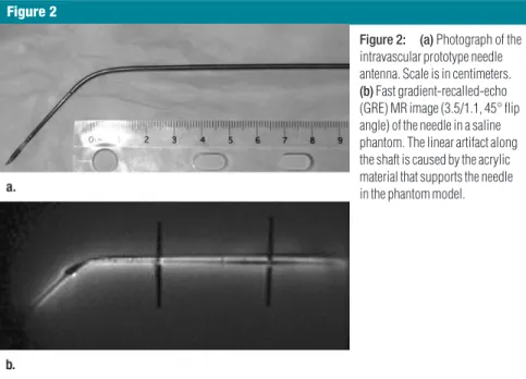
Figure 2
Figure 2: (a)Photograph of the intravascular prototype needle antenna. Scale is in centimeters. (b)Fast gradient-recalled-echo (GRE) MR image (3.5/1.1, 45° flip angle) of the needle in a saline phantom. The linear artifact along the shaft is caused by the acrylic material that supports the needle in the phantom model.
the needle was reintroduced into the femoral vein sheath to perform the sec-ond puncture.
A puncture was defined as advance-ment of the needle from the IVC to the SMV. A pass was defined as an attempt to puncture the SMV. In addition to success rates, the optimal imaging pa-rameters and the number of passes re-quired for each successful procedure were noted.
Results
Success Rates
MR imaging– guided puncture of the SMV was successfully performed in all six pigs. All procedures were performed with real-time MR imaging by using free-breathing techniques and without electrocardiographic gating. Punctures were performed with no change in car-diac rhythm or rate and with no se-quelae. Ten punctures were performed in six swine. All 10 procedures were successful (100%), with direct puncture of the SMV and no traversal of any ret-roperitoneal organs and/or vessels, as demonstrated with an electrocardio-graphically gated fast spin-echo double-inversion black-blood sequence (Fig 4). MR portograms were immediately
ob-tained after all punctures (Fig 5). Two punctures were made in four animals. After removal of the needle from the first puncture, delayed imaging did not demonstrate retroperitoneal bleeding in any of the pigs.
Imaging Parameters
Because of the mobility of the SMV, real-time imaging of all punctures was necessary to reorient the needle toward the SMV in order to enter the vessel. During real-time GRE sequences, four to eight frames per second were possi-ble throughout the procedure. Routine transverse imaging after each puncture helped confirm that the needle entered the SMV without traversing other retro-peritoneal structures (Fig 4).
Three standard views were used with the real-time sequence for each puncture. A sagittal view of the IVC (real-time GRE) was used to track the entry of the needle and provide direc-tionality and orientation of the needle (Fig 3a). A short transverse view of the abdomen (fast SPGR and real-time GRE) was used to identify the relation-ship of the needle to the IVC, SMV, and retroperitoneal structures (Fig 3b, 3c). Finally, an oblique sagittal view along the needle tip (real-time GRE) was used to monitor the path of the needle as it
exited the IVC and entered the SMV. During the procedure, the intervention-alist was able to immediately change any of these prescribed planes. In all punctures, an immediate return of blood was noted from the needle hub; this was followed by the obtaining of a confirmatory portogram (Fig 5). The typical duration of each puncture was approximately 10 – 40 minutes. This pe-riod began with the insertion of the nee-dle into the femoral vein and ended once the needle penetrated the SMV and the confirmatory portogram was obtained. Repeat thin-section imaging of this area after each puncture demon-strated no retroperitoneal bleeding.
Number of Passes
For each puncture attempt, it was nec-essary to perform an average of one to three separate passes from the IVC once the active needle was outside the IVC. With each pass, multiplanar im-aging helped confirm that retroperito-neal structures were not injured or traversed. Repeat delayed imaging in animals that underwent a second puncture showed no bleeding into the retroperitoneum, no vascular hemato-mas, and no change in orientation of the vasculature after removal of the intravascular needle.
Figure 3
Figure 3: Real-time puncture of SMV from IVC with intravascular needle. (a) Sagittal real-time GRE MR image (3.4/1.2, 45° flip angle). The needle is introduced through a femoral vein and oriented to the SMV. Arrowhead⫽ tip of needle, arrow ⫽ IVC. (b) Transverse real-time GRE MR image (4.4/1.3) shows needle being ad-vanced to the SMV. Arrowhead points to the needle, long arrow points to the IVC, and short arrow points to the SMV. (c) Transverse MR image obtained with fast spoiled gradient-recalled acquisition in the steady state (SPGR) (6.0/1.5, 60° flip angle) as needle enters the SMV; note indentation of needle on posterior wall of SMV.
Discussion
Because of the invasiveness of transca-val punctures and the potential for in-jury to various retroperitoneal struc-tures, we developed an active MR intravascular needle that can be com-pletely visualized in an MR imaging en-vironment and used to perform MR im-aging– guided extravascular punctures. In addition to monitoring the needle, we were able to visualize all viscera and pertinent vascular structures to per-form this procedure. The use of this needle, in combination with real-time and fast GRE MR imaging, enabled ade-quate temporal and spatial resolution so that active tracking of the needle during transcaval punctures was possible. The results of this study demonstrate that MR imaging– guided puncture of the SMV is feasible in a swine model.
Because of anatomic concerns, di-rect percutaneous puncture of the SMV or splenic vein currently cannot be per-formed safely with conventional radio-graphic guidance. Because of the ana-tomic variability in the relationship of these vessels to the IVC and the close proximity of adjacent vessels and or-gans (eg, aorta, superior mesenteric ar-tery, and pancreas), the advancement of a needle into the retroperitoneum cannot be performed solely under
con-ventional radiographic guidance. Both the portal and splenic veins lie in the anterior pararenal space of the retro-peritoneum, adjacent to the pancreas. The main trunk of the SMV is embed-ded in extraperitoneal fat within the mesentery and located anterior to these retroperitoneal structures. The IVC is
also located in the retroperitoneum, but posterior to the SMV, splenic vein, and portal venous system (5). Because these vessels are also surrounded by bowel, a conventional percutaneous approach is precluded. In addition, the portal, splenic, and SMV systems are isolated from the systemic circulation and
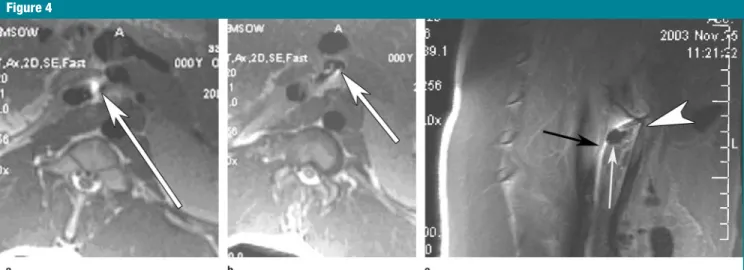
can-Figure 4
Figure 4: (a)Transverse fast spin-echo MR image (1900/4.5, 90° flip angle) shows that the intravascular needle (area of high signal intensity indicated by arrow) has exited the IVC. The SMV is located anteriorly, and the needle (arrow) is seen heading toward the SMV. (b) Transverse MR image (2000/18.0) shows the needle in the SMV (arrow). (c) Sagittal fast spin-echo MR image (2000/18, 90° flip angle) demonstrates the needle (black arrow) superior to a loop of bowel (white arrow) and puncturing the SMV (arrowhead). Notice that the SMV is displaced anteriorly with the puncture.
Figure 5
Figure 5: (a, b)MR Portograms obtained (a) in the vascular and parenchymal phases and (b) in the hepatic parenchymal phase after the administration of gadopentetate dimeglumine by using a projection image (fast SPGR, 6/1.3, 90° flip angle, 1.5 frames per second).
not be readily visualized with conven-tional contrast material injections.
Because of these anatomic con-straints, the percutaneous puncture of the mesenteric or splenic venous system has undergone limited study. Kaminou et al (6) first described a percutaneous approach to the splenic vein from the IVC by using only radiographic fluoros-copy. In a swine model, the portal vein was directly punctured; then, a wire basket was placed in the splenic vein to serve as a target. Next, a second needle puncture was performed from the IVC into the wire basket inside the splenic vein. With this procedure, a shunt was successfully created in four of five pigs. In all four successful punctures, how-ever, necropsy demonstrated that the needle and stent were placed through the pancreas. In addition to being fairly cumbersome and necessitating exten-sive fluoroscopy, the inability to visual-ize retroperitoneal structures limited the usefulness of this technique.
In a more recent study, Vivas et al (7) attempted to create an extrahepatic portal caval shunt in a canine model. With use of jejunal veins from the mes-entery that were exposed after a lapa-rotomy, the SMV and portal vein were catheterized. After placement of an-other catheter in the IVC, a blind punc-ture was made from the portal vein into the IVC. With this access, a covered prosthesis was placed to simulate a por-tal caval shunt. Because of the lack of visibility and poor placement of the prosthesis, however, six of the 10 ani-mals died after the procedure owing to substantial retroperitoneal bleeding that resulted from multiple vascular punctures. Both of these studies further reinforce the importance of visualizing the retroperitoneum during these inva-sive procedures.
In our study, all punctures were per-formed in a controlled fashion because all vasculature and viscera were readily visualized. The real-time GRE sequence enables rapid data acquisition with di-rect manipulation of section prescrip-tion, flip angle, section thickness, and field of view—all in a real-time setting.
With use of this system, adequate visu-alization of both the vasculature and vis-cera was possible without the need for contrast material. This system provided several advantages: (a) All vessels were completely visualized with the trans-verse imaging plane because of the T2 and/or T1 effects the GRE sequence has on blood; (b) rapid multiplanar capabil-ities enabled us to monitor the transca-val punctures; and (c) there was ade-quate temporal resolution to perform real-time manipulation of the needle. The use of this system provided sub-stantial improvement for obtaining mul-tiplanar views to identify a needle tra-jectory.
We did, however, notice difficulty in identifying vascular structures with the real-time GRE sequence alone. Because there was increased signal from other adjacent tissue, it was difficult to prop-erly identify the vasculature. Therefore, the adjunctive use of a transverse SPGR sequence with real-time temporal reso-lution helped properly identify the vas-culature for a proper needle trajectory. The limitations of this study are re-lated to safety. Because this is a first-generation device, radiofrequency safety issues have not yet been fully an-alyzed. Insulation is known to improve the possible radiofrequency safety prob-lems, and it is also possible to limit the delivered radiofrequency power level with the pulse sequences used. We plan to measure the safety index—the ex-pected temperature increase caused by the needle for a given unit of specific absorption rate distribution—and ad-just the power level accordingly (8). Fi-nally, although MR imaging did not de-pict any retroperitoneal bleeding or nontarget punctures, pathologic analy-sis of the vessels and retroperitoneum structures was not performed; there-fore, the safety of this procedure is not known.
In conclusion, we found that with use of only MR imaging guidance and an active MR intravascular needle system, we were repeatedly able to successfully puncture the SMV from the IVC in a highly controlled manner with direct
vi-sualization of all components including the needle, IVC, SMV, and surrounding abdominal organs. The multiplanar ca-pability of MR imaging, combined with its excellent soft-tissue resolution, en-abled us to identify a needle trajectory for direct, accurate punctures of the SMV.
Practical application: Because of
anatomic concerns, direct percutaneous puncture of the SMV or splenic vein currently cannot be performed with conventional radiographic guidance. MR imaging guidance enabled us to si-multaneously visualize and puncture the SMV. In addition, a minimally invasive means of accessing the portal, mesen-teric, and splenic venous system may create unique treatment opportunities such as the creation of a mesoportoca-val shunt or MR imaging– guided trans-vascular delivery of agents to target or-gans such as the liver, pancreas, or spleen.
References
1. Lardo AC. Real-time magnetic resonance imaging: diagnostic and interventional appli-cations. Pediatr Cardiol 2000;21(1):80 –98. 2. Ocali O, Atalar E. Intravascular magnetic
res-onance imaging using a loopless catheter an-tenna. Magn Reson Med 1997;37(1):112– 118.
3. Atalar E, Kraitchman DL, Carkhuff B, et al. Catheter-tracking FOV MR fluoroscopy. Magn Reson Med 1998;40(6):865– 872. 4. Serfaty JM, Atalar E, Declerck J, et al.
Real-time projection MR angiography: feasibility study. Radiology 2000;217(1):290 –295. 5. McLoughlin RF, Rankin RN. Portacaval space
anatomy: potential implications for percuta-neous portacaval shunts. J Vasc Interv Radiol 1996;7(5):761–767.
6. Kaminou T, Rosch J, Yamada R, et al. Percu-taneous retroperitoneal splenorenal shunt: an experimental study in swine. Radiology 1998; 206(3):799 – 802.
7. Vivas I, Bilbao JI, Martinez-Cuesta A, et al. Percutaneous extrahepatic portacaval shunt with covered prostheses: feasibility study. J Vasc Interv Radiol 2003;14(12):1543–1552. 8. Yeung CJ, Susil RC, Atalar E. RF safety of
wires in interventional MRI: using a safety index. Magn Reson Med 2002;47(1):187–193.