CT and MR characteristics of hibernoma: six cases
Memduh Dursun
a,⁎
, Ayaz Agayev
a, Baris Bakir
a, Harzem Ozger
b, Levent Eralp
b,
Mustafa Sirvanci
c, Koray Guven
a, Mehtap Tunaci
aa
Department of Radiology, Istanbul Faculty of Medicine, Istanbul University, 34390 Capa, Istanbul, Turkey
b
Department of Orthopaedics and Traumatology, Istanbul Faculty of Medicine, Istanbul University, 34390 Capa, Istanbul, Turkey
c
Department of Radiology, Faculty of Medicine, Florance Nighthingale Hospital, University of Kadir Has, 80220 Sisli, Istanbul, Turkey Received 10 June 2007; accepted 11 July 2007
Abstract
Purpose: The purpose of this study was to describe the computed tomography (CT) and magnetic resonance (MR) imaging findings of hibernoma. Materials and methods: We retrospectively evaluated imaging findings of CT and MR examinations of six patients (three men and three woman, aged 27–48 years) with histopathological diagnosis of hibernoma. Results: On CT examination, the lesions were slightly hyperdense, and on T1-and T2-weighted MR images, they were isointense or slightly hypointense compared to the subcutaneous fat. All of these lesions showed contrast enhancement and one out of the six lesions had internal linear septations. Conclusion: Hibernoma has a wide spectrum of CT and MR imaging findings, which should be considered in differential diagnosis, especially with other lipomatous lesions.
© 2008 Elsevier Inc. All rights reserved.
Keywords: Hibernoma; Computed tomography; Magnetic resonance imaging
1. Introduction
Hibernomas that were originally described in 1906 by Merkel [1], who named them “pseudolipomas,” are rare soft tissue tumors of brown fat origin. In 1914, Gery derived the name hibernoma from the tumor's histological similarity to brown fat in hibernating animals. They occur chiefly in adults and they are more commonly seen in the interscapular–periscapular area; in the neck; in axillary regions; and in the thigh, intrathoracic, and retroperitoneal regions[2,3].
Awareness of this entity and imaging appearances allow its inclusion in the differential diagnosis. In the past decade, multiple case reports and two series have described computed tomography (CT) or magnetic resonance (MR) imaging characteristics of these tumors[3–9]. The purpose
of our study was to describe imaging findings of hibernoma on CT and MR imaging modalities in six patients.
2. Materials and methods 2.1. Patient population
We retrospectively reviewed imaging findings in six patients (three men and three woman aged 27–48 years; mean age, 35 years) with histopathological diagnosis of hibernoma. All patients were examined over the past 10 years in two institutions. A patient underwent CT and MR examinations, four patients underwent MR imaging, and one patient underwent only CT examination prior to operation. Histopathological diagnosis was made by the detection of typical hibernoma cells, characterized by multiple large multivacuolations, granular cytoplasm, and eccentric vesicular nuclei with single prominent nucleoli on light microscopy by core needle biopsy in all patients. All six patients underwent curative excisional biopsy with marginal borders.
⁎ Corresponding author. Department of Radiology, Istanbul Faculty of Medicine, Istanbul University, Capa 34390, Istanbul, Turkey. Tel.: +90 212 533 75 05; fax: +90 212 631 0728.
E-mail address:[email protected](M. Dursun).
0899-7071/08/$– see front matter © 2008 Elsevier Inc. All rights reserved. doi:10.1016/j.clinimag.2007.07.001
2.2. CT and MR Imaging
CT examination was performed on a four-detector row CT and a spiral CT scanner. In one patient, pre- and postcontrast images and, in the other patient, only none-nhanced images were obtained in CT examinations. T1-weighted echo, axial–coronal, T2-weighted spin-echo axial, short tau inversion recovery (STIR) axial, and contrast-enhanced T1 weighted and/or fat-suppressed axial and coronal images were obtained on 1- and 1.5-Tesla scanners—two different MR scanners with different TR, TE, field of view, and matrix.
2.3. Image interpretation
CT and MR imaging characteristics were evaluated by three experienced musculoskeletal radiologists (M.D., A.A.,
M.S.), with consensus on imaging findings. Radiological features assessed included lesion size, location, and internal structure. CT attenuation and MR signal characteristics of T1-weighted, T2-weighted, and STIR images were evaluated and compared to subcutaneous fat, and contrast enhance-ment patterns were noted. The patients' age and sex were also recorded.
3. Results
The location of the lesions was evaluated. In four patients, the lesion was located on the thigh: three of them at the intermuscular zone, and one, intramuscularly. In one patient, the lesion extended from the pelvis to the gluteal region through the sciatic notch and the mass displaced rectum laterally (Fig. 1). In another patient, the lesion was located in the right paravertebral muscle group (Fig. 2). The smallest lesion, measuring 2×4×7 cm, was located at the iliopsoas muscle (Fig. 3); the biggest lesion, which was extending from the pelvis to the gluteal region, measured 8×9×18 cm. Fig. 1. Patient 1. (A) On contrast-enhanced, fat-suppressed T1-weighted
axial image, the mass (arrowheads) extended to the pelvis through the sciatic notch and displaced rectum laterally and shows slightly contrast enhance-ment. (B) T1-weighted coronal image shows the mass in the right gluteal region extending trough the sciatic notch to the pelvis (arrowheads). U, uterus; O, ovary.
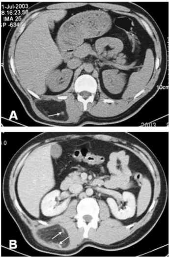
Fig. 2. Patient 2. (A) On unenhanced CT images, the lesion is slightly hyperdense compared with subcutaneous fat; also, vascularity is detected inside the mass (arrow). (B) On postcontrast examinations, the lesion shows enhancement and vascular structures are also seen (arrows).
In two patients who underwent CT examination, density of the lesions was higher than subcutaneous fat (Figs. 2 and 3). Contrast enhanced images were obtained in one of two patients in whom the lesion showed slight enhancement, and linear septations with internal vasculature were noted (Figs. 2B and 3D).
On T1-weighted MR images, signal intensities were slightly hypointense compared to subcutaneous fat in three patients (Figs. 4A and 5A), hypointense in one patient (Fig. 3A), and isointense in another patient. On T2-weighted images, the lesions' signal intensities were isointense with subcutaneous fat in three patients (Figs. 1B and 6A) and slightly hypointense in two patients (Figs. 3B and 4B). On STIR sequences, the lesions were slightly hyperintense in three patients (Figs. 5B and 6B), dominantly hypointense with some hyperintense regions in one patient, and hyperintense in one patient (Fig. 1A). In contrast-enhanced T1-weighted sequences, three lesions showed significant contrast enhancement, and two showed slight enhancement (Figs. 1A and 5B).
The internal structure of four lesions showed linear hypointense septations (Figs. 1B, 1B, 4A, 5A, and 6A). In two patients, contrast-enhanced CT examination and fat-suppressed, contrast-enhanced MR images showed promi-nent vascular structures within the septations of the lesions (Figs. 2B and 5B). Only one of six patients demonstrated heterogeneous internal structure in CT and MR images Fig. 3. Patient 3. (A) T1-weighted axial image shows a heterogenous
hypointense mass which located in the left iliopsoas muscle (arrow). (B) T2-weighted axial image shows a heterogenous slightly hypointense mass (arrow). (C) On postcontrast examination, T1-weighted, fat-suppressed axial sequence demonstrates significant contrast enhancement (arrow). (D) On unenhanced CT examination, a mass lesion, hyperdense compared with subcutaneous fat (arrow).
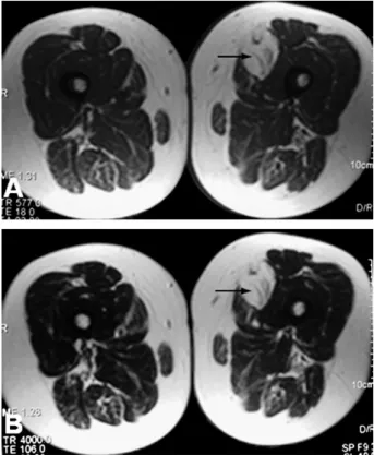
Fig. 4. Patient 4. (A) T1-weighted axial image shows a mass which isointense with subcutaneous fat also containing internal septations (arrow). (B) T2-weighted axial image shows the same imaging findings of the T1-weighted sequence (arrow).
(Fig. 3).Table 1summarizes CT and MR imaging findings of the patients.
4. Discussion
Hibernoma is a rare, benign soft tissue tumor of brown fat origin. The name hibernoma was derived from the tumor's histological similarity to the brown fat in hibernating animals
[3]. It is well accepted that two types of adipose tissue exist: white adipose and brown adipose. Brown adipose tissue was first described by Wilsh in 1670[10,11]. Although the exact function of brown fat is uncertain, it is believed to be important in nonshivering thermogenesis in hibernating animals and in the newborn. In humans, brown fat markedly decreases after 8 weeks of life, although small quantities remain in all age groups[11]. When brown fat is confluent over a large region and associated with hypervascularity and arteriovenous shunting microscopically and with imaging studies, this is typical for hibernoma[4]. Hibernomas seem to form in the areas with remaining brown fat, such as the scapular region, neck, axilla, chest wall, mediastinum, perirenal areas, thigh, buttock, popliteal fossa, scalp, breast, periureteric region, and scrotum [11–13]. In our cases, in four patients, the lesion was localized at the thigh; in one patient, at the back; and in one patient, extending from the pelvis to the gluteal region through the sciatic notch.
Hibernomas have occurred in almost all age groups, with a peak incidence in the third decade. Several studies have shown a slight female predilection [11]. Hibernomas generally present as a slow-growing, pliable, subcutaneous mass, which may be mobile and is usually painless. Localized warmth over the mass may be apparent due to its hypervascular nature[3]. In our study, five of six patients presented with a slow-growing painless mass, and 1 patient presented with sciatalgia due to the extension of the lesion through the sciatic notch.
Core needle biopsy has not been recommended in cases of suspected hibernoma due to the tumor's hypervascularity
[3,11], and at least one case report documents excessive
bleeding during a percutaneous biopsy of a hibernoma that resulted in termination of the procedure[3,14]. In our study, core needle biopsies were performed in all patients preoperatively, and no bleeding complication was noted in
Fig. 6. Patient 6: (A) T2-weighted axial image shows a mass with internal septations slightly hypointense compared with subcutaneous fat. (B) In STIR coronal image, the mass is slightly hyperintense compared to subcutaneous fat.
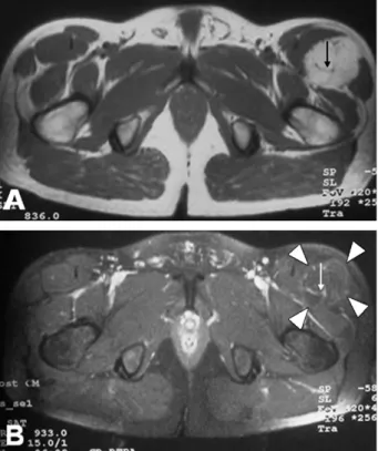
Fig. 5. Patient 5. (A) On T1-weighted axial images, septations (arrow) are seen on the mass located at the anterior portion of the thigh. (B) Fat-suppressed postcontrast image shows signal loss (arrowheads) and slight contrast enhancement of the mass, and vascularity is shown in the internal septations (arrow).
any patient. Complete excision is generally regarded as curative, although local recurrence has been noted after incomplete surgical resection[3].
Radiographic, sonographic, angiographic, and scinti-graphic appearances of this tumor have been described. Radiographs may show a mass with radiolucency consistent with a lipomatous neoplasm. No underlying osseous involvement is present[11]. The sonographic appearance of hibernoma has been a uniformly hyperechoic mass[15]. At angiography, these tumors have rich vascularity and occa-sional arteriovenous shunting[15,16]. Hibernomas have also been detected incidentally on technetium 99m tetrofosmin myocardial perfusion studies performed in patients with chest pain, appearing as mass-like areas of increased activity. Active uptake has also been reported as an F-18 fluorodeox-yglucose positron emission tomography finding[17].
On precontrast CT, a low-attenuation mass with septae is characteristic. After intravenous contrast administration, enhancement is seen within the septae and may also be present more diffusely within the mass[11]. In our series, one of the two patients who underwent CT examination had a hyperdense lesion compared to the subcutaneous fat in precontrast images; in contrast-enhanced images, the lesion showed slight enhancement and showed internal septations. In the second patient, the lesion was slightly hyperdense, compared to subcutaneous fat in precontrast images, and had a heterogeneous internal structure.
On MR images, high T1-weighted signal intensity has been the most commonly reported finding, although inter-mediate signal intensity has also been reported[15]. In our cases, three lesions were isointense with subcutaneous fat, and two were slightly hypointense. On T2-weighted images, most authors agree that the lesion is nearly isointense with subcutaneous fat, although hypointensity has been noted[5–9,11,18]. In our patients, 4 lesions were isointense with subcutaneous fat, and one was hypointense, which are concordant with literature. Mild heterogeneity and internal septations were noted on many sequences, which are probably due to vascular tissue intermixed with the fatty components of the tumor[3]. In 5 patients, we noted the presence of internal
septations, and two of these showed vascularity in the septations. In one patient, the lesion had a heterogenous internal structure, and no septations were noted. One previous case also reported diffuse hyperintensity throughout the lesion with STIR images[7]. In our cases, the STIR sequence gave the most variable results. In one case, the lesion was significantly hypointense; in three cases, slightly hyper-intense; and in one case, the lesion contained hypo- and hyperintense regions. Although it is not diagnostically important, a previous study shows that the MR signal char-acteristics vary with cellular composition and vascularity[9]. Contrast enhancement is usually present[5], although in one case, no contrast enhancement was evident on either CT or MR imaging[18]. Four of our cases showed significant enhancement, and two cases showed slight enhancement.
The differential diagnosis in hibernoma includes benign processes such as angiolipoma, spindle cell lipoma, pleomorphic lipoma, lipoblastoma, and elastofibroma, as well as malignant processes including liposarcoma, clear cell sarcoma, and alveolar soft part sarcoma[4].
5. Conclusion
Awareness of hibernoma and its imaging appearances is important because of its hypervascular nature, and core needle biopsy should be avoided. Hibernoma should be considered in the differential diagnosis of clinically slow-growing, painless masses which are slightly hyperdense compared to the subcutaneous fat in CT examination and isointense or slightly hypointense compared to the sub-cutaneous fat in T1- and T2-weighted MR images, showing contrast enhancement and containing linear septations on CT or MR examination.
References
[1] Merkel H. On pseudolipoma of the breast. Beitr Pathol Anat 1906;39: 152–7.
[2] Weiss SW, Goldblum JR. Benign lipomatous tumors. In: Weiss SW, Goldblum JR, editors. Soft tissue tumors. 4th ed. St. Louis (MO): Mosby, 2001. pp. 571–640.
Table 1
CT and MR imaging findings Patient no./
age (y)/sex
Clinical
history Location Size (cm) CT density
T1-weighted signal T2-weighted signal Fat suppression STIR Contrast enhancement Internal structure 1/34/female Pain, sciatalgia Pelvis 8×9×18 – Slightly hypointense
Isointense Yes Hypo-slightly hyperintense Slightly Linear septation 2/29/male Painless swelling Back 4×7×9 Slightly hyperdense – – – – Yes Linear septation 3/27/male Painless swelling
Thigh 2×4×7 Hyperdense Hypointense Slightly hypointense
Yes Hyperintense Yes Slightly
heterogeneous 4/38/female Painless swelling Thigh 3×4×8 – Slightly hypointense Slightly hypointense Yes Slightly hyperintense Yes Linear septation 5/48/female Painless swelling
Thigh 4×4×8.5 – Isointense Isointense Yes Slightly
hyperintense Yes Linear septation 6/35/male Painless swelling Thigh 5×9×15 – Slightly hypointense
Isointense Yes Slightly hyperintense
Slightly Linear septation
[3] Kallas KM, Vaughan L, Parviz H, Resnick D. Hibernoma of the left axilla; a case report and review of MR imaging. Skeletal Radiol 2003;32:290–4.
[4] Anderson SE, Schwab C, Stauffer E, Banic A, Steinbach LS. Hibernoma: imaging characteristics of a rare benign soft tissue tumor. Skeletal Radiol 2001;30:590–5.
[5] Seynaeve P, Mortelmans L, Kockx M, Van Hoye M. Case report 813. Hibernoma of the left thigh. Skeletal Radiol 1994;23:137–8. [6] Deseran MW, Seeger LL, Doberneck SA, Eckardt JJ. Hibernoma of the
right gracilis muscle. Skeletal Radiol 1994;23:301–2.
[7] Atilla S, Eilenberg SS, Brown JJ. Hibernoma: MRI appearance of a rare tumor. Magn Reson Imaging 1995;12:335–7.
[8] Peer S, Kuhberger R, Dessl A, Judmaier W. MR imaging findings in hibernoma. Skeletal Radiol 1997;26:507.
[9] Ritchie DA, Aniq H, Davies AM, et al. Hibernoma—correlation of histopathology and magnetic-resonance-imaging features in 10 cases. Skeletal Radiol 2006;35:579–89.
[10] Rasmussen AT. The so-called hibernating gland. J Morphol 1923;38: 147–205.
[11] Alvine G, Rosenthal H, Murphey M, Huntrakoon M. Hibernoma. Skeletal Radiol 1996;25:493–6.
[12] Seemayer TA. On the ultrastructure of hibernoma. Cancer 1975;36: 1785–93.
[13] Kindblom LG. Intermuscular intramuscular lipomas and hibernomas, a clinical, roentgenologic, histologic and prognostic study of 46 cases. Cancer 1974;33:754.
[14] Lung RJ, Lapidus S, Miller SH, Graham WP. Hibernoma: report of two cases. J Surg Oncol 1977;9:563–6.
[15] Baskurt E, Padgett DM, Matsumoto JA. Multiple hibernomas in a 1-month-old female infant. AJNR Am J Neuroradiol 2004;25:1443–5. [16] Balestreri L, Canzonieri V. Case report: axillary hibernoma: radi-ological and pathradi-ological findings of a rare tumor. Clin Radiol 1998;53:853–5.
[17] Chatterton BE, Mensforth D, Coventry BJ, Cohen P. Hibernoma: intense uptake seen on Tc-99m tetrofosmin and FDG positron emission tomographic scanning. Clin Nucl Med 2002;27:369–70.
[18] Cook MA, Stern M, de Silva RD. MRI of hibernoma. J Comput Assist Tomogr 1996;20:333–5.