The Azoarcus Group I Intron Ribozyme Misfolds and Is
Accelerated for Refolding by ATP-dependent RNA Chaperone
Proteins
*
□SReceived for publication, July 29, 2011, and in revised form, August 26, 2011Published, JBC Papers in Press, August 30, 2011, DOI 10.1074/jbc.M111.287706
Selma Sinan‡§1, Xiaoyan Yuan‡, and Rick Russell‡2
From the‡Department of Chemistry and Biochemistry, Institute for Cellular and Molecular Biology, University of Texas at Austin, Austin, Texas 78712 and the§Balıkesır University, Science and Art Faculty, Department of Biology, Molecular Biology,
Balıkesır 10000, Turkey
Background:Group I introns are valuable for studying RNA folding and chaperone proteins.
Results:A catalytic activity assay was developed and used to demonstrate two prominent phases for Azoarcus ribozyme folding. The slow phase displays hallmarks of a misfolded intermediate.
Conclusion:This RNA accumulates a misfolded intermediate and interacts productively with RNA chaperones. Significance:Delineating misfolding and chaperone roles is crucial for understanding how cellular RNAs fold.
Structured RNAs traverse complex energy landscapes that include valleys representing misfolded intermediates. In Neuro-spora crassa and Saccharomyces cerevisiae, efficient splicing of mitochondrial group I and II introns requires the DEAD box proteins CYT-19 and Mss116p, respectively, which promote folding transitions and function as general RNA chaperones. To test the generality of RNA misfolding and the activities of DEAD box proteins in vitro, here we measure native folding of a small group I intron ribozyme from the bacterium Azoarcus by mon-itoring its catalytic activity. To develop this assay, we first meas-ure cleavage of an oligonucleotide substrate by the prefolded ribozyme. Substrate cleavage is rate-limited by binding and is readily reversible, with an internal equilibrium near unity, such that the amount of product observed is less than the amount of native ribozyme. We use this assay to show that approximately half of the ribozyme folds readily to the native state, whereas the other half forms an intermediate that transitions slowly to the native state. This folding transition is accelerated by urea and increased temperature and slowed by increased Mg2ⴙ
concen-tration, suggesting that the intermediate is misfolded and must undergo transient unfolding during refolding to the native state. CYT-19 and Mss116p accelerate refolding in an ATP-depen-dent manner, presumably by disrupting structure in the inter-mediate. These results highlight the tendency of RNAs to mis-fold, underscore the roles of CYT-19 and Mss116p as general RNA chaperones, and identify a refolding transition for further dissection of the roles of DEAD box proteins in RNA folding.
Structured RNAs participate in diverse cellular processes, from protein synthesis and regulation to the maintenance of chromosome ends. To achieve their functional structures, RNAs traverse complex folding landscapes that typically include multiple intermediates and alternative structures. Indeed, nearly every RNA that has been studied in vitro has been shown to populate alternative, misfolded structures at equilibrium and/or as kinetic intermediates during folding (1–3). In vivo, nearly all processes that involve structured RNAs also require ATP-dependent RNA helicases, the largest group of which is the DEAD box proteins (4). These proteins are pro-posed to use ATP binding and hydrolysis to disrupt RNA struc-tures, thereby accelerating folding transitions and conforma-tional changes (5–7).
Group I and II introns have been particularly valuable for studies of RNA folding. These RNAs include multiple domains that fold locally and assemble into structures that catalyze exci-sion of the intron from the precursor RNA. They are apparently prone to misfolding in vivo, because the DEAD box proteins CYT-19 and Mss116p are required for efficient folding and splicing of multiple introns in the mitochondria of Neurospora
crassaand Saccharomyces cerevisiae, respectively (8, 9). The
relative simplicity and tractability of intron RNAs have facili-tated detailed studies of RNA folding and RNA chaperone activity by DEAD box proteins (3, 5).
In particular, the folding of group I introns and their ribozyme derivatives has been dissected using biophysical and biochemical approaches (2, 3, 10). When folding is initiated by the addition of Mg2⫹, RNAs fold from extended conformations
that possess secondary structure but lack defined tertiary struc-ture to give compact, highly ordered strucstruc-tures. A ribozyme derived from the Tetrahymena thermophila group I intron was shown by chemical footprinting and small angle x-ray scatter-ing (SAXS)3to fold through a series of intermediates, becoming compact and structured on time scales ranging from millisec-*This work was supported, in whole or in part, by National Institutes of Health
Grant GM70456 (to R. R.). This work was also supported by Welch Founda-tion Grant F-1563 (to R. R.).
□S The on-line version of this article (available at http://www.jbc.org) contains
supplemental Table S1 and Figs. S1–S3.
1Supported by a fellowship from the Scientific and Technological Research
Council of Turkey.
2To whom correspondence should be addressed: Dept. of Chemistry and
Biochemistry, 1 University Station A4800, University of Texas at Austin, Austin, TX 78712. Tel.: 512-471-1514; Fax: 512-232-3432; E-mail: rick_ [email protected].
3The abbreviations used are: SAXS, small angle x-ray scattering; MOPS,
3-(N-morpholino)-propanesulfonic acid.
by guest on August 7, 2019
http://www.jbc.org/
Group I introns have also been used to probe the mecha-nisms of DEAD box proteins as general RNA chaperones. Early studies showed that CYT-19 promotes a conformational tran-sition in a mutant of the Tetrahymena ribozyme (8), and later work showed that CYT-19 accelerates folding transitions of the ribozyme, including refolding of the long-lived misfolded con-formation to the native state, by disrupting structure without specifically recognizing misfolded structure (20, 21). This non-specific activity nevertheless gives accumulation of the native state because it is more stable and unfolded by CYT-19 less efficiently (21, 22). This basic mechanism has been suggested to underlie much of the general RNA chaperone activity by DEAD box proteins (5, 23, 24), but DEAD box protein-assisted folding has been studied for only a relatively small number of RNAs (24 –33), and gaps in our understanding of the intrinsic folding processes for some of these RNAs limit the mechanistic insights into the roles of DEAD box proteins.
A second group I intron that has been studied extensively is from a tRNA gene in the bacterium Azoarcus. The Azoarcus intron is particularly interesting because it has a relatively sim-ple architecture with minimal peripheral structure (see Fig. 1), yet it forms the canonical group I intron core structure with high stability and carries out self-splicing efficiently (34, 35). SAXS and footprinting studies indicated that this RNA acquires structure considerably faster than the Tetrahymena ribozyme, with fewer detectable intermediates. Indeed, in early studies the ribozyme appeared to fold to completion in milliseconds (36, 37), although further work has indicated transient formation of intermediates for at least a fraction of the population (38 – 40). Here we develop and use a catalytic activity assay to study the folding kinetics of the Azoarcus ribozyme, complementing work using physical approaches. We find that approximately half of the population folds readily to the native state, whereas the other half misfolds. Refolding to the native state is on the time scale of an hour at 25 °C and is accelerated by the DEAD box proteins CYT-19 and Mss116p. This RNA and its mis-folded conformation should be useful in efforts to understand how RNAs misfold and how DEAD box proteins function as RNA chaperones.
EXPERIMENTAL PROCEDURES
Preparation of RNA—The L-3 Azoarcus ribozyme was
tran-scribed in vitro from a plasmid that includes a promoter for T7 RNA polymerase immediately upstream from the coding sequence and an EarI restriction site adjacent to the 3⬘ termi-nus. The plasmid was digested to completion with EarI, and in
vitrotranscription was performed as described (41). The RNA
transcript was purified using a Qiagen RNeasy column as described (16). Substrate and product oligonucleotides were
binding protein and purified as described (23, 27, 42). After purification, proteins were dialyzed against storage buffer (20 mMTris-Cl, pH 7.5, 500 mMKCl, 1 mMEDTA, 0.2 mMDTT,
50% glycerol), divided into aliquots, flash frozen, and stored at ⫺80 °C.
Protein concentrations were determined by the Bradford assay (Bio-Rad) using bovine serum albumin as a standard. The values were in good agreement with absorbance measurements at 280 nm using extinction coefficients calculated from the amino acid sequences (data not shown).
General Kinetics Methods—For experiments measuring
sub-strate cleavage and ligation, the ribozyme was prefolded at 37 °C (50 mMNa-MOPS, pH 7.0, 10 mMMgCl2, 100M
guanos-ine). Cleavage reactions were then initiated at 25 °C by adding 2-fold excess substrate over the ribozyme, including trace32
P-labeled substrate. For reactions with prebound substrate ( sup-plemental Fig. S1C), the prefolding step included the substrate but not guanosine, and the incubation was 60 min to ensure complete binding. Guanosine was then added at 25 °C to initi-ate the cleavage reaction.
Reaction time points (2l) were quenched by adding two volumes of 90% formamide, 100 mMEDTA solution with 0.01%
(w/v) bromphenol blue, and 0.01% (w/v) xylene cyanol. Radio-labeled substrate and product were separated by 20% polyacryl-amide, 7 M urea gel electrophoresis; quantitated by using a
phosphorimaging device; and analyzed by ImageQuant (GE Healthcare). The results are presented as the averages and standard errors of at least two independent determinations.
Continuous Catalytic Activity Assay—To initiate the folding
and substrate cleavage reactions simultaneously, the ribozyme was added to a solution that included substrate and Mg2⫹. Final concentrations were 0.4Mribozyme, 0.8Msubstrate
includ-ing trace32P-labeled substrate, and 10 m
MMg2⫹(50 mM
Na-MOPS, pH 7.0, 100Mguanosine, 25 °C). Portions of the reac-tion (2l) were withdrawn at various times and quenched and processed as described above.
Discontinuous Catalytic Activity Assay—Folding reactions
were performed essentially as described (18). Solution condi-tions for stage 1 (folding) were 50 mMNa-MOPS, pH 7.0, 10 mM
MgCl2, and 1.6Mribozyme at 25 °C unless otherwise
indi-cated. At various times, portions of the reaction were added to stage 2 to measure the fraction of native ribozyme by catalytic activity. The conditions for stage 2 were 50 mMNa-MOPS, pH
7.0, 50 mMMgCl2, and 100Mguanosine at 25 °C. Under these
conditions, substrate cleavage is nearly 100-fold faster than the slow phase of folding (see Figs. 2B and 6C), allowing the cleav-age reaction to give a “snapshot” of the fraction of native ribozyme at the time of transfer to stage 2. Control experiments showed that using 10-fold higher guanosine concentration (1
by guest on August 7, 2019
http://www.jbc.org/
mM) gave modestly larger bursts at all folding times, as expected
from the results in Fig. 3, but did not affect the measured rate constants for native state formation (data not shown). For reac-tions with CYT-19 or Mss116p, the protein was destroyed prior to the measurement of catalytic activity by the inclusion of pro-teinase K (1 mg/ml) in stage 2.
For all of the reactions, the substrate was added in 2-fold excess (0.8 Msubstrate, 0.4 Mribozyme). In early
experi-ments, complete time courses were collected to determine the rate constant and amplitude of the rapid phase of substrate cleavage. In later experiments, a single time point was taken at 15 min. This time is sufficient to allow completion of the rapid phase while minimizing the contribution from the slower phase, which reflects further accumulation of native ribozyme and multiple catalytic turnovers. Time points were quenched and processed as above.
RESULTS
Development of a Catalytic Activity Assay for Folding—To
use catalytic activity to follow Azoarcus ribozyme folding quan-titatively, it was necessary to determine the catalytic properties of the prefolded ribozyme (18). Lacking exons, the ribozyme binds and cleaves a 9-nucleotide substrate that mimics the 5⬘ splice site (Fig. 1). We sought to identify conditions under
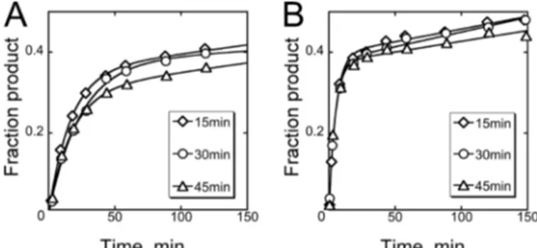
which substrate cleavage is rapid and could therefore reflect the progress of folding. Further, we sought to understand how the burst amplitude in a substrate cleavage reaction, i.e. the amount of substrate cleaved rapidly before slow product release limits subsequent turnovers, relates to the amount of native ribozyme. As a starting point, we prefolded the ribozyme with 10 mMMg2⫹for various times at 37 °C (15– 45 min) and then
initiated reactions at 25 °C by adding a 2-fold excess of the sub-strate. A fraction of the substrate was cleaved in minutes, with a rate constant of 0.08⫾ 0.03 min⫺1(Fig. 2A), in a reaction that was rate-limited by substrate binding (supplemental Fig. S1).4
The progress curves did not depend on preincubation time across the range tested, suggesting that ribozyme folding is complete within 15 min at 37 °C.
Based on previous results with other group I and II introns, we also tested higher Mg2⫹concentration with the idea that it could increase the rate of the reaction without also increasing the rate of folding and would therefore be useful in folding experiments. Indeed, reactions at 50 mMMg2⫹, again after pre-folding at 37 °C and 10 mMMg2⫹, gave an observed rate
con-stant for cleavage of 0.29⫾ 0.05 min⫺1(Fig. 2B),⬃4-fold larger than that at 10 mMMg2⫹. Therefore, we used 50 mMMg2⫹for substrate cleavage in most subsequent experiments.
The burst of cleaved substrate corresponded to 52⫾ 4% of the ribozyme population at 10 mMMg2⫹and 60⫾ 2% at 50 mM
Mg2⫹after accounting for the 2-fold excess of substrate (Fig. 2).
The ratio of product to ribozyme did not depend on the ribozyme and substrate concentrations (supplemental Fig. S1),
4We found that the rate constant for this burst of substrate cleavage
increased linearly with substrate concentration. The dependence of the rate constant on concentration gave a (kcat/KM)Svalue of 6.5⫻ 104M⫺1
min⫺1(supplemental Fig. S1A), similar to a value determined previously for reactions at 20 °C and 15 mMMg2⫹(57), and this rate constant was con-firmed by single turnover measurements across a wider range of ribozyme concentrations (supplemental Fig. S1B). The rate-limiting step under these conditions is substrate binding, because reactions in which substrate was preincubated with the ribozyme and then substrate cleavage was initiated by guanosine addition gave substantially larger rate constants ( supple-mental Fig. S1C).
FIGURE 1. Azoarcus ribozyme construct. All of the experiments herein used the L-3 ribozyme construct and its 9-nucleotide substrate, as shown (35). Within the ribozyme-substrate complex, paired regions P1 to P10 are labeled, and two canonical tetraloop-receptor tertiary interactions are indicated by arrows between the loop L2 and helix P8 and between L9 and P5. The oligo-nucleotide substrate is shown in lowercase letters, and the site of cleavage is indicated by an arrow from a G, representing the guanosine nucleophile.
FIGURE 2. Prefolding of the Azoarcus ribozyme. A, the ribozyme (0.4M) was folded at 37 °C and 10 mMMg2⫹for 15, 30, or 45 min, and then substrate (0.8M) was added at 25 °C and 10 mMMg2⫹. The plot shows the time course of substrate cleavage monitored by the appearance of product. Multiple independent determinations gave average rate constants of 0.08 min⫺1and amplitudes of 0.26, with no systematic dependence of either parameter on preincubation time. B, substrate cleavage reaction at 25 °C and 50 mMMg2⫹ after prefolding ribozyme as in A. Multiple determinations gave average rate constants of 0.29 min⫺1and amplitudes of 0.30, again with no systematic dependence on preincubation time. To calculate the ratio of product to ribozyme (see “Results”), the amplitudes were multiplied by two to account for the 2-fold excess of substrate over the ribozyme.
by guest on August 7, 2019
http://www.jbc.org/
indicating that the ribozyme was fully bound to the substrate. The substoichiometric product formation could indicate that a fraction of the ribozyme was inactive, because it was either damaged or misfolded. Alternatively or in addition, it was pos-sible that the cleavage products were religated faster than they were released from the ribozyme. This behavior would give ref-ormation of substrate and guanosine and fref-ormation of an inter-nal equilibrium between substrate and products (KINTin Fig.
3A) (43, 44), reminiscent of the self-splicing reaction by the intact intron (34). In this case, the amount of product could be substantially less than the amount of active ribozyme. To deter-mine the amount of native ribozyme from the amount of prod-uct produced, it would be necessary to determine this internal equilibrium value.
Therefore, we first varied the guanosine concentration in cleavage reactions with the idea that, if substrate cleavage and ligation reach equilibrium and guanosine binding is readily reversible, lower guanosine concentrations will result in smaller product bursts because incomplete guanosine binding will shift the equilibrium toward the substrate (Fig. 3A). Indeed, we found a strong dependence of the burst amplitude on guanosine concentration (Fig. 3B). There was little or no effect on the rate constant, as expected for rate-limiting substrate binding. We tested this model further by generating a large burst by using high guanosine concentration and then diluting the reaction to a lower guanosine concentration. As predicted from this model, upon dilution we observed a re-equilibration with rapid formation of the substrate and loss of the product (Fig. 3C).
Finally, to provide additional constraints for global modeling of the cleavage and ligation reactions and determination of the value of the internal equilibrium, we performed a series of
reac-tions analogous to those in Fig. 3B but starting from the prod-ucts (0.8 Mof each product, 2-fold in excess of ribozyme).
These reactions gave bursts of substrate formation, and as expected, the bursts were smaller with high guanosine concen-trations and largest in the absence of added guanosine (Fig. 3D). Analogous reactions at 10 mMMg2⫹are shown in
supplemen-tal Fig. S2.
All of the results in Fig. 3 andsupplemental Fig. S2were fit using global models, with one set of rate and equilibrium con-stants for reactions at 50 mMMg2⫹and a second set for
reac-tions at 10 mMMg2⫹(supplemental Table S1). The value of the
internal equilibrium is approximately unity at 10 mM Mg2⫹
(1.2) and rises to 2.1 at 50 mMMg2⫹, accounting for the larger
bursts of product formation at 50 mMMg2⫹(Fig. 2). To
calcu-late the amount of native ribozyme from the amount of product produced, it is necessary to account for the ribozyme that is active but remains substrate-bound as this internal equilibrium is reached. Multiplying the burst amplitudes for prefolded ribozyme with saturating guanosine concentration (Fig. 3B and supplemental Fig. S2A) by the correction factor ((Kint⫹ 1)/Kint) gives calculated values of active ribozyme that are the same within error as the total amount of ribozyme.
Together, the results above indicate that the ribozyme folds to completion in less than 15 min at 37 °C and that most or all of the ribozyme is present in the native state at equilibrium under these conditions. This result indicates that the native confor-mation is more stable than any accessible misfolded conforma-tions. In the sections below, we use the information on the catalytic reaction and equilibrium folding to measure the kinet-ics of native ribozyme folding.
Monitoring Ribozyme Folding by Catalytic Activity—To
measure the time dependence of native ribozyme formation FIGURE 3. Substrate cleavage and ligation by prefolded ribozyme. A, schematic of reaction steps used in modeling results in subsequent panels. KINT
represents the internal equilibrium between substrate cleavage and ligation. E, Azoarcus ribozyme; G, guanosine; S, ribozyme substrate CAUACGGCC; 5⬘P, reaction product CAU; 3⬘P, reaction product GACGGCC; S, substrate CAUACGGCC. B, varying guanosine concentration in substrate cleavage reactions at 25 °C, 50 mMMg2⫹after prefolding the ribozyme as above. Guanosine was absent (red) or present at 1M(blue), 2M(orange), 10M(cyan), 50M(green), 200M
(pink), or 1000M(black). C, dilution of guanosine (10-fold, circles) after allowing formation of internal equilibrium for substrate cleavage reactions. Initial guanosine concentrations are 100M(purple) or 200M(pink). Also shown are results from parallel reactions in which the guanosine concentration was held constant during the 10-fold dilution (triangles). D, ligation reactions starting from products (25 °C, 50 mMMg2⫹). Guanosine concentrations are shown using the same colors as in B. The curves in B–D reflect global fits using Kinetic Explorer (Ref. 62; seesupplemental Table S1for rate and equilibrium constants) (Refs. 63 and 64). Reactions equivalent to those in B and D but performed at lower Mg2⫹concentration (10 mM) are shown insupplemental Fig. S2. Reactions analogous to those in B andsupplemental Fig. S2Abut with the substrate prebound to the ribozyme are shown insupplemental Fig. S1.
by guest on August 7, 2019
http://www.jbc.org/
using catalytic activity, we first performed experiments analo-gous to those above but without a folding preincubation. Thus, we added substrate simultaneously with Mg2⫹, so that
ribozyme folding and substrate cleavage would occur in the same reaction (termed a continuous assay; Ref. 18). If the entire ribozyme population folded to the native state with at least as large a rate constant as the substrate is cleaved by the folded ribozyme, the progress of substrate cleavage would be identical with or without a folding preincubation. On the other hand, if some of the ribozyme did not reach the native state rapidly, less substrate would be cleaved rapidly in the reaction without the prefolding step.
When Mg2⫹and substrate were added simultaneously to the ribozyme at 25 °C, a burst of product was formed with a rate constant of⬃0.1 min⫺1, the expected rate constant for binding-limited substrate cleavage. Thus, a fraction of the ribozyme folded rapidly to the native state (Fig. 4). However, a parallel reaction in which the ribozyme was prefolded in 10 mMMg2⫹
(37 °C, 10 min) displayed a larger burst, in the range observed previously with the same guanosine concentration (Fig. 2A). The amplitude from the folding reaction was 65% that of the prefolded control, indicating that approximately two-thirds of the ribozyme folded to the native state with a rate constant larger than that for substrate binding and cleavage, whereas the remaining third did not reach the native state on the time scale of substrate cleavage. Pretreatment of the unfolded ribozyme by incubating it at 50 °C for 10 min (39) gave a small but detect-able increase in the fraction of ribozyme that folded rapidly to the native state upon addition of Mg2⫹and substrate (Fig. 4), suggesting that the initial conformation of the ribozyme influ-ences the folding outcome for this small subpopulation (13, 15, 45) but that the slow folding pathway remains extensively populated.
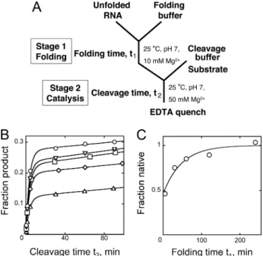
To explore the slow folding further, we used a discontinuous assay (18), in which portions of a folding reaction (termed stage 1) are transferred at various times to stage 2, where the
condi-tions allow rapid substrate cleavage, while minimizing further native folding (18). In stage 2 (25 °C, 50 mMMg2⫹), the
sub-strate is added, and the fraction of the subsub-strate that is cleaved rapidly provides a measure of the fraction of the ribozyme that was native at the time of the transfer (Fig. 5A). When folding (stage 1) was performed under the same conditions as the experiment in Fig. 4 (25 °C, 10 mMMg2⫹), the shortest
accessi-ble folding times gave bursts that corresponded to⬃50% native ribozyme (Fig. 5, B and C). This value is similar but slightly lower than indicated by the continuous assay, suggesting that substrate binding during folding may give a small bias for native folding. As folding time was increased, the burst amplitude increased slowly (k⫽ 0.02 min⫺1), ultimately approaching the level of prefolded ribozyme and indicating 90 –100% native ribozyme. We also explored whether the fraction of ribozyme that folds slowly is affected by conditions of the folding reac-tion. When folding was performed at 15 or 37 °C, the same fraction was observed to reach the native state rapidly as at 25 °C. Further, when these reactions were transferred to 25 °C after a 5-min incubation, they gave the same rate constant for the slow accumulation of native ribozyme (0.02 min⫺1), indi-cating that the same intermediate is formed at all three temper-atures (data not shown).
Together, these experiments indicate that although much of the ribozyme folds to the native state in a few minutes or less, a substantial fraction populates one or more long-lived interme-diates before ultimately reaching the thermodynamically FIGURE 4. Continuous folding and catalytic activity assay. The folding and
catalytic reactions were initiated simultaneously by adding the ribozyme (0.4 M) to 10 mMMg2⫹and 0.8Msubstrate at 25 °C (circles). Also shown is a reaction in which the ribozyme was preincubated for 10 min at 50 °C in the absence of Mg2⫹before Mg2⫹and substrate were added together (triangles) and a reaction in which the ribozyme was prefolded for 30 min at 37 °C in 10 mMMg2⫹and then the substrate was added (squares). The burst amplitude of this prefolded ribozyme reaction corresponded to 24% of the input substrate (48% of the ribozyme with 2-fold substrate excess). After corrections for the internal equilibrium for cleavage and ligation (Keq⫽ 1.2 under these
condi-tions; seesupplemental Table S1) and the subsaturating guanosine concen-tration (100M; KD⫽ 110–120M; seesupplemental Table S1), this burst
amplitude corresponds to a homogeneous population of native ribozyme.
FIGURE 5. Slow accumulation of native ribozyme. A, schematic of the dis-continuous assay for Azoarcus ribozyme folding. The ribozyme folds in stage 1, and portions of the reaction are transferred to stage 2 to measure the fraction of native ribozyme. B, the ribozyme was folded in stage 1 (25 °C, 10 mMMg2⫹) for 5 min (triangles), 30 min (diamonds), 60 min (squares), 120 min (inverted triangles), or 240 min (circles) before being transferred to stage 2 (50 mMMg2⫹). The larger bursts with increased folding time (t1) indicate
accumu-lation of the native ribozyme on this time scale. C, burst amplitude from reac-tions in B plotted against folding time, t1, at 25 °C and 10 mMMg2⫹. The y axis
values are normalized by the apparent end point, which corresponded to 53⫾ 4% of the substrate, nearly the same as obtained by prefolding the ribozyme at 37 °C. by guest on August 7, 2019
http://www.jbc.org/
favored native state with a half-life of⬃30 min under these conditions. The simple first order transition to the native state most simply suggests that the slow folding results from forma-tion of a single intermediate or an ensemble of rapidly convert-ing intermediates, and work below provides further support for this conclusion.
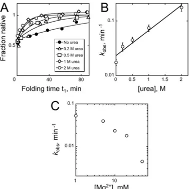
A Kinetically Trapped Intermediate—To explore further the
long-lived intermediate and its folding transition to the native state, we measured how changes in solution conditions affect the rate of the slow folding transition (17, 19, 46). The long-lived intermediate was formed by a short incubation under con-stant conditions (5 min at 25 °C, 10 mMMg2⫹), and then
con-ditions were changed for stage 1 by adding urea or by varying Mg2⫹concentration or temperature, and at various times
ali-quots were trapped in stage 2 (25 °C, 50 mMMg2⫹). The
frac-tion of native ribozyme was determined by catalytic activity as above. We found that the denaturant urea strongly accelerated the folding transition to the native state, giving an m value of ⫺0.7 ⫾ 0.1 kcal mol⫺1 M⫺1 and indicating that transient
unfolding of the long-lived intermediate is required (Fig. 6, A and B). Increased Mg2⫹concentration slowed the transition, suggesting that the intermediate is structured and stabilized by Mg2⫹(Fig. 6C). Higher temperatures increased the rate, with
nitude of the change was interpreted to indicate participation of only a small fraction of the ribozyme, we found that half of the population formed the intermediate, the same within error as under our standard conditions (supplemental Fig. S3C). Refold-ing to the native state gave a rate constant of 0.29 min⫺1, the same within error as that detected by SAXS. Thus, the two approaches likely monitor the same folding transition, and the small change observed by SAXS most likely reflects that the misfolded intermediate is compact and resembles the fully folded structure.
Together these results indicate that, under a wide range of conditions, approximately half of the ribozyme misfolds to an intermediate that refolds slowly to the native state in a process that requires transient unfolding. Misfolding was demon-strated previously for the pre-tRNA containing this intron, but its refolding is accelerated much less by urea and is accelerated, not slowed, by increased Mg2⫹concentration (46). Thus, the
pre-tRNA most likely forms a different misfolded conforma-tion, at least under the conditions of this previous work, con-sistent with the authors’ interpretation of non-native interac-tions with exon sequences. On the other hand, our results are reminiscent of misfolding by the Tetrahymena group I intron ribozyme (17, 47), raising the possibility of common features in the folding and misfolding of these two RNAs (see “Misfolding of the Azoarcus Ribozyme” under “Discussion”).
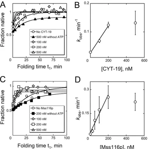
Acceleration of Refolding by DEAD Box Proteins—We also
investigated the effects of the DEAD box proteins CYT-19 and Mss116p on Azoarcus ribozyme refolding. Using the discontin-uous assay, we first folded the ribozyme to a mixture of the native and misfolded conformations and then added CYT-19 or Mss116p in the presence or absence of ATP. At various times, further folding was blocked by increasing Mg2⫹concentration
to 50 mM, and the fraction of native ribozyme was determined
by catalytic activity as above. At relatively low concentrations of protein, just slightly in excess of the ribozyme, both DEAD box proteins accelerated the formation of native ribozyme (Fig. 7). These accelerations required ATP, because omitting nucleo-tide or including the nonhydrolyzable analog AMP-PNP (2 mM) gave no detectable acceleration and decreased the end point somewhat, perhaps by trapping folding intermediates (Fig. 7, A and C, and data not shown). The efficiency of the reaction was moderately larger for Mss116p than for CYT-19, qualitatively consistent with previous work in which splicing was monitored for several different group I and group II introns (24, 27). At higher protein concentrations, the folding rates reached a pla-teau and possibly decreased for Mss116p, most likely reflecting stable binding to RNA folding intermediates and/or ATP-de-pendent disruption of the native state (24, 33).
FIGURE 6. Effects of urea and Mg2ⴙ on refolding of the misfolded ribozyme. A, refolding progress in the presence of urea. The misfolded
ribozyme was first formed by incubating the ribozyme in the absence of urea (25 °C, 10 mMMg2⫹, 5 min), and then the ribozyme (8M) was transferred to a solution that included the desired urea concentration. At various times, portions of the reaction were withdrawn and diluted 20-fold into stage 2 to determine the fraction of native ribozyme. This dilution was much larger than for standard reactions to eliminate possible effects of urea on the cleavage reaction. The y axis values are normalized by the end point in the absence of urea, which corresponded to complete formation of the native ribozyme. B, dependence of refolding rate on urea concentration. The slope of the log-linear dependence gave a urea m value of 0.7⫾ 0.1 kcal mol⫺1M⫺1. C, dependence of refolding rate on Mg2⫹concentration. As in A, the misfolded ribozyme was first formed by incubating the ribozyme for 5 min under stand-ard folding conditions, and then the ribozyme was transferred to solutions containing different Mg2⫹concentrations for refolding.
by guest on August 7, 2019
http://www.jbc.org/
DISCUSSION
Here, we designed and used assays to monitor native folding of the Azoarcus group I intron ribozyme by measuring the onset of its catalytic activity. We found that a substantial fraction of the ribozyme population folds to a long-lived intermediate that displays experimental hallmarks of a misfolded conformer and is accelerated for refolding by the DEAD box chaperone pro-teins Mss116p and CYT-19. These results have important par-allels to previous findings for other group I introns and high-light the pervasive nature of RNA misfolding and RNA chaperone activity (15, 17, 48 –50).
Catalytic Activity as a Probe of Azoarcus Ribozyme Folding—
Catalytic activity provides a powerful probe that is complemen-tary to physical approaches because it can readily distinguish the native state from all inactive conformations, regardless of how similar they are structurally. For the Azoarcus ribozyme, catalytic activity has been used previously to measure the Mg2⫹
dependence of equilibrium folding and cleavage (51), and it has recently been used to measure the relative folding progress, providing quantitative information on folding rates and relative measures of the extent of native folding (40).
By delineating the properties of the substrate cleavage and ligation reactions, our results allow the amplitudes of product
formation, as well as the rate constants, to be interpreted quan-titatively. Slow release of both products leads to the formation of an internal equilibrium between cleavage and ligation, such that the amount of product formed rapidly is less than the amount of native ribozyme. This conclusion is qualitatively consistent with previous work in which the maximal cleavage burst reflected 40% of the substrate in single turnover reactions (51).
With regard to the slow release of products, it is striking that the 5⬘ product (CAU) is released in tens of minutes because it would be expected to dissociate from base pairs with its partner strand, the internal guide sequence of the ribozyme, in millisec-onds (52, 53). Slow dissociation is also supported by pulse-chase experiments, which gave a rate constant of 0.1 min⫺1at 50 °C5
and previous work that established a Kmvalue of 50 nM(54).
Tight binding presumably stems from strong tertiary interac-tions between the product-containing duplex and the ribozyme body. These contacts are inherent to group I introns and posi-tion the reactive groups in the active site, but their strength can vary widely (55–57). It is notable that substrate binding is also quite slow (⬍106
M⫺1min⫺1),⬃100-fold slower than binding
5X. Yuan and R. Russell, unpublished results.
FIGURE 7. Acceleration of ribozyme refolding by DEAD box proteins CYT-19 and Mss116p. A and B, progress curves and protein concentration depend-ence for ribozyme refolding in the presdepend-ence of CYT-19. C and D, progress curves and concentration dependdepend-ence for refolding with Mss116p. The dependdepend-ences of observed rate constant on CYT-19 and Mss116p concentrations gave second order rate constants of 4.8 (⫾ 0.5) ⫻ 105
M⫺1min⫺1and 1.2 (⫾ 0.2) ⫻ 106 M⫺1
min⫺1, respectively, from linear fits within the concentration regimes that gave folding activation. The apparent breakpoints at higher protein concentrations presumably reflect the intersection of activation and inhibition, as observed for other ribozymes (24, 33). In A and C, the y axis values are normalized by the endpoints in the absence of protein. The reactions included 2 mMATP-Mg2⫹unless otherwise indicated.
by guest on August 7, 2019
http://www.jbc.org/
folding steps for at least a fraction of the population (39, 40). By tracking formation of the native state directly, our results build on these findings by showing that it is a large fraction, approx-imately half of the ribozyme, that reaches the native state slowly, on the time scale of an hour under our conditions. Our results are consistent with a recent study in which footprinting and kinetic modeling suggested two dominant pathways, pop-ulated approximately equally, which give fully folded ribozyme with rate constants of⬃2 and 0.2 min⫺1at 37 °C (40). This study provided rate information on the faster process, which was not probed in our work beyond giving a lower limit of⬃0.1 min⫺1(Fig. 4), and it gave a rate constant for the slower phase that is consistent with our measurements at 37 °C ( supplemen-tal Fig. S3). Our results demonstrate that both of these path-ways result in native state formation and that all or nearly all of the ribozyme is present in the native state at the completion of these two folding phases, indicating that the native state is sub-stantially more stable in solution than accessible alternative conformations.
Our results also give new insights into the nature of the slow folding pathway. The rate-limiting folding transition is acceler-ated by urea and increased temperature and decreased by increasing Mg2⫹ concentration, indicating that it involves a transient loss of structure and therefore reflects refolding of one or more misfolded intermediates. Under all of the condi-tions tested, we observed single exponential kinetics for native state formation, most simply suggesting that a single interme-diate or ensemble of interconverting intermeinterme-diates limits fold-ing to the native state. This misfolded intermediate is probably highly structured, because the urea dependence for its transi-tion to the native state corresponds to the transient exposure of ⬃10 base pairs (59). This interpretation is also supported by prior work indicating only small differences between the native state and the intermediate as monitored by hydroxyl radical footprinting (36, 37, 40) or by SAXS (39). These prior results also suggest that the intermediate bears a strong resemblance to the native state and most likely includes substantial native structure. The ribozyme is stabilized by two canonical tetraloop-receptor interactions (34, 35) (Fig. 1) and forms native tertiary contacts cooperatively (37, 51, 60). Some of these tertiary contacts may be able to form and stabilize the misfolded structure.
The properties of the misfolded conformation and its transi-tion to the native state resemble those of the Tetrahymena ribozyme, which also partitions between pathways in folding, giving predominantly a misfolded conformation that includes extensive native structure, refolds slowly, and gives qualita-tively similar dependences on temperature and Mg2⫹and urea
concentrations. This misfolded conformation involves
struc-kcal mol M (17), compared with 0.7 kcal mol M for the
Azoarcusribozyme; Fig. 6B). Nevertheless, there may be many
folding intermediates with these properties, and further work will be necessary to address whether the misfolded forms of the two introns have related structures and physical origins.
Accelerated Refolding by DEAD Box Proteins—In keeping
with their physiological roles as general RNA chaperone pro-teins (8, 9), both CYT-19 and Mss116p accelerate refolding of the misfolded Azoarcus ribozyme to the native state. For both proteins, detectable acceleration requires ATP, suggesting that the acceleration involves ATP-dependent disruption of struc-ture within the misfolded intermediate. This disruption may include unwinding of ribozyme helices, because both proteins have been shown to use ATP to unwind short helices efficiently (20, 24, 27, 42, 61). Because the misfolded ribozyme appears to be compact and extensively structured, it is likely that tertiary contacts must also be disrupted, either directly or indirectly, and the required unfolding may include native structure as well as non-native structure. The observed net conversion of mis-folded to native structure presumably arises because of the greater stability of the native structure (21), as indicated by its accumulation at equilibrium in the absence of protein. We expect that this folding transition of the Azoarcus ribozyme will be useful for further probing of the origins of RNA misfolding and the mechanisms of DEAD box proteins in RNA folding. Acknowledgments—We thank Matthew Kanke for performing early experiments on ribozyme refolding by CYT-19, Ken Johnson for the Kinetic Explorer simulation program, and Inga Jarmoskaite and other members of the Russell lab for helpful comments on the manuscript.
REFERENCES
1. Treiber, D. K., and Williamson, J. R. (1999) Curr. Opin. Struct. Biol. 9, 339 –345
2. Shcherbakova, I., Mitra, S., Laederach, A., and Brenowitz, M. (2008) Curr. Opin. Chem. Biol. 12,655– 666
3. Russell, R. (2008) Front Biosci. 13, 1–20
4. Fairman-Williams, M. E., Guenther, U. P., and Jankowsky, E. (2010) Curr. Opin. Struct. Biol. 20,313–324
5. Pan, C., and Russell, R. (2010) RNA Biol. 7, 667– 676 6. Jankowsky, E. (2011) Trends Biochem. Sci. 36, 19 –29 7. Jarmoskaite, I., and Russell, R. (2011) WIREs RNA 2, 135–152
8. Mohr, S., Stryker, J. M., and Lambowitz, A. M. (2002) Cell 109, 769 –779 9. Huang, H. R., Rowe, C. E., Mohr, S., Jiang, Y., Lambowitz, A. M., and
Perlman, P. S. (2005) Proc. Natl. Acad. Sci. U.S.A. 102, 163–168 10. Woodson, S. A. (2010) Annu. Rev. Biophys. 39, 61–77
11. Sclavi, B., Sullivan, M., Chance, M. R., Brenowitz, M., and Woodson, S. A. (1998) Science 279, 1940 –1943
12. Russell, R., Millett, I. S., Doniach, S., and Herschlag, D. (2000) Nat. Struct. Biol. 7,367–370
by guest on August 7, 2019
http://www.jbc.org/
13. Russell, R., Zhuang, X., Babcock, H. P., Millett, I. S., Doniach, S., Chu, S., and Herschlag, D. (2002) Proc. Natl. Acad. Sci. U.S.A. 99, 155–160 14. Kwok, L. W., Shcherbakova, I., Lamb, J. S., Park, H. Y., Andresen, K.,
Smith, H., Brenowitz, M., and Pollack, L. (2006) J. Mol. Biol. 355, 282–293 15. Laederach, A., Shcherbakova, I., Jonikas, M. A., Altman, R. B., and
Bre-nowitz, M. (2007) Proc. Natl. Acad. Sci. U.S.A. 104, 7045–7050 16. Russell, R., and Herschlag, D. (1999) J. Mol. Biol. 291, 1155–1167 17. Russell, R., Das, R., Suh, H., Travers, K. J., Laederach, A., Engelhardt, M. A.,
and Herschlag, D. (2006) J. Mol. Biol. 363, 531–544
18. Wan, Y., Mitchell, D., 3rd, and Russell, R. (2009) Methods Enzymol. 468, 195–218
19. Wan, Y., Suh, H., Russell, R., and Herschlag, D. (2010) J. Mol. Biol. 400, 1067–1077
20. Tijerina, P., Bhaskaran, H., and Russell, R. (2006) Proc. Natl. Acad. Sci. U.S.A. 103,16698 –16703
21. Bhaskaran, H., and Russell, R. (2007) Nature 449, 1014 –1018
22. Johnson, T. H., Tijerina, P., Chadee, A. B., Herschlag, D., and Russell, R. (2005) Proc. Natl. Acad. Sci. U.S.A. 102, 10176 –10181
23. Del Campo, M., Tijerina, P., Bhaskaran, H., Mohr, S., Yang, Q., Jankowsky, E., Russell, R., and Lambowitz, A. M. (2007) Mol. Cell 28, 159 –166 24. Del Campo, M., Mohr, S., Jiang, Y., Jia, H., Jankowsky, E., and Lambowitz,
A. M. (2009) J. Mol. Biol. 389, 674 – 693
25. Solem, A., Zingler, N., and Pyle, A. M. (2006) Mol. Cell 24, 611– 617 26. Mohr, S., Matsuura, M., Perlman, P. S., and Lambowitz, A. M. (2006) Proc.
Natl. Acad. Sci. U.S.A. 103,3569 –3574
27. Halls, C., Mohr, S., Del Campo, M., Yang, Q., Jankowsky, E., and Lambow-itz, A. M. (2007) J. Mol. Biol. 365, 835– 855
28. Bifano, A. L., and Caprara, M. G. (2008) J. Mol. Biol. 383, 667– 682 29. Fedorova, O., Solem, A., and Pyle, A. M. (2010) J. Mol. Biol. 397, 799 – 813 30. Bifano, A. L., Turk, E. M., and Caprara, M. G. (2010) J. Mol. Biol. 398,
429 – 443
31. Zingler, N., Solem, A., and Pyle, A. M. (2010) Nucleic Acids Res. 38, 6602– 6609
32. Karunatilaka, K. S., Solem, A., Pyle, A. M., and Rueda, D. (2010) Nature
467,935–939
33. Potratz, J. P., Del Campo, M., Wolf, R. Z., Lambowitz, A. M., and Russell, R. (2011) J. Mol. Biol. 411, 661– 679
34. Tanner, M., and Cech, T. (1996) RNA 2, 74 – 83
35. Adams, P. L., Stahley, M. R., Kosek, A. B., Wang, J., and Strobel, S. A. (2004) Nature 430,45–50
36. Rangan, P., Masquida, B., Westhof, E., and Woodson, S. A. (2003) Proc. Natl. Acad. Sci. U.S.A. 100,1574 –1579
37. Chauhan, S., and Woodson, S. A. (2008) J. Am. Chem. Soc. 130, 1296 –1303
38. Chauhan, S., Behrouzi, R., Rangan, P., and Woodson, S. A. (2009) J. Mol. Biol. 386,1167–1178
39. Roh, J. H., Guo, L., Kilburn, J. D., Briber, R. M., Irving, T., and Woodson, S. A. (2010) J. Am. Chem. Soc. 132, 10148 –10154
40. Mitra, S., Laederach, A., Golden, B. L., Altman, R. B., and Brenowitz, M. (2011) RNA 17, 1589 –1603
41. Zaug, A. J., Grosshans, C. A., and Cech, T. R. (1988) Biochemistry 27, 8924 – 8931
42. Grohman, J. K., Del Campo, M., Bhaskaran, H., Tijerina, P., Lambowitz, A. M., and Russell, R. (2007) Biochemistry 46, 3013–3022
43. Golden, B. L., and Cech, T. R. (1996) Biochemistry 35, 3754 –3763 44. Karbstein, K., Carroll, K. S., and Herschlag, D. (2002) Biochemistry 41,
11171–11183
45. Pan, J., Thirumalai, D., and Woodson, S. A. (1997) J. Mol. Biol. 273, 7–13 46. Rangan, P., Masquida, B., Westhof, E., and Woodson, S. A. (2004) J. Mol.
Biol. 339,41–51
47. Russell, R., and Herschlag, D. (2001) J. Mol. Biol. 308, 839 – 851 48. Pan, J., and Woodson, S. A. (1998) J. Mol. Biol. 280, 597– 609 49. Zhang, L., Xiao, M., Lu, C., and Zhang, Y. (2005) RNA 11, 59 – 69 50. Jiang, Y. F., Xiao, M., Yin, P., and Zhang, Y. (2006) RNA 12, 561–566 51. Chauhan, S., Caliskan, G., Briber, R. M., Perez-Salas, U., Rangan, P.,
Thi-rumalai, D., and Woodson, S. A. (2005) J. Mol. Biol. 353, 1199 –1209 52. Freier, S. M., Kierzek, R., Jaeger, J. A., Sugimoto, N., Caruthers, M. H.,
Neilson, T., and Turner, D. H. (1986) Proc. Natl. Acad. Sci. U.S.A. 83, 9373–9377
53. Serra, M. J., and Turner, D. H. (1995) Methods Enzymol. 259, 242–261 54. Strauss-Soukup, J. K., and Strobel, S. A. (2000) J. Mol. Biol. 302, 339 –358 55. Narlikar, G. J., and Herschlag, D. (1996) Nat. Struct. Biol. 3, 701–710 56. Testa, S. M., Haidaris, C. G., Gigliotti, F., and Turner, D. H. (1997)
Bio-chemistry 36,15303–15314
57. Kuo, L. Y., Davidson, L. A., and Pico, S. (1999) Biochim. Biophys. Acta
1489,281–292
58. Herschlag, D., and Cech, T. R. (1990) Biochemistry 29, 10159 –10171 59. Shelton, V. M., Sosnick, T. R., and Pan, T. (1999) Biochemistry 38,
16831–16839
60. Moghaddam, S., Caliskan, G., Chauhan, S., Hyeon, C., Briber, R. M., Thi-rumalai, D., and Woodson, S. A. (2009) J. Mol. Biol. 393, 753–764 61. Yang, Q., Del Campo, M., Lambowitz, A. M., and Jankowsky, E. (2007)
Mol. Cell 28,253–263
62. Johnson, K. A., Simpson, Z. B., and Blom, T. (2009) Anal. Biochem. 387, 20 –29
63. McConnell, T. S., Cech, T. R., and Herschlag, D. (1993) Proc. Natl. Acad. Sci. U.S.A. 90,8362– 8366
64. Mei, R., and Herschlag, D. (1996) Biochemistry 35, 5796 –5809
by guest on August 7, 2019
http://www.jbc.org/
Alerts:
When a correction for this article is posted
•
When this article is cited
•
to choose from all of JBC's e-mail alerts
Click here
Supplemental material:
http://www.jbc.org/content/suppl/2011/08/30/M111.287706.DC1 http://www.jbc.org/content/286/43/37304.full.html#ref-list-1This article cites 64 references, 14 of which can be accessed free at
by guest on August 7, 2019
http://www.jbc.org/