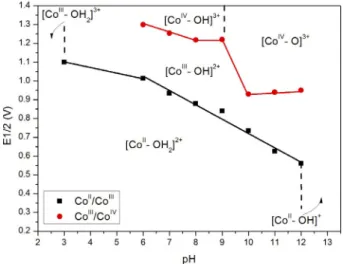
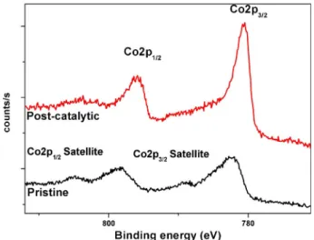
Metal Dicyanamides as Efficient and Robust
Water-Oxidation Catalysts
Satya Vijaya Kumar Nune,
[a]Aysun Tekin Basaran,
[a]Emine 3lker,
[a, b]Rupali Mishra,
[a]and
Ferdi Karadas*
[a, c]Introduction
Fossil-based fuels have been used extensively for centuries,
but the reserves of these fuels are being depleted rapidly.[1]
Moreover, the use of these fuels has a serious impact on the environment, such as hazardous greenhouse gas emissions
and the change in the atmospheric equilibrium,[2] which
em-phasizes the necessity for more reliable, clean, and environ-mental friendly power sources.[3]Given the high energy density
of hydrogen (143.0 MJkg@1), hydrogen-based fuels are one of
the most promising alternatives[4] for clean and renewable
energy without any waste.[5]Water splitting, which involves the
production of H2and O2 from water, is considered to be the
bottleneck in the hydrogen economy.[6]The conversion of solar
energy into chemical energy through the splitting of water molecules in photosynthesis is a well-defined process that uses chlorophyll. The whole procedure works on the association of an electron acceptor and an electron donor.[7]
Over the past few decades, many research groups have aimed to perfect the art of mimicking the photosynthesis pro-cess of splitting water to generate energy.[8]Both
electrocata-lytic[9] and photocatalytic[10] routes have been studied
exten-sively with a wide range of catalysts, which include semicon-ductors,[11] transition metal oxides,[12] metal–organic
frame-works (MOFs),[13] perovskite-type compounds,[14] and
amor-phous and porous catalysts.[15] Over the past few years,
non-oxide systems have been emphasized not only because of their high stabilities in both acidic and basic media but also because of their high catalytic activities obtained per metal
site. Various Co-based non-oxide systems such as cyanide-[16]
and cyanamide-based systems[17] are efficient and robust
water-oxidation catalysts (WOCs). In 2014, Gal#n-Mascarjs et al. reported the application of various metal hexacyanome-talates as heterogeneous WOCs, which are stable even under extremely acidic conditions and had quantum yields in the range 50–80 %.[16a]A similar study was reported in 2015 by
Fu-kuzumi et al. who used hetero-polynuclear cyanide systems with Co and Pt ions.[18] In 2016, the use of
pentacyanoferrate-coordinated poly(4-vinylpyridine) as a precursor to obtain a cya-nide-based coordination polymer with a high current density
at low overpotentials was reported by our group.[19]
Further-more, Patzke et al. reported the application of cobalt carbodii-mide for both photochemical and electrochemical water oxida-tion in neutral and basic media in 2015.[17]Overall, these
stud-ies suggest that heterogeneous WOCs with Co ions exhibit ex-cellent turnover frequencies (TOFs) if the metal ions are sur-rounded by N donor atoms rather than O atoms as in the case of oxides. The electrocatalytic studies performed on
homoge-neous single-site WOCs, [Co(Py5)(OH2)](ClO4)2 (Py5=
2,6-[bis(bis-2-pyridyl)-methoxymethane]pyridine)[20] and cobalt
Non-oxide cobalt-based water-oxidation electrocatalysts have received attention recently for their relative ease of prepara-tion, they are stable both in acidic and basic media, and they have higher turnover frequencies than cobalt oxides. Recent studies show that one of the main bottlenecks in the imple-mentation of non-oxide systems to water splitting is the low number of active metal sites, which is in the order of
nmol cm@2. Herein, a new series of non-oxide water-oxidation
catalysts has been introduced to the field. Cobalt dicyanamides
are observed to have around four times higher surface active sites and better catalytic performances than cyanide-based sys-tems. Long-term catalytic studies (70 h) at an applied potential of 1.2 V and electrochemical studies performed in solutions in pH values of 3.0–12.0 indicate that the compounds are robust and retain their structures even under harsh conditions. More-over, the addition of Ni impurities to cobalt dicyanamides is a feasible method to improve their catalytic activities.
[a] Dr. S. V. K. Nune, A. T. Basaran, Prof. Dr. E. 3lker, Dr. R. Mishra, Prof. Dr. F. Karadas Department of Chemistry Bilkent University 06800, Ankara (Turkey) E-mail: karadas@fen.bilkent.edu.tr [b] Prof. Dr. E. 3lker
Department of Chemistry, Faculty of Arts & Sciences Recep Tayyip Erdogan University
53100, Rize (Turkey) [c] Prof. Dr. F. Karadas
UNAM-Institute of Materials Science and Nanotechnology Bilkent University
Ankara, 06800 (Turkey)
Supporting information and the ORCID identification number(s) for the author(s) of this article can be found under http://dx.doi.org/10.1002/ cctc.201600976.
hangman porphyrins,[9a] which involve single Co atoms
sur-rounded by N donor atoms, also support this.
In our current investigation, we are interested in expanding the portfolio of heterogeneous WOCs that contain metal ions surrounded by N donor atoms. The type of ligand plays a criti-cal role in the stability and the catalytic performance of the compounds. Our current research focuses on the introduction
of a new N-donor bridging group, dicyanamide (N(CN)2@), to
this field. Metal dicyanamide systems have been explored to
a great extent for their interesting structures,[21] magnetic
properties,[22] and selectivity in gas adsorption.[23] For the first
time, we report the application of a cobalt dicyanamide matrix in electrochemical WOCs. Moreover, a 1D coordination com-pound rather than a 3D extended network was chosen as a catalyst of interest to improve the number of active metal sites as the main drawback of non-oxide Co-containing WOCs is the relatively low number of active metal sites on the surface because of the larger distance between the metal sites com-pared to that in oxide-based systems.
Results and Discussion
Synthesis and characterizationSingle-crystal XRD studies performed on fine crystals of the compounds reveal that they are all isostructural and crystallize in a monoclinic system with the space group P21/n. The crys-tallographic structure and refinement parameters are given in
Table S1. The asymmetric unit of the M(dca)2 (DCA
=mide) structure contains half of a metal(II) site, two dicyana-mide groups, and one DMF molecule. Each metal ion shows
a distorted octahedral MN4O2coordination environment, which
results from the coordination of four N atoms of different DCA groups and two O atoms from DMF molecules (Figure 1). The crystal structure could be described as a 1D ladder-like double-chain coordination polymer (Figure 2). Neighboring metal ions in each chain are connected to each other through two dicyanamide groups, which results in a distance of 7.3 a
between the CoIIcenters. Two ligands adopt a certain
configu-ration to connect two metal centers in such a way as to form rectangular units. All of the M@N and M@O distances are within the normal range of statistical errors (Table S2). The supramolecular framework is stabilized by H···N interactions (2.555(5)–2.686(5) a), which originate from the H atoms of the coordinated DMF molecules and the central N atom of the DCA groups (Figure S1 and S2).
Powder XRD studies of the as-synthesized metal dicyana-mide clusters (Co, Fe, and Ni) confirm that the series of com-pounds is isostructural with varying degrees of crystallinity. The XRD patterns, which exhibit 2q positions that almost over-lap each other, are presented in Figure 3.
The IR spectra of the Co and Ni derivatives exhibit sharp and strong stretching bands, whereas that of the Fe cluster shows broad and weak bands (Figure 4). The nsym&asym(C /N) stretches
were observed in the range n˜ =2360–2184 cm@1, the n
asym(C@
N) stretch was at around n˜=1380–1364 cm@1, and the n
sym(C@
N) stretch was at around n˜= 938 cm@1, all of which can be
at-tributed to the cyanide groups in the DCA fragments.[23a]
Strong bands at n˜=1108–1015 cm@1 correspond to the C@N
group, whereas bands at around n˜= 1647–1640 and 2970–
2934 cm@1can be assigned to the C=O stretches and aliphatic
C@H stretching vibrations of DMF, respectively.[24]The presence
of a broad stretch at n˜=3500–3250 cm@1 in the spectrum of
Figure 1. Fragment of the crystal structure of Co(dca)2(dmf)2, which depicts
the MN4O2coordination sphere of the metal site. Thermal ellipsoids are
pro-jected at the 50% probability level. Hydrogen atoms are not shown for clarity.
Figure 2. 1D chain structure of [Codca2]. Color code: Co= purple; O =red; C =gray; N=blue. Thermal ellipsoids are projected at the 50% probability level. Hydrogen atoms are not shown for clarity.
the Fe derivative can caused by the adsorption of excess mois-ture, which can also be a reason for the relatively poor crystallinity of the Fe derivative.
Electrochemical studies
Cyclic voltammograms (CVs) of M(dca)2(dmf)2 (M= Co, Fe, and
Ni) deposited on fluorine-doped tin oxide (FTO) electrodes, re-ferred to as [Mdca2] henceforth, were recorded in a phosphate
buffer solution with 1m KNO3 as the electrolyte over 0–1.5 V
vs. Ag/AgCl (Figure 5). The prominent feature of the CVs of [Mdca2] is the increase in the anodic current above ~1 V, which can be attributed to catalytic oxygen evolution. The CVs of the three derivatives reveal that [Codca2] shows a signifi-cantly higher catalytic activity than the Fe and Ni derivatives. Hence further studies were performed with [Codca2]. The CV of [Codca2] shows a quasireversible redox couple with a signifi-cant oxidation peak at 0.95 V and a reduction peak at 0.83 V
vs. Ag/AgCl (E1/2=0.89 V, Ec@Ea=120 mV), which can be
as-signed to the Co2++/Co3++redox couple. A second oxidation
pro-cess was observed at around 1.35 V vs. Ag/AgCl, which can be assigned to the formation of CoIVspecies.[20]The current
densi-ty in the catalytic region refers only to Faradaic currents as the measured capacitive currents were insignificant (Figure S16). CVs with different scan rates were recorded in the range of 0.5–1.1 V vs. Ag/AgCl to determine the coverage of the redox-active Co centers on the electrode, the so-called surface con-centration (Figure S3). The surface concon-centration (G) was
calcu-lated as 5.80 nmol cm@2 from the slope and is approximately
four times higher than that reported for cobalt hexacyano-ferrates (G= ~ 1.4 nmolcm@2).[16a] The relatively high surface
concentration of [Codca2] can be explained by a comparison of the distances between the neighboring Co atoms in these systems. The Co sites are much closer to each other in the [Codca2] system (Co@Co=7.3 a) than in cobalt hexacyano-ferrates (Co@Co&10 a).
For a detailed assessment of the catalytic activity of the modified electrodes, chronoamperometry measurements were performed at different applied potentials by using a two-com-partment cell with a glass frit separator in pH 7.0 K2HPO4and
KH2PO4 (KPi) buffer solution with 1m KNO3 as the electrolyte.
The plot of the logarithm of the steady current densities versus overpotential shows a linear relationship between 323
and 483 mV with a slope of 94 mVdec@1. The current density is
1 mAcm@2 at h=580 mV (Figure S4), whereas it is above
600 mV for cobalt hexacyanoferrates. The improvement in the catalytic performance can be attributed to the increase in the surface concentration. The surface concentration was also used to compare the TOFs of other non-oxide Co-based systems. A
TOF of 2V10@3 was obtained at an overpotential of 358 mV
(Figure S5), which is higher than that obtained for a cobalt oxide film at pH 7.0 (h= 410 mV).[25]A self-assembled IrO
2
col-loid on an FTO electrode and an electrodeposited IrO2-coated
indium tin oxide (ITO) electrode in an aqueous medium were
reported have TOF values of 2.3V104 (pH 5.3) and 1.64 V
104h@1 (pH 6.3) at 1.3 V vs. Ag/AgCl, respectively, whereas
a TOF value of 2.00V104h@1(pH 7) for [Codca2] was obtained
under the same potensiostatic conditions.[26]
Figure 3. XRD patterns of [Mdca2].
Figure 4. FTIR spectra of [Mdca2].
Figure 5. CVs of [Mdca2]-modified FTO electrodes recorded in 50 mm KPi buffer solution with 1m KNO3as electrolyte at pH 7.0 with a 50 mV s@1
sweep rate and a blank scan recorded under the same conditions without the catalyst (black line). The arrow shows the scanning direction.
The stability of [Codca2] was tested over approximately six days by using chronoamperometry under same experimental conditions at 1.2 V vs. Ag/AgCl (Figure 6). During this period, electrolysis was performed for four days without interruption and then continued for two more days. The current density de-creases until approximately 1 h and then it inde-creases for a short period, probably because of the change in the mor-phology of the electrode surface, which leads to a change in the surface concentration. Upon further electrolysis, the cur-rent density decreases until it stabilizes at around
0.15 mA cm@2 after 144 h, which implies the stable
per-formance of [Codca2] during water electrolysis and the lack of degradation of the FTO electrode surface. Furthermore, CVs re-corded before and after the long-term stability measurement show a similar behavior, which verifies the stability of [Codca2] (Figure 6, inset). Moreover, the stability of [Codca2] was investi-gated in acidic and basic media. The Pourbaix diagram ob-tained by performing CVs at different pH values shows that the Co2++/3++and Co3++/4++redox bands shift to higher potentials
gradually as the pH decreases (Figure 7). CVs recorded at dif-ferent pH values also show that current density obtained at high anodic potentials increases significantly with an increase of the pH, which is expected from the electrocatalytic water oxidation process that involves proton-coupled electron transfer (Figure S6).
The Faradaic efficiency of the process was evaluated by
using bulk electrolysis for 3 h. O2evolution was measured by
using an oxygen-sensing instrument and compared with the
theoretical amount calculated from Faraday’s law for a 4e@
redox process (Figure 8). The amount of dissolved O2
mole-cules detected during bulk electrolysis matches the theoretical
amount of evolved O2 with an efficiency of 100%. This
con-firms that no competing redox reactions take place and that the current density is quantitative for oxygen production.
Characterization of electrodes
X-ray photoelectron spectroscopy (XPS) was performed on the electrodes before (pristine electrode) and after the bulk elec-trolysis (postcatalytic electrode; Figure 9) to study possible changes in the oxidation state and the composition of
[Codca2]. The Co2p3/2 signal at a binding energy (BE) of
782.78 eV and that of Co2p1/2at BE= 798.78 eV were observed
as broad peaks with a high full width at half maximum (FWHM; >4 eV) in the pristine electrode, which corresponds
well with standard CoII(BE=782.28 and 798.38 eV,
respective-ly).[27] Additionally, scalable satellite bands were observed 4–
8 eV above the principle signals. The postcatalytic electrode ex-hibits a slight shift to lower binding energies (~2.5 eV) with
a Co2p3/2signal at BE=780.48 eV and a Co2p1/2signal at BE=
Figure 6. Current profile of long-term electrolysis performed for 6 days at 1.2 V (vs. Ag/AgCl) at pH 7.0 of a [Codca2]-modified electrode.
Figure 7. Pourbaix diagram that shows the pH/potential characteristics of the precatalytic features of the [Codca2] system. The features denoted as & represent the first precatalytic feature, whereas those represented as * refer to the second precatalytic feature. As reduction bands are not ob-served particularly in the acidic region and for the second precatalytic fea-tures, E1/2values are estimated from the midpoint of the oxidation bands.
Figure 8. Catalytic oxygen evolution recorded (blue) during bulk electrolysis by using an oxygen-sensing probe and theoretical plot assuming Faradaic behavior (black).
795.88 eV.[28] The signals are relatively sharper with a lower
FWHM (~3 eV), and the satellite bands, though identifiable, are less distinctive. Earlier studies reported that these changes can be attributed to the partial oxidation of the surface metal sites.[19]FTIR spectra of the pristine and postcatalytic electrodes
show no visible changes that can be attributed to structural or compositional changes in the catalyst. Hence, the partial oxida-tion of the surface Co sites is not permanent and is reversible.[19]
Furthermore, the XPS O1s signals of the pristine and postca-talytic samples were analyzed to study the nature of the partial oxidation (Figure S7). The O1s signal at a BE higher than
530 eV corresponds to oxygen species such as @OH[29]
ad-sorbed onto the surface of the catalyst, which indicates the ab-sence of Co@O species during the course of the electrolysis.[17]
The slight shift in the O1s position and the relatively higher in-tensity can be attributed to the partial replacement of DMF on the surface with water because of their miscibility. Moreover, grazing-incidence X-ray diffraction (GI-XRD) analysis of the pris-tine and postcatalytic electrodes (Figure S17) reveals that there is no distinguishable change in the crystalline phase of [Codca2]. Hence it can be concluded that no cobalt oxides are formed, which confirms that the partial oxidation of surface Co sites is not permanent.
Metal-doped cobalt dicyanamides
The introduction of a secondary metal ion such as Ni by the partial substitution of metal sites to Co-based heterogeneous WOCs has been studied previously for both oxide and non-oxide systems to enhance the water-oxidation performance of the catalysts.[30] This strategy has also been employed for the
catalysts studied here, not only to obtain a detailed map of the catalytic performances of mixed-metal dicyanamides but also to investigate the origin of the effect of doping on the catalytic activity. Five mixed-metal dicyanamides with different stoichiometric ratios of Co, Fe, and Ni, which are formulated as Co0.5Ni0.5(dca)2(dmf)2, Co0.5Fe0.5(dca)2(dmf)2, Co0.9Ni0.1(dca)2(dmf)2,
Co0.9Fe0.1(dca)2(dmf)2, and Co0.9Ni0.05Fe0.05(dca)2(dmf)2, were
syn-thesized using the same synthetic protocol applied for [Mdca2] (Co, Ni, and Fe). The compounds were characterized by using XRD and IR spectroscopy, which showed that the compounds are isostructural (Figures S8–S9).
The current densities of eight compounds were recorded after 600 s under an applied potential of 1.2 V. The data are represented in the contour plot displayed in Figure 10. Rela-tively lower current densities are obtained for the Fe deriva-tives (Figure S10). The contour plot exhibits the highest current
density if the Ni concentration is supported at 10–50 % for binary compounds, whereas [Nidca2] exhibits lower current densities than [Codca2] and the mixed Co/Ni derivatives. CVs of the Co/Ni dicyanamides show a similar trend in which [Co0.9Ni0.1dca2] exhibits the highest current at high anodic
po-tentials (Figure 11). Moreover, chronoamperometric studies performed on the derivatives indicate that all three catalysts have similar Tafel slopes, and the best catalytic performance is
Figure 9. XPS spectra of the Co2p region of the surface of the pristine and postcatalytic electrodes.
Figure 10. Contour plots of the current densities of the eight metal dicyan-amide derivatives.
Figure 11. Cyclic voltammograms of [Codca2], [Nidca2], [Co0.9Ni0.1dca2], and
[Co0.5Ni0.5dca2] recorded in 50 mm KPi buffer solution with 1m KNO3as
elec-trolyte at pH 7.0 with a 50 mVs@1sweep rate and a blank scan recorded
observed for [Co0.9Ni0.1dca2] (Figure 12). A current density of
1 mAcm@2 can be achieved only with an overpotential of
511 mV for [Co0.9Ni0.1dca2], whereas it is 580 mV for [Codca2].
Even though the comparison of the catalytic activities of [Codca2] and [Nidca2] indicates clearly that Co sites serve as better catalytic sites than Ni ones, the partial substitution of Co atoms with Ni leads to an increase in the catalytic activity. The unexpected improvement in the catalytic activity can be well correlated with surface concentrations of catalysts that are obtained by performing CV on the Co2++/Co3++band at different
scan rates (Table 1 and Figure S11). The change of TOF with re-spect to the overpotential is almost identical, which suggests that the origin of the catalytic activity is the same active site and that the partial substitution of Co sites with Ni ions has a significant effect on the morphology of the catalyst and, thus, on the number of electroactive Co sites on the surface (Figure S12).
Detailed structural characterization and electrochemical
studies were performed on [Codca2], [Co0.5Ni0.5dca2], and
[Co0.9Ni0.1dca2] to investigate the effect of partial addition of
nickel to the microstructure of the samples. The compositions of these clusters were confirmed by using energy-dispersive X-ray (EDX) analysis (Table S3). XRD patterns indicate that all of the compounds are isostructural, and a broadening is observed as the amount of Ni increases, which can be attributed to a de-crease in the degree of crystallinity. An estimation of the crys-tallite sizes by applying the Scherrer formula shows that the
crystallite sizes decrease to 602 and 584 a, respectively, for [Co0.9Ni0.1dca2] and [Co0.5Ni0.5dca2] compared to that of
[Codca2] (>1000 a). SEM images obtained for the clusters (Fig-ure S13) show a significant change in the morphology of the particles with the addition of Ni. These three compounds were
also characterized by using N2sorption analysis to deduce the
surface area. The samples were subjected to BET surface area
measurements by studying the N2 adsorption and desorption
isotherms at 77 K (Figures S14 and S15). All of the samples appear to exhibit mesoporous behavior with varying surface areas. [Codca2] appears to have the lowest surface area, whereas [Co0.5Ni0.5dca2] exhibits a relatively higher surface area
with a more uniform porosity, which corresponds well with the crystallite size calculations from the XRD results and the mor-phological changes observed by using SEM. Such a correlation was not achieved for the Fe derivatives because of their hygro-scopic nature, which is confirmed by using FTIR spectroscopy and SEM. Overall, the study shows clearly that the addition of Ni sites to cobalt dicyanamide leads to an increase in the sur-face area and the number of metal atoms on the sursur-face, how-ever, with the expense of substituting some of the electroac-tive Co sites with less acelectroac-tive Ni sites. The highest surface con-centration was obtained for [Co0.9Ni0.1dca2], although the
high-est surface area was achieved for [Co0.5Ni0.5dca2].
Conclusions
Recently, the investigation of new non-oxide heterogeneous water-oxidation catalysts has become a growing theme as re-search on both single Co complexes with pyridil groups and
cyanide-based systems show that {CoN6} matrices have better
turnover frequencies (TOFs) than {CoO6} ones. Heterogeneous
systems based on cyanide and cyanamide bridging groups have been reported recently. In this study, a new family of 1D coordination compounds with dicyanamide bridging groups was reported. Single-crystal and powder XRD studies indicate that all compounds, Co, Fe, and Ni derivatives and mixed-metal dicyanamides, reported in this study are isostructural.
Cobalt dicyanamides are efficient water-oxidation catalysts
with a TOF value of 2V10@3at h=358 mV, and an
overpoten-tial of only 580 mV is required to produce a current density of
1 mAcm@2. This overpotential could further be reduced to
510 mV by introducing 10% Ni impurities. The catalyst of inter-est is robust in a wide pH range (3.0–12.0). It also retains its structural integrity during long-term catalytic tests (70 h).
We have shown that the estimation of surface concentration using the slope of the linear fit between the peak currents and scan rate is a viable method that could correlate the cata-lytic performance of compounds with identical crystalline structures.
In summary, the rich and diverse chemistry of metal dicyana-mides has promising applications in the field of water oxida-tion. They have several key advantages as water-oxidation cat-alysts: i) they have relatively high surface active sites, ii) they have higher TOFs than cobalt oxides, iii) they are robust at high anodic potentials and in acidic and basic media, and iv) the structure of the final product is highly sensitive to the
Figure 12. Tafel plots obtained for [Codca2] (black), [Co0.9Ni0.1dca2] (red), and
[Co0.5Ni0.5dca2] (blue). Measurements performed in 50m KPi buffer solution
with 1m KNO3as electrolyte.
Table 1. Structural parameters of the catalysts.
M(dca)2(dmf)2 Surface SBET Pore Crystallite
concentration size Size
[nmol cm@2] [m2g@1] [a] [a]
[Codca2] 5.80 2.0 98.9–298.79 >1000 [Co0.9Ni0.1dca2] 6.64 10.2 157.525–230.178 602
synthetic protocol, which enables the easy tuning of the struc-ture. Our preliminary studies show that cobalt dicyanamides with different crystal structures could be prepared by using dif-ferent solvents during synthesis. Such flexibility in the synthe-sis will be used in the future to establish a correlation with the structure and catalytic application by a systematic investiga-tion of the catalytic activities of similar cobalt dicyanamides.
Experimental Section
Co(NO3)2·6H2O, FeSO4·7H2O, Ni(NO3)2·6H2O, NaN(CN)2, and all the solvents were of analytical grade, procured from Sigma–Aldrich, and used without any further processing. Millipore deionized water
(resistivity: 18 mWcm@1) was used for all the experiments.
Typically, dicyanamide solution in DMF was added slowly to metal salt in water under constant stirring in a stoichiometric ratio of 2:1
(dca/metal).[24]The resulting pink suspension was stirred overnight
followed by filtration using suction. The collected precipitate was then washed several times with water/methanol and dried over-night in the oven at 60 8C. IR: n˜=2979(w), 2925(w), 2333(m),
2247(sh), 2184(vs), 1236(m), 932 cm@1(m).
Instrumentation
Elemental analyses were performed by using a Thermo Scientific FLASH 2000 CHNS/O analyzer. FTIR spectra were measured by using a Bruker ALPHA Platinum-ATR spectrometer in the wave
number range 4000–400 cm@1. XRD patterns were recorded by
using a Panalytical X’PertPro Multipurpose X-Ray Diffractometer
(MPD) employing CuKaX-ray radiation (l=1.5418 a). GI-XRD
pat-terns were recorded by using a Panalytical X’Pert3 MRD Material
Research Diffractometer (MRD) employing CuKa X-ray radiation
(l=1.5418 a) at an incident (w) angle of 0.58. Single-crystal XRD was performed by using a Rigaku MicroMax 007HF model
equipped with monochromatic MoKaradiation. SEM imaging was
performed at beam voltage 5 kV, and EDX analysis was performed at 30 kV by using a FEI-Quanta 200 FEG ESEM. XPS studies were performed by using a Thermo Scientific K-Alpha X-Ray
Photoelec-tron Spectrometer system operated with a AlKamicrofocused
mon-ochromator source (hn=1486.6 eV, 400 mm spot size) along with a flood gun for charge neutralization, a pass energy of 200 eV was used for the survey scan and 30 eV was used for individual ele-ment scans. A Micromeritics Tristar 3000 surface area and porosity
analyzer was used to perform N2 adsorption studies at 77 K to
obtain the surface area. Origin Pro 8.5 was used to plot and ana-lyze all of the graphs.
Preparation of catalyst-modified electrodes
FTO electrodes (1V2 cm, 2 mm slides with 7 Wsq@1surface
resistiv-ity and ~80% transmittance) were washed by sonication for 10 min in basic soapy solution, deionized water, and isopropanol followed by annealing at 4008C for 30 min. The catalyst was coated onto the FTO electrode by a drop-casting method. A mix-ture of 5 mg catalyst, 500 mL water, 500 mL DMF, and 20 mL Nafion was sonicated for 30 min to make a stable suspension. Then, 50 mL of the sonicated suspension of the catalyst was then dropped onto
a clean FTO electrode (1 cm2). The electrode was then dried in an
oven at 808C for 10 min and left under desiccation until further use for CV and bulk electrolysis studies. The electrode was rinsed with deionized water before use.
Electrochemical studies
Electrochemical experiments were performed at RT by using a Gamry Instruments Interface 1000 Potentiostat/Galvanostat. A conventional three-electrode electrochemical cell was used with a Ag/AgCl electrode (3.5m KCl) as the reference electrode, Pt wire as the counter electrode, and FTO as the working electrode. Before the preparation of an electrode, FTO slides were cleaned described
previously.[16a] Buffer solutions were prepared using K
2HPO4 and
KH2PO4(KPi) and then adjusted by adding H3PO4or KOH to obtain
the desired pH. CVs were recorded with a scan rate of 50 mVs@1in
50 mm KPi (pH 7) that contained 1m KNO3as the electrolyte
be-tween 0 and 1.5 V (vs. Ag/AgCl). For double-layer capacitance
de-terminations, the scan rate was varied between 10 and 300 mVs@1
over a small window in which no Faradaic current was observed.
The electrochemical double-layer capacitance (CDL) was extracted
from ic=nVCDL, in which n is the scan rate and icis the current
density for the specific curve at 0.15 V. All experiments were
per-formed under a N2atmosphere.
Bulk water electrolysis and Tafel analysis
Bulk water electrolysis studies were performed by using a two-compartment cell with a glass frit separator. The Pt wire counter electrode was placed in one compartment, and the FTO working electrode and Ag/AgCl reference electrode were placed in the other compartment. The electrolysis experiments were performed
in KPi buffer (pH 7.0) solution that contained 1m KNO3as the
sup-porting electrolyte. Tafel data were collected under the same con-ditions at different applied potentials using the steady current den-sity of an equilibrium time of 600 s. Oxygen evolution was deter-mined by using a YSI 5100 oxygen-sensing instrument equipped with a dissolved oxygen field probe inserted into the anodic com-partment.
Acknowledgements
The authors thank the Science and Technology Council of Turkey, TUBITAK (Project No: 114Z473) for financial support. A.T.B. thanks TUBITAK (2210-C) for the scholarship. E.U. thanks TUBITAK for support (Project No: 1929B011500059).
Keywords: amides · cobalt · electrochemistry · n ligands · water splitting
[1] a) J. Goldemberg, Science 1995, 269, 1058 – 1059; b) N. S. Lewis, D. G. Nocera, Proc. Natl. Acad. Sci. USA 2006, 103, 15729 – 15735; c) M. Hoel, S. Kverndokk, Resour. Energy Econ. 1996, 18, 115–136; d) M. Hççk, X. Tang, Energy Policy 2013, 52, 797– 809.
[2] a) M. I. Hoffert, K. Caldeira, A. K. Jain, E. F. Haites, L. D. D. Harvey, S. D. Potter, M. E. Schlesinger, S. H. Schneider, R. G. Watts, T. M. L. Wigley, D. J. Wuebbles, Nature 1998, 395, 881 –884; b) T. M. L. Wigley, R. Richels, J. A. Edmonds, Nature 1996, 379, 240 –243; c) A. K. Jain, H. S. Kheshgi, M. I. Hoffert, D. J. Wuebbles, Global Biogeochem. Cycles 1995, 9, 153 –166; d) S. H. Schneider, L. H. Goulder, Nature 1997, 389, 13–14; e) C. Witha-gen, Resour. Energy Econ. 1994, 16, 235 –242.
[3] S. Chu, A. Majumdar, Nature 2012, 488, 294– 303.
[4] D. J. Durbin, C. Malardier-Jugroot, Int. J. Hydrogen Energy 2013, 38, 14595 –14617.
[5] L. Schlapbach, A. Zettel, Nature 2001, 414, 353– 358.
[6] P. F. Smith, L. Hunt, A. B. Laursen, V. Sagar, S. Kaushik, K. U. D. Calvinho, G. Marotta, E. Mosconi, F. De Angelis, G. C. Dismukes, J. Am. Chem. Soc. 2015, 137, 15460–15468.
[7] a) J. Barber, Chem. Soc. Rev. 2009, 38, 185 –196; b) W. Wang, Z. Wang, Q. Zhu, G. Han, C. Ding, J. Chen, J.-R. Shen, C. Li, Chem. Commun. 2015, 51, 16952 –16955.
[8] a) D. G. Nocera, Acc. Chem. Res. 2012, 45, 767–776; b) K. N. Ferreira, T. M. Iverson, K. Maghlaoui, J. Barber, S. Iwata, Science 2004, 303, 1831 – 1838; c) G. Ananyev, L. Zaltsman, C. Vasko, G. Dismukes, Biochim. Bio-phys. Acta Bioenerg. 2001, 1503, 52– 68.
[9] a) D. K. Dogutan, J. R. McGuire, D. G. Nocera, J. Am. Chem. Soc. 2011, 133, 9178 –9180; b) K. M. Macounov#, N. Simic, E. Ahlberg, P. Krtil, J. Am. Chem. Soc. 2015, 137, 7262– 7265.
[10] a) Y. Gao, X. Ding, J. Liu, L. Wang, Z. Lu, L. Li, L. Sun, J. Am. Chem. Soc. 2013, 135, 4219 –4222; b) A. A. Ismail, D. W. Bahnemann, Sol. Energy Mater. Sol. Cells 2014, 128, 85– 101.
[11] a) K. Maeda, J. Photochem. Photobiol. C 2011, 12, 237– 268; b) D. J. Martin, P. J. T. Reardon, S. J. A. Moniz, J. Tang, J. Am. Chem. Soc. 2014, 136, 12568 – 12571.
[12] a) M. D. Symes, D. A. Lutterman, T. S. Teets, B. L. Anderson, J. J. Breen, D. G. Nocera, ChemSusChem 2013, 6, 65–69; b) A. M. Ullman, C. N. Brod-sky, N. Li, S.-L. Zheng, D. G. Nocera, J. Am. Chem. Soc. 2016, 138, 4229 – 4236.
[13] a) K. Meyer, M. Ranocchiari, J. A. van Bokhoven, Energy Environ. Sci. 2015, 8, 1923 –1937; b) B. You, N. Jiang, M. Sheng, S. Gul, J. Yano, Y. Sun, Chem.Mater. 2015, 27, 7636 – 7642.
[14] a) F. He, F. Li, Energy Environ. Sci. 2015, 8, 535 –539; b) Gurudayal, D. Sabba, M. H. Kumar, L. H. Wong, J. Barber, M. Gr-tzel, N. Mathews, Nano Lett. 2015, 15, 3833 –3839.
[15] B. Li, F. Li, S. Bai, Z. Wang, L. Sun, Q. Yang, C. Li, Energy Environ. Sci. 2012, 5, 8229 –8233.
[16] a) S. Pintado, S. Goberna-Ferrjn, E. C. Escudero-Ad#n, J. R. Gal#n-Mas-carjs, J. Am. Chem. Soc. 2013, 135, 13270 – 13273; b) S. Goberna-Ferrjn, W. Y. Hern#ndez, B. Rodr&guez-Garc&a, J. R. Gal#n-Mascarjs, ACS Catal. 2014, 4, 1637– 1641; c) H. T. Bui, D. Y. Ahn, N. K. Shrestha, M. M. Sung, J. K. Lee, S.-H. Han, J. Mater. Chem. A 2016, 4, 9781 – 9788.
[17] D. Ressnig, M. Shalom, J. Patscheider, R. Mor8, F. Evangelisti, M. Anto-nietti, G. R. Patzke, J. Mater. Chem. A 2015, 3, 5072 –5082.
[18] Y. Yamada, K. Oyama, R. Gates, S. Fukuzumi, Angew. Chem. 2015, 127, 5705 –5709.
[19] M. Aksoy, S. V. K. Nune, F. Karadas, Inorg. Chem. 2016, 55, 4301 –4307.
[20] a) D. J. Wasylenko, R. D. Palmer, E. Schott, C. P. Berlinguette, Chem. Commun. 2012, 48, 2107 –2109; b) D. J. Wasylenko, C. Ganesamoorthy, J. Borau-Garcia, C. P. Berlinguette, Chem. Commun. 2011, 47, 4249 – 4251.
[21] a) D. Mal, R. Sen, E. Rentschler, K. Okamoto, Y. Miyashita, S. Koner, Inorg. Chim. Acta 2012, 385, 27– 30; b) J. L. Manson, D. W. Lee, A. L. Rheingold, J. S. Miller, Inorg. Chem. 1998, 37, 5966 –5967; c) J. L. Manson, C. R. Kmety, Q. Huang, J. W. Lynn, G. M. Bendele, S. Pagola, P. W. Stephens, L. M. Liable-Sands, A. L. Rheingold, A. J. Epstein, Chem. Mater. 1998, 10, 2552 –2560; d) J. W. Raebiger, J. L. Manson, R. D. Sommer, U. Geiser, A. L. Rheingold, J. S. Miller, Inorg. Chem. 2001, 40, 2578 –2581. [22] a) D. Rajan, P. A. Quintero, K. A. Abboud, M. W. Meisel, D. R. Talham,
Poly-hedron 2013, 66, 142 –146; b) J. L. Manson, A. M. Arif, C. D. Incarvito, L. M. Liable-Sands, A. L. Rheingold, J. S. Miller, J. Solid State Chem. 1999, 145, 369–378.
[23] a) A. Tekin, O. Karalti, F. Karadas, Microporous Mesoporous Mater. 2016, 228, 153– 157; b) L. Tabrizi, H. Chiniforoshan, P. McArdle, H. Tavakol, B. Rezaei, M. M. Dehcheshmeh, Polyhedron 2014, 69, 84– 89.
[24] S. R. Batten, P. Jensen, C. J. Kepert, M. Kurmoo, B. Moubaraki, K. S. Murray, D. J. Price, J. Chem. Soc. Dalton Trans. 1999, 2987 –2997. [25] Y. Surendranath, M. W. Kanan, D. G. Nocera, J. Am. Chem. Soc. 2010, 132,
16501 –16509.
[26] a) M. Yagi, E. Tomita, T. Kuwabara, J. Electroanal. Chem. 2005, 579, 83– 88; b) T. Kuwabara, E. Tomita, S. Sakita, D. Hasegawa, A. Koji Sone, M. Yagi, J. Phys. Chem. C 2008, 112, 3774 –3779; c) M. Yagi, E. Tomita, S. Sakita, T. Kuwabara, K. Nagai, J. Phys. Chem. B 2005, 109, 21489– 21491. [27] N. S. McIntyre, M. G. Cook, Anal. Chem. 1975, 47, 2208 –2213.
[28] M. C. Biesinger, B. P. Payne, A. P. Grosvenor, L. W. M. Lau, A. R. Gerson, R. S. C. Smart, Appl. Surf. Sci. 2011, 257, 2717 –2730.
[29] S. C. Petitto, E. M. Marsh, G. A. Carson, J. Mol. Catal. A 2008, 281, 49– 58. [30] X. Zou, J. Su, R. Silva, A. Goswami, B. R. Sathe, T. Asefa, Chem. Commun.
2013, 49, 7522 –7524.
Manuscript received: August 5, 2016 Revised: September 26, 2016
Accepted Article published: October 10, 2016 Final Article published: December 1, 2016