IMMUNOTHERAPEUTIC APPLICATIONS OF
NUCLEIC ACID BASED TLR AGONISTS AND
ANTAGONISTS
A THESIS
SUBMITTED TO THE DEPARTMENT OF MOLECULAR BIOLOGY AND GENETICS
AND THE GRADUATE SCHOOL OF ENGINEERING AND SCIENCE OF BILKENT UNIVERSITY
IN PARTIAL FULFILLMENT OF THE REQUIREMENTS FOR THE DEGREE OF
DOCTOR FO PHILOSOPHY
BY
FUAT CEM YAĞCI
JANUARY, 2014
I certify that I have read this thesis and that in my opinion it is fully adequate, in scope and in quality, as a thesis for the degree of Doctor of Philosophy.
Assoc. Prof. Dr. İhsan Gürsel Assoc. Prof. Dr. Mayda Gürsel (Advisor) (Co-Advisor)
I certify that I have read this thesis and that in my opinion it is fully adequate, in scope and in quality, as a thesis for the degree of Doctor of Philosophy.
Prof. Dr. Kamil Can Akçalı
I certify that I have read this thesis and that in my opinion it is fully adequate, in scope and in quality, as a thesis for the degree of Doctor of Philosophy.
Prof. Dr. Tamer Yağcı
I certify that I have read this thesis and that in my opinion it is fully adequate, in scope and in quality, as a thesis for the degree of Doctor of Philosophy.
Assist. Prof. Dr. Ali Osmay Güre
I certify that I have read this thesis and that in my opinion it is fully adequate, in scope and in quality, as a thesis for the degree of Doctor of Philosophy.
Assoc. Prof. Dr. Rengül Çetin Atalay
Approved for the Graduate School of Engineering and Science
Prof. Dr. Levent Onural Director of the Graduate School
ABSTRACT
IMMUNOTHERAPEUTIC APPLICATIONS OF NUCLEIC ACID BASED TLR AGONISTS AND ANTAGONISTS
Fuat Cem Yagci
Ph.D. in Molecular Biology and Genetics Supervisor: Assoc. Prof. Dr. İhsan Gürsel
January, 2014
Recent evidence suggests that genetic material is not just the blueprint of life. Depending on the type of the source that genomic material is extracted or the type of motif that DNA harbors DNA could be immunostimulatory or immunosuppressive to innate immune system cells. Unmethylated CpG motifs, from bacterial genome is recognized by TLR9 expressing cells as “non-self” and initiates an orchestrated immune activation characteristics of Th1-biased immunity. Conversely, mammalian genome rich in G-runs motifs, such as telomeric ends expressing TTAGGG repeats downregulate Th1-biased immune responses and contributes to restore over exuberant immune response. Several TLR agonists/antagonists candidates are currently under preclinical/clinical trials to prevent or treat cancer, infectious disease, allergic disorders and autoimmune or autoinflammatory diseases. In this thesis, we attempted to broaden the application of nucleic acid based TLR therapy by investigating their beneficial effects either as vaccine adjuvants against Foot and Mouse Disease or to test as immunosuppressive agents that may control autoimmune uveitis. We showed that an immunosuppressive ODN namely A151 downregulated severity of an established animal model of uveitis, endotoxin induced uveitis (EIU), up to more than 60% histologically, or more than 80% based on cytokine production such as MIP3α, IL6, IFNγ from uveitic animals. In order to compare effects of this biological immunosuppressive agent, next we studied effects of a broad-spectrum immunosuppressive drug, namely cyclosporine A in an emulsion form, commercially known as Restasis™. Our results indicated that Restasis™ had significantly lower capacity to reduce disease severity and downregulate in vivo chemokine or cytokine levels compared to A151. The second theme of this thesis was to demonstrate effective utilization of CpG ODN as an immunostimulatory agent. The adjuvant effect of CpG ODN 1555-PS in Foot and Mouth Disease (FMD) vaccine formulations were tested. In this study, CpG ODNs were formulated either with commercially used monovalent vaccine or mixed with free Serotype-O Ag. Data suggested that, in mice, inclusion of CpG ODN as an adjuvant, spared the Ag by 6 fold and the vaccine dependent virus neutralization titers were not only higher but also long lasting compared to commercial monovalent vaccine. CpG inclusion in the FMD vaccine helped to generate 1.5-2 fold more cell mediated immunity 24 h after booster injection. Implicating that virus infection could be more effectively controlled by the novel approach. In the last part of this thesis, effects of CpG ODNs as prophylactic agents for newborn broiler chicken were tested in vivo. Our results suggested that CpG ODN pretreatment not only significantly reduced mortality rates
of great potential to be developed as therapeutic agents either in the clinic or for controlling health of industrially important animals.
Keywords: Innate immunity, TLR, suppressive ODN A151, Cyclosporine A EIU, FMD, vaccine, adjuvant, chicken, prophylactic agent.
ÖZET
NÜKLEİK ASİT TEMELLİ TLR AGONİST VE
ANTAGONİSTLERİNİN İMMÜNÖTERAPÖTİK UYGULAMALARI
Fuat Cem Yağcı
Moleküler Biyoloji ve Genetik, Doktora Tezi Tez Yöneticisi: Doç. Dr. İhsan Gürsel
Ocak, 2014
Yapılan son çalışmalar genetik materyalin yaşamın sadece mavi baskısı olmadığını göstermektedir. Genetik materyal, elde edildiği kaynağa ve barındırdığı DNA örgesine göre immün-stimülan ya da immün baskılayıcı özellik göstermektedir. Bakteri genomu kökenli metillenmemiş CpG örgeleri, TLR9 eksprese eden hücreler tarafından yabancı madde olarak algılanıp Th1 tipte bağışıklık yanıtına neden olmaktadır. Memeli genomu kökenli G-zengini sekanslar, telomerik TTAGGG örgesi gibi, ise aksine Th1 tip immün yanıtı baskılamakta ve immün homeostazın sağlanmasını desteklemektedir. Çeşitli TLR ulağı agonist/antagonistleri preklinik/klinik test fazlarında kanserin, bulaşıcı hastalıkların, alerjik hastalıkların ve otoimmün ya da otoenflammatuvar hastalıkların tedavisi için aday ajanlar olarak denenmektedir. Bu tezde, nükleik asit bazlı TLR terapisinin kullanım alanları ŞAP hastalığına karşın aşı formülasyonlarında adjuvant olarak kullanımları ve otoimmün üveit hastalığının kontrolünde immün baskılayıcı ajan olarak kullanımları test edilerek genişletilmeye çalışılmıştır. Yaptığımız çalışmalarda A151 isimli immün baskılayıcı özellikteki nükleik asidin endotoksin kaynaklı üveiti histolojik olarak %60 gerilettiğini, aynı zamanda üveitli hayvanlardaki MIP3α, IL6 ve IFNγ gibi pro-enflammatuvar sitokinleri de %80 oranında baskıladığını gösterdik. Bu immün baskılayıcı ajanın biyolojik etkilerini geniş spektrumlu immün baskılayıcı ilaç olan Restasis™ (emülsiyon formda siklosporin A) ile karşılaştırdık. Yaptığımız bu çalışmada A151’e göre Restasis™ hem hastalık şiddetini gidermede hem de in vivo sitokin ve kemokin düzeylerini dengelemekte daha düşük etkinlik göstermiştir. Tezin ikinci bölümünde ise metillenmemiş CpG DNA örgülerinin (CpG ODN) immün stimülan ajan olarak kullanılması yer almaktadır. Yapılan çalışmada 1555-PS tipi CpG ODN ŞAP aşısı formülasyonlarında adjuvant olarak kullanılmıştır. Bu çalışmada CpG ODN’ler ticari monovalent ŞAP aşısına katılarak ya da serbest Serotip-O antijeni ile formüle edilerek test edilmiştir. CpG ODN katılımının aşı formülasyonlarında kullanılan antijen ihtiyacını 6 kat azalttığı ve virüs nötralizasyon sonuçlarına göre hem daha güçlü hem de daha uzun süreli koruma sağladığı fare çalışmalarında görülmüştür. ŞAP aşı formülasyonlarına CpG ODN katılımı güçlendirici (ikincil) enjeksiyondan 24 saat sonra hücresel bağışık yanıtı 1,5-2 kat arttırmıştır. Sonuçlarımız, bu özgün yaklaşımın virüs enfeksiyonunun çok daha efektif bir şekilde kontrol altına alınabileceğini göstermektedir. Bu tezin son bölümünde CpG ODN’lerin yeni doğan besi piliçlerinde profilaktik ajan olarak kullanımları in vivo etkileri test edilmiştir. Sonuçlarımız CpG ODN kullanımının sadece mortalite oranlarını düşürmekle kalmadığını (> 2.0 %) aynı zamanda endüstriyel açıdan önemli bu hayvanların büyüme performanslarını da olumlu yönde
gerekse endüstriyel açıdan önemli hayvanların korunmasında terapötik amaçlı ajan olarak geliştirilmesi için yüksek potansiyele sahip olduğunu göstermektedir.
Anahtar kelimeler: Doğal bağışıklık, polisakkarit, nanokompleks, ilaç salım sistemi, TLR, CpG ODN, kanser immünöterapi, aşı, adjuvan.
Acknowledgements
First and foremost, I would like to thank and express my deepest gratitude to my academic parents, Assoc. Prof. Dr. İhsan Gürsel and Assoc. Prof. Dr. Mayda Gürsel for giving me the opportunity to work with them and for providing endless support, knowledge, encouragement and patience during my PhD studies. Being a member of their lab is the greatest honor of my lifetime.
I also would like to thank to past and present IG and MG Group members, Gizem, Tamer, Arda, Banu, Gözde, Begüm, İhsan, Mehmet, Fadime, Kübra, Kutay, Erdem, Hande, Reshad Fırat, Bilgi, Esin, Soner and Mine for their help, precious friendship and understanding during my PhD studies.
I would like to thank veterinarians Burcu, Emre and Gamze for their help during animal experiments and technicians Abdullah Amca and Turan Amca for solving every technical problems I have encountered.
I also would like to thank my friend Teoman for helping me during writing process of this thesis.
Moreover, I would like to thank to all of my dearest friends Emre O, Işıl, Elif, Zeynep, Pelin, Çiğdem, Ece, Melda, Merve, Emre Y, Tolga and all other MBG family members for their support and friendship throughout my PhD education.
Without my parents, their encouragement and endless love, none of these outstanding things in my life would have been achievable. Here, I would like to express my deepest love to my mom and dad Gönül and Ali Yağcı for their everlasting support and faith in me throughout my life.
In addition to my parents, I also would like to thank to my brothers Can and Uğur and my sister Sevda for their invaluable love and support.
Finally, I would like to express my deepest love and thankfulness to my wife Gökçen for her everlasting support and faith. Her love, care and support is the most
significant motivation for me, which makes my accomplishments meaningful.
This work is supported by TÜBİTAK (SBAG 106S102, SBAG 108S316) and SANTEZ Grant (#STZ-0448-2009-2)
Contents
1. Introduction ... 1
1.1 Immune System ... 1
1.2 Innate Immune System ... 5
1.3 Pattern Recognition Receptors ... 6
1.4 Toll-Like Receptors ... 9
1.4.1 Cell Surface Toll-Like Receptors ... 10
1.4.1.1 TLR1, TLR2 and TLR6 ... 10
1.4.1.2 TLR4 ... 11
1.4.1.3 TLR5 ... 11
1.4.1.5 TLR11 ... 12
1.4.2 Intracellular Toll-Like Receptors ... 13
1.4.2.1 TLR3 ... 13
1.4.2.2 TLR7 and TLR8 ... 14
1.4.2.3 TLR9 ... 14
1.4.2.4 Trafficking of intracellular TLRs ... 16
1.4.2.5 Endosomal Cleavage of Intracellular TLRs ... 17
1.5 TLR Signalling ... 18
1.5.1 MyD88-dependent pathway ... 20
1.5.2 TRIF-dependent pathway ... 21
1.6 Effects of DNA on Immune System ... 21
1.6.1 Immunostimulatory CpG ODN ... 22
1.6.2 CpG DNA as adjuvants for vaccines ... 24
1.6.3 Immunosuppressive ODN ... 25
1.7 Endotoxin Induced Uveitis ... 35
1.8 Cyclosporin A ... 36
1.9 Foot and Mouth Disease... 37
1.10 Immunostimulatory Effects of CpG ODNs in Chicken ... 38
1.11 Aim and Strategy ... 39
2.1. Materials and Methods Used in EIU Experiments ... 42
2.1.1. The Maintenance of the Animals ... 42
2.1.2. Induction of Endotoxin Induced Uveitis ... 42
2.1.3. Clinical Evaluation and Histopathological Investigation ... 45
2.1.4. Spleen and Ocular Cell Preperation ... 46
2.1.5. Cell Number Detection with Cell Count ... 46
2.1.6. Cytokine and IgM ELISAs and NO Assays ... 47
2.1.7. Analysis of Cell-surface Molecule Expression by FACS ... 48
2.1.8. Determination of the Gene Expression ... 48
2.1.8.1. Total RNA Isolation from the Cells ... 48
2.1.8.2. cDNA Synthesis ... 49
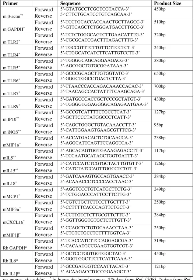
2.1.8.3 Primers ... 49
2.1.8.4. Semi-Quantitative RT-PCR ... 51
2.1.8.5. Agarose Gel Electrophoresis ... 51
2.2. Materials and Methods Used in Foot and Mouth Disease Experiments ... 52
2.2.1. Mice ... 52 2.2.2. CpG Oligodeoxynucleotiedes ... 52 2.2.3 Preparation of vaccines ... 54 2.2.4. Immunization ... 54 2.2.5. Ig ELISA ... 56 2.2.6. IFNγ ELISA ... 56
2.2.7. Virus Neutralization Assay ... 57
2.2.8. Statistical Analysis ... 57
2.3. Materials and Methods Used in Chicken Experiments ... 58
2.3.1 Chicken Housing ... 58
2.3.2 Immunization ... 58
2.3.3 FCR and EPEF Calculation ... 58
3. Results ... 59
3.1 Immunotherapeutic Use of Suppressive ODN in Endotoxin Induced Uveitis . 59 3.2 Immunosuppressive Effects of Restasis in EIU ... 68
4. Discussion ... 93
4.1 Immunotherapeutic Use of Suppressive ODN in Endotoxin Induced Uveitis . 93 4.2 Immunosuppressive Effects of Restasis in EIU ... 95
4.3 Use of CpG ODN as a Vaccine Adjuvant Against Foot And Mouth Disease . 97 4.4 Prophylactic Use of CpG ODNs in Broiler Chickens ... 100
5. References ... 103
6. Appendices ... 126
List of Figures
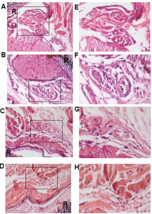
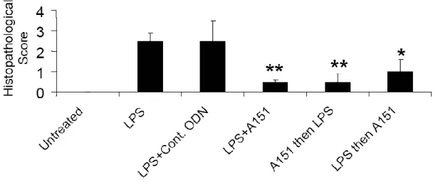
Figure 1.1. Interactions of Innate and Adaptive Immunity ... 6 Figure 1.2. PAMP recognition by cell-surface TLRs ... 13 Figure 1.3. PAMP recognition by intracellular TLRs and the role of UNC93B1 on their trafficking from ER to endolysosome ... 17 Figure 1.4. TIR domain containing adaptors in TLR signaling ... 19 Figure 1.5. MyD88 and TRIF dependent pathways ... 20 Figure 2.1. Injection and bleeding schedule of mice used in immunization studies. . 55 Figure 3.1. Effect of A151 treatment on clinical EIU. ... 60 Figure 3.2. Histopathological changes showing retinal perivascular infiltration in eyes of mice following LPS and/or suppressive ODN treatment. ... 61 Figure 3.3. A151 mediated changes in histopathological scoring following EIU ... 62 Figure 3.4 Suppressive ODN A151 administration after LPS challenge
downregulated IL1β expressions in iris. ... 63 Figure 3.5. Suppressive ODN A151 administration after LPS challenge
downregulated IL6 expressions in cornea. ... 64 Figure 3.6. A) Suppressive ODN A151 treatment after 100 µg LPS challenge
significantly downregulates MIP3α, IP10, iNOS, MIP1β and CXCL16 mRNA expression levels on the eyes of mice. ... 65 Figure 3.7. Suppressive ODN A151 administration significantly suppressed IL6 release from murine splenocytes. ... 67 Figure 3.8. Inhibitory effect of suppressive ODN on LPS mediated immune
activation. ... 68 Figure 3.9. Effect of Restasis treatment on clinical EIU... 69 Figure 3.10. Restasis treatment pre- or post 20 µg LPS challenge significantly
downregulated several TLR mRNA expression levels ... 70 Figure 3.11. Restasis treatment pre- or post 20 µg LPS challenge significantly
Figure 3.12. Restasis treatment pre- or post 20 µg LPS challenge significantly
diminished IL6 levels from murine splenocytes ... 72
Figure 3.13. Restasis treatment pre- or post high dose LPS challenge significantly downregulated A) MCP1, B) IP10 expression levels of mice eyes ... 74
Figure 3.14. Restasis administration after LPS treatment downregulated IL1β and IL6 expressions of rabbit cornea and vitreous ... 76
Figure 3.15. Immunostimulatory effects of different CpG ODNs. ... 79
Figure 3.16. Anti Serotype-O total IgG responses of immunized mice ... 80
Figure 3.17. Anti Serotype-O IgG1 responses of immunized mice ... 81
Figure 3.18. Anti Serotype-O IgG2a responses of immunized mice ... 82
Figure 3.19. FMDV neutralizing Ab Titers of immunized mice at post booster 2 weeks ... 84
Figure 3.20. FMDV neutralizing Ab titers of immunized mice at post booster 8 weeks ... 85
Figure 3.21. FMDV neutralizing Ab titers of immunized mice at post booster 20 weeks ... 86
Figure 3.22. Rapid serum IFNγ response of immunized mice ... 88
Figure 3.23. Mortality percentages of chickens at the end of 41 days. ... 89
Figure 3.24 Body weights of chickens at the end of 41 days ... 90
Figure 3.25. FCRs values of chickens at the end of 41 days. ... 91
List of Tables
Table 1.1. Components of the Immune System ... 3
Table 1.2. PAMP Detection by TLRs and other PRRs ... 9
Table 1.3.Chromosomal localizations of TLRs. ... 10
Table 1.4. Immunostimulatory effect of CpG ODN ... 22
Table 1.5. Use of CpG ODN as an adjuvant with various peptides and vaccines ... 25
Table 1.6. Effect of Suppressive ODN in CpG induced immune activation ... 26
Table 1.7. Effect of Suppressive ODN on IFNγ production induced by several immune activators ... 27
Table 1.8. Different types of immunosuppressive ODNs used for several immunotherapeutic applications ... 30
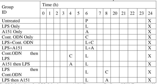
Table 2.1. EIU induction and A151 treatment schedule of mouse experiments ... 44
Table 2.2: EIU induction and Restasis treatment schedule of mouse experiments ... 44
Table 2.3. EIU induction and A151 treatment schedule of rabbit experiments ... 45
Table 2.4. EIU induction and Restasis treatment schedule of rabbit experiments ... 45
Table 2.5. Oligonucleotide PCR primers used in mouse and rabbit experiments .... 50
Table 2.6. List of CpG ODNs used in FMD study ... 53
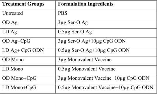
Table 2.7. Injection groups and formulation ingredients used throughout immunization studies ... 55
Table 3.1. Comparative effects of biological and non-biological immunosuppressive agents to control established LPS induced EIU. ... 77
Table 3.2. Number of immunized mice developed detectable virus neutralization titers for a duration of 20 weeks. ... 87
Abbreviations
Ab Antibody
AIM2 Absent in melanoma 2
APC Antigen presenting cell
ASC Apoptosis-associated speck-like protein
BAL Bronchoalveolar lavage
BHK-21 Baby Hamster Kidney -21
BCR B-cell receptor
BEI Binary ethyleneimine
bp Base pairs
BRB Blood-retinal barrier
BSA Bovine serum albumin
CARD Caspase activation and recruitment
domain
CD Cluster of differentiation
cDNA Complementary Deoxyribonucleic Acid
CIA Collagen-induced arthritis
ck Chicken
CLR C-type lectin receptors
CMV Cytomegalovirus CpG Unmethylated cytosine-guaniosine motifs CRP C-reactive protein CsA Cyclosporin A CXCL CXC-chemokine ligand DC Dendritic cell
ddH2O Double distilled water
DNA Deoxyribonucleic acid
dsRNA Double-stranded RNA
EAE Experimental autoimmune
encephalomyelitis
EAU Experimental Autoimmune Uveitis
EDTA Ethylenediaminetetraacetic acid
EIU Endotoxin induced Uveitis
ELISA Enzyme Linked-Immunosorbent Assay
EPEF European Efficiency of Productivity
Factor
ER Endoplasmic reticulum
EtOH Ethanol
FACS Fluorescence-activated cell sorting
FASL Fas ligand
FBS Fetal Bovine Serum
FcR Fc Receptor
FCR Feed Conversion Ratio
Flt3 Fms-like tyrosine kinase 3
FMD Foot and Mouth Disease
FMDV Foot and Mouth Disease Virus
GPI Glycophosphatidylinositol
GVHD Graft versus Host Disease
HBV Hepatitis-B Virus
HEK Human embryonic kidney
HSV Herpes Simplex Virus
hu Human
IFN Interferon
Ig Immunoglobulin
IP-10 Interferon gamma-induced protein 10
IRAK IL-1 receptor-associated kinase
IRBP interphotoreceptor retinoid-binding
protein
IRF3 Interferon-regulatory factor 3
IVA Influenza A Virus
KO Knock out
KC Kerationcyte chemoattractant
LAL Limulus amebocyte lysate
ICAM-1 Intercellular Adhesion Molecule 1
IFI16 Gamma-interferon-inducible protein
IκK Inhibitor kappa B kinase
INH ODN Inhibitory oligonucleotides
IRS Immunoregulatory sequences
LBP LPS-binding protein
LITAF Lipopolysaccharide-induced tumor
necrosis factor-alpha factor
LGP Laboratory of genetics and physiology 2
LPS Lipopolysaccharide
LRR Leucine-rich repeats
LTA Lipotheicoic Acid
mAb Monoclonal antibody
MALP Mycoplasmal lipopeptide
MAP Mitogen-activated protein
MAPK Mitogen-activated protein kinase
MCMV Mouse cytomegalovirus
MCP Monocyte Chemoattractant Protein
MD Myeloid differentiation protein
MDA 3,4-methylenedioxyamphetamine
MHC Major Histocompatibility Complex
MIP Macrophage Inflammatory Protein
mu Murine
MyD-88 Myeloid differentiation primary response
gene (88)
NALP NAcht leucine-rich-repeat protein 1
NFAT Nuclear factor of activated T-cells
NZB/W New Zealand Black/White
NF-κB Nuclear factor-kappa B
NK Natural killer
NKG2D Natural-killer group 2, member D
receptor
NLR Nucleotide-binding oligomerization
domain like proteins or receptors
NO Nitric oxide
NOD Nucleotide-binding Oligomerization
Domain
ODN Oligodeoxynucleotide
PAMP Pathogen Associated Molecular Patterns
PBS Phosphate buffered saline
PBMC Peripheral Blood Mononuclear Cells
PCR Polymerase chain reaction
pDC Plasmacytoid dendritic cells
PGN Peptidoglycan
pIC (polyIC) Polyriboinosinic polyribocytidylic acid
PNPP Para-nitrophenyl phosphate
PRAT4 Protein Associated with TLR4
PRR Pattern Recognition Receptors
PS CpG ODN Phosphorothioate backbone modified
CpG ODN
PS Phosphorothioate
RIG Retinoic acid-inducible gene
RLR Retinoic acid-inducible gene-I-like
receptors
RNA Ribonucleic acid
RPMI Roswell Park Memorial Institute
RSV Respiratory Syncytial Virus
RT-PCR Reverse transcriptase PCR
RT Room temperature
SA-AKP Streptavidin Alkaline-phosphatase
SAP Serum amyloid protein
SAT Southern African Territories
SLE Systemic Lupus Erythematosus
SOCS Suppressor of cytokine signaling
STAT Signal transducer and activator of
transcription
ssDNA Single stranded DNA
ssRNA Single-stranded RNA
TAK1 TGF- β -activated kinase 1
TBK TANK-binding kinase
TCR T-cell receptor
TH T-helper
TIR Toll/IL-1 receptor
TIRAP Toll/IL1 receptor-associated protein
TLR Toll-like Receptor
TNF Tumor Necrosis Factor
TPA 7,12-dimethylbenz(a)anthracene
(DMBA)/12-O-tetradecanoylphorbol-l3-acetate
TRAM TRIF-related adaptor molecules
TRIF TIR domain containing adaptor inducing
IFNβ
UV Ultraviolet
VKH Vogt-Koyanagi-Harada syndrome
Chapter 1
1.
Introduction
1.1 Immune System
All living organisms from bacteria to human have evolved several strategies to combat invading pathogens. In higher organisms the mechanisms involved in providing protection collectively constitute the immune system [1]
In mammals, the epithelial surfaces of the body establish the first lines of defense against invading pathogens. Epithelial cells of the skin, gastrointestinal, respiratory and urogenital tracts are joined by tight junctions and provide a physical barrier between the internal milieu and the external world. Moreover, movement of mucus by cilia and longitudinal flow of air and fluids through internal body surfaces prevents adhesion of pathogens to the epithelium. Other then these physical protection mechanisms, several chemical and enzymatic factors such as antimicrobial peptides, salivary enzymes and low pH of the stomach also contribute to protection from infections. In addition to these defenses, most epithelial surfaces are in contact
with microbial flora which consists of non-pathogenic bacteria, fungi and viruses that compete with the pathogenic ones.[2, 3]
Mammalian immune system consists of two interrelated arms: the evolutionarily conserved, immediate, but non-specific innate immunity and the highly specific but comparably late-acting adaptive immune system. These two systems are interlinked and work together to enable the recognition and elimination of invading pathogens. The innate and the adaptive immune system use different strategies to recognize and eliminate infection. While the innate immune system uses several germ-line encoded receptors to detect invading pathogens, the adaptive immune system uses randomly generated and highly specific receptors for detection of infection. [1]
One unifying feature of both the innate and the adaptive immune system is their ability to discriminate self from non-self. In immunology, “self-molecules” are defined as the host’s own molecules that can be distinguished from other foreign substances. In contrast, “non-self molecules” are those that are recognized as “foreign” by the immune system. By means of this ability to discriminate “self” from “non-self”, the immune system can detect various pathogens such as viruses, bacteria and parasitic worms and distinguish them from normal cells and tissues of the host [4].
Table 1.1. Components of the Immune System (Adapted from[5])
Attribute
Innate Immunity
Adaptive Immunity
Response time Minutes/hours Highly specific;
discriminates even minor differences in molecular structure; details of microbial or nonmicrobial structure recognized with high specificity
Diversity A limited number of germ
line-encoded receptors
Highly diverse; a very large number of receptors arising from genetic recombination of receptor genes
Memory responses None Persistent memory, with
faster response of greater magnitude on subsequent infection
Self/nonself discrimination
Perfect; no microbe-specific pattern in host
Very good; occasional failures of self/nonself discrimination result in autoimmune disease Soluble components of
blood or tissue fluids
Many antimicrobial peptides and proteins
Antibodies
Major cell types Phagocytes (monocytes, macrophages,
neutrophils), natural killer (NK) cells, dendritic cells
T cells, B cells, antigen-presenting cells
Recognition of microbial pathogens by innate immune cells is mediated by germline-encoded pattern recognition receptors (PRRs). PRRs recognize molecular structures that are expressed by a diverse group of pathogens, collectively known as pathogen associated molecular patterns (PAMPs). Following PAMP recognition, PRRs initiate a series of signaling cascades that generate an initial response against the pathogen [6]. PAMP recognition leads to the maturation and activation of antigen presenting cells (APCs) and allows effective presentation of microbial antigens to cells of the adaptive immune system..[7]
Adaptive Immunity can be divided into two subsections: humoral immunity and cell mediated immunity. Humoral immunity involves the production of antibodies by B cells that can opsonize or neutralize pathogens in blood or other body fluids. On the other hand, cell mediated immunity is responsible for eradication of cancer cells or cells that are infected with intracellular pathogens via cytotoxic T cells. Another class of T cells called T helper cells do not have cytotoxic or phagocytic activity rather they activate other immune cells such as B cells, cytotoxic T cells, macrophages and decide the type of immune response either Th1 or Th2, Th17 or Th9 biased [8, 9] B cells and T cells express B cell receptors (BCR) and T cell receptors (TCR), respectively. These receptors are generated via DNA recombination during the development of these cells and are able to recognize huge numbers of pathogens. Each TCR and BCR consist of a constant and a variable region. Variable region of these receptors are encoded via different gene segments. By random assembly of these gene segments during creation of each receptor, a huge receptor repertoire can be produced. If a pathogen invades the body, T and B cells with corresponding receptors become activated and induce humoral and cellular immune responses. In contrast to the innate immune system, the adaptive immune system has memory When the same pathogen re-invades the body, memory B and T cells expand monoclonally and induce a more rapid and robust immune response [10].
1.2 Innate Immune System
Our bodies are constantly exposed to infectious microorganisms present in our environment. In spite of this frequent exposure, disease occurance is quite low. Most of the microorganisms that can cross the protective epithelial surfaces are detected and eliminated within minutes or hours by defence mechanisms that do not require a prolonged period of induction since they do not rely on the clonal expansion of antigen specific lymphocytes. [2, 11]
Innate immune system is an evolutionarily ancient part of the immune system. Since plants and animals share the same modules, it seems that it arose before the split of these two kingdoms. Defects in innate immunity are almost always lethal, further supporting the evidence that this system is evolutionarily conserved [2, 12].
Innate immune system is composed of many types of cells such as Mast cells, natural killer cells, eosinophils, basophils; and the phagocytic cells including macrophages, neutrophils and dendritic cells (DCs) [13, 14].
If a pathogen can breach the epithelial layer and start to replicate in tissues, it would be recognized by the phagocytes such as DCs, In the classical view of the immune system, DCs engulf invading pathogens, digest their proteins into small peptides and then present these digested peptides on their surface in association with major histocompatibility complex (MHC) proteins. DCs then migrate to the regional lymph nodes and present the antigens to naïve T cells. Once a T cells with a corresponding T-cell receptor recognizes the MHC:peptide complex displayed on the surface of the DC, it becomes activated and starts proliferating (Figure 1.1)
This view is an incomplete one as recent evidence suggests that a DC can only prime a T cell if the DC itself is activated in the first place. Such a prerequisite for DC activation explains why DCs that capture self-antigens from dead or dying cells do
not activate T cells under steady-state conditions (i.e, when there is no infection). For efficient T cell activation to occur, the DCs must first be activated following recognition of PAMPS via the PRRs they express. This recognition leads to a series of signaling events that culminates in induction of co-stimulatory molecule expression on the surface of the DC and provides the “second signal” necessary to fully activate the naive T cell [10].
Figure 1.1. Interactions of Innate and Adaptive Immunity (Adapted from:[10])
1.3 Pattern Recognition Receptors
Innate immune cells express various PRRs that enable them to recognize PAMPs from different origins. Several classes of PRRs such as Toll-like Receptors (TLRs), Retinoic acid-inducible gene (RIG)-I-like receptors (RLRs) and Nucleotide-binding oligomerization domain (NOD)-like receptor (NLRs) have been identified in mammals, each specialized to sense the presence of pathogen at a unique subcellular
(NK) cells and professional antigen presenting cells (APCs) and induces release of several inflammatory and proinflammatory cytokines and chemokines such as TNF, IL1IP10, MIP1, MIP3, MCP Regulated upon activation, normal T-cell expressed, (RANTES), IL6, IL12, IL15, IL18, IFNγ and type I interferons (IFN and IFN; important for anti-viral response) that are pivotal for host defense. [2, 11, 14-17]
Toll-like Receptors (TLRs) are the first identified and most well characterized class of PRRs that can recognize a wide range of PAMPs [6, 13, 14, 18]. TLRs are type I transmembrane proteins containing 3 domains. Ectodomains of TLRs consists of leucine-rich repeats (LRRs) which mediates the recognition of PAMPs. Transmembrane and intracellular Toll interleukin 1 (IL-1) receptor (TIR) domains are required for downstream signal transduction. To date 12 different functional TLRs in mice and 10 different functional TLRs in human are identified [19]. Each TLR detects distinct PAPMs originating from viruses, mycoplasma, bacteria, fungi and parasites such as lipoproteins (TLR1,TLR2,TLR6), double stranded (ds) RNA (TLR3), lipopolysaccaride (LPS) (TLR4), flagellin (TLR5), single stranded (ss) RNA (TLR7, TLR8) and DNA (TLR9) [6, 13, 19].
TLRs are expressed on various cell types including immune and non-immune cells such as B cells, macrophages, NK cells, DCs, epithelial cells, endothelial cells and fibroblasts. However, cellular localization of TLRs differs according to the specific ligands they recognize. While TLR1, TLR2, TLR4, TLR5, TLR6 are localized on the plasma membrane, nucleic acid sensing TLRs such as TLR3, TLR7, TLR8 and TLR9 are localized in endosomes [6, 10, 13, 14].
PAMP recognition with corresponding TLRs initiates several downstream signaling pathways that lead to the production of inflammatory cytokines, type I IFNs, chemokines and antimicrobial peptides. These modulators cause recruitment of neutrophils, induction of IFN stimulated genes and direct killing of the pathogen.
Furthermore, activation of TLR signaling leads to the activation of DCs, thereby contributing to the induction of adaptive immune responses [6, 19]
Although TLRs can recognize several PAMPs that are extracellular or present in endosomes, other non-TLR PRRs such as membrane-bound C-type lectin receptors (CLRs)[20], cytosolic proteins such as NOD-like receptors (NLRs) [21] and RIG-I-like receptors (RLRs) [22], also contribute to the recognition of PAMPs. CLRs are membrane bound proteins that can induce inflammatory responses upon recognition of PAMPs from different origins such as bacteria and fungus. NLRs are a large group of cytosolic proteins that contains more than 20 members. NLRs respond to various types of PAMPs to trigger an innate immune response. NLRs such as NALP1 and NALP3 form inflammasomes with the contribution of ASC and Caspase-1 and mediate the processing of pro- IL1β to mature IL1β [21, 23]. RLRs are RNA helicases that can recognize RNA species in various types of cells. RLRs such as RIG-1, MDA5 and LGP2 recognize RNA in the cytoplasm and coordinate antiviral responses via type I IFN induction [24]
In case of an infection with an intact pathogen, several classes of PRRs recognize several molecular structures expressed on that pathogen which leads to multiple activation of PRRs due to multiple PAMP exposure. Moreover, different PRRs can recognize the same PAMP. The nature and the strength of the generated innate immune response depends on the coordination between TLRs and other non-TLR PRRs. In conclusion, TLRs in concert with other PRRs orchestrates innate immune responses against various pathogens such as bacteria, fungi, viruses and other protozoan parasites [6]. Table 1.2 shows PAMPs originating from different pathogens and their relevant PRRs expressed by immune cells.
Table 1.2. PAMP Detection by TLRs and other PRRs (Adapted from [6])
Species PAMPs TLR Usage PRRs Involved
in Recognition
Bacteria, mycobacteria
LPS TLR4
Lipoproteins, LTA PGN TLR2/1, TLR2/6 NOD1, NOD2, NALP3, NALP1
flagellin TLR5 IPAF, NAIP5
DNA TLR9 AIM2
RNA TLR7 NALP3
Viruses
DNA TLR9 AIM2, DAI, IFI16
RNA TLR3, TLR7, TLR8 RIG-1, MDA5, NALP3 structural protein TLR2, TLR4 Fungus
zymosan, β glucan TLR2, TLR6 Dectin-1, NALP3
Mannan TLR2, TLR4 DNA TLR9 RNA TLR7 Parasites tGTPI-mutin (Trypanosoma) TLR2 glycoinositolphospholipids, (Trypanosoma) TLR4 DNA TLR9
hemozoin (Plasmodium) TLR9 NALP3
Profiling-like molecule
(Toxoplasma gondii) TLR11
1.4 Toll-Like Receptors
Toll protein was first identified in fruitflies (Drosophila) as a protein that is involved in defining dorsoventral polarity during embryogenic development.[25] Following this, its role in mounting an effective immune response against the fungus
Aspergillus fumigatus was also described, causing researches to focus on the
identification of its homologues in mammals. In 1997, Janeway and his colleagues reported that “A human homologue of the Drosophila Toll protein signals activation
of adaptive immunity.” and named the homologue as Toll-like Receptors [26]. As mentioned in the previous section, to date several TLRs have been identified in mouse and human and each recognize a distinct PAMP. Table 1.3 shows the chromosomal locations of the genes encoding TLRs [6, 13, 19] (Table 1.2 and Table
1.3).
Table 1.3.Chromosomal localizations of TLRs.
Chromosome TLR Mouse Human TLR1 5 37.0 cM 4p14 TLR2 3 E3 4q32 TLR3 8 B2 4q35 TLR4 4 33.0 cM 9q32-q33 TLR5 1 98.0 cM 1q41-q42 TLR6 5 37.0 cM 4 4p14 TLR7 X F5 Xp22.3 TLR8 X F5 Xp22 TLR9 9 F1 3p21.3 TLR10 N/A 4p14 TLR11 14 C1 N/A TLR12 4 D2.2 N/A TLR 13 X D N/A
TLRs can be devided into two groups with regard to their cellular localizations: One group is expressed on the cell surface and include TLR1, TLR2, TLR4, TLR5, TLR6, TLR11 responsible for recognition of mainly microbial membrane components such as proteins, lipids and lipoproteins [19] (Figure 1.3). The other group consists of TLR3, TLR7, TLR8 and TLR9 which are expressed in the intracellular compartments and recognize nucleic acid ligands [19] (Figure 1.3).
1.4.1 Cell Surface Toll-Like Receptors
1.4.1.1 TLR1, TLR2 and TLR6
can be explained by its ability to form heterodimers with TLR1, TLR6 or with non-TLR molecules such as CD36, CD14 and dectin-1. non-TLR2-non-TLR1 heterodimers and TLR2-TLR6 heterodimers recognize triacyl-lipopeptide from Gram negative bacteria and diacyl-lipopeptide from mycoplasma, respectively. [13, 27, 28] Thus, TLR1 and TLR6 functionally associate with TLR2 and discriminate between diacyl or triacyl lipopeptides [29]. TLR2 also recognizes zymosan with association with dectin-1, a C-type Lectin Receptor. Furthermore, a type of class II scavenger receptor called CD36 functions as a co-receptor in sensing LTA and MALP-2 molecules of Staph.
Aureus by the TLR2-TLR6 heterodimer [30].
1.4.1.2 TLR4
TLR4 is the receptor that recognizes lipopolysaccharide (LPS), a component of the outer membrane of Gram-negative bacteria. LPS is a potent immunostimulatory molecule and causes septic shock [31]. The recognition of LPS by TLR4 is dependent on the complex formation of TLR4 with a recognition subunit, MD2 (myeloid differentiation protein-2) and the membrane bound GPI (glycosylphosphatidylinositol) anchored CD14 [32].Additionaly, LPS binding protein (LBP) is another important protein that plays a role in LPS recognition [33]. LBP acts as a soluble or plasma membrane protein that binds LPS. CD14 binds LBP and delivers LPS-LBP complex to the TLR4-MD2 complex. MD2 binds to lipid A (active component of LPS) and this binding activates the dimerization of two TLR4 receptors and TLR4 signalling pathway [24]. In addition to LPS, TLR4 also recognizes glycoinositolphospholipids from Trypanosoma [34] and the fusion protein from RSV (respiratory syncytial virus) [13, 35].
1.4.1.3 TLR5
TLR5 recognizes flagellin, which is the monomer of an important structural protein of pathogenic and non-pathogenic bacteria called the flagella [36]. Flagellin induces mucosal immune responses by acting on TLR5 expressed on epithelial cells or
macrophages. The amino acid sequence of flagellin is highly conserved and hence is a target of innate immunity. TLR 5 is expressed in the digestive tract, the urinary tract and the respiratory tract, all constituting surfaces that are prone to infection by flagellated bacteria. In intestinal eptihelia, TLR 5 is expressed only in the basolateral surface and not on the apical surface of the cells. This polarized expression pattern, enables TLR5 to recognize only invading bacteria that have breached the epithelial barrier but not the non-invading commensals. TLR5 activation induces chemokine expression from epithelial cells and thereby contributes to inflammatory responses and leukocyte accumulation at the site of infection [10].
1.4.1.5 TLR11
TLR11 recognizes profilin-like molecules of Toxoplasma Gondii, an intracellular protozoan parasite [37]. The active component of this profilin-like molecule has a role in parasite motility and invasion [38]. In addition to its role in recognition of profilin-like molecules originating from Toxoplasma Gondii, TLR11 also contributes to protection from uropathogenic bacteria as mice deficient for TLR11 were shown to be highly susceptible to infection with such bacteria [39]. Moreover, TLR11 expression in the kidney and bladder is higher than that of TLR5, suggesting that this receptor is likely to sense uropathogenic bacteria-derived products, although no such “specific ligand” has been identified thus far [24, 39]. Interestingly, TLR11 is expressed only in mice but not in humans which is attributed to the presence of at least one premature stop codon [24].
Figure 1.2. PAMP recognition by cell-surface TLRs (Adapted from [19])
1.4.2 Intracellular Toll-Like Receptors
TLR3, TLR7, TLR8, TLR9 are the receptors specialized for sensing foreign nucleic acids. These receptors are expressed within the intracellular compartments such as endosomes, lysosomes and the endoplasmic reticulum (ER) [40, 41]. Upon ligand recognition, these receptors initiate an anti-viral innate immune response characterized by production of type I IFNs and inflammatory cytokines [24] (Figure 1.2).
1.4.2.1 TLR3
TLR3 recognizes viral ds RNA from etiher ds RNA viruses or ssRNA viruses that are producing dsRNA during their replication [42-44]. TLR3 also recognizes a synthetic analogue of ds RNA known as polyI:C. TLR3 is expressed both in immune cells such as conventional DCs, macrophages, B cells, NK cells and non-immune cells such as uterine, airway, vaginal, cervical, corneal, biliary, and intestinal epithelial cells, which function as efficient barriers to infection [13]. Interestingly,
TLR3 is not expressed in plasmocytoid DCs (pDCs) which are potent type I IFN producers. In addition, TLR3 is highly expressed in the brain [13, 45]. Viral RNA recognition via TLR3 triggers production of type I IFNs and inflammatory cytokines. Nevertheless, despite such a response, TLR3’s role in protection against viral infections is controversial. Some reports indicate that TLR3 deficient mice are more susceptible to infections with mouse cytomegalovirus (MCMV) and West Nile Viruses (WNV) [46, 47]. In contrast, other reports indicate that TLR3 plays a role in pathogenesis rather that protection. For example, TLR3 deficient mice were shown to have increased survival rates following infection with WNV or influenza A virus (IVA) [43, 48]. Moreover, it has been shown that TLR3 deficiency does not affect anti-viral responses of CD4+ and CD8+ T cells following infection with MCMV, reoviruses and lymphocytic choriomeningitis virus [49].
1.4.2.2 TLR7 and TLR8
TLR7 and TLR8 recognizes ssRNA, synthetic antiviral imidazoquinoline compounds (R848, Imiquimod, etc.) and some guanine nucleotide analogs (loxoribine etc. ) [6, 50]. TLR7 and TLR 8 genes show high homology to each other and both are expressed on X chromosome. Although both genes are expressed in mouse, mouse TLR8 seems to be non-functional [13]. TLR7 and TLR8 are located in endosomal compartments .In general, viruses gain entry into cells via receptor mediated endocytosis and reach the phagolysosomes. Phagolysosomes have a highly acidic environment and contain degradative enzymes that hydrolyse virus coat proteins, leading to the exposure of viral RNA to TLRs. In contrast, host’s ssRNA cannot enter into these compartments due to the activity of RNases in plasma [51]. TLR 7 is highly expressed in pDCs and triggers the secretion of huge amounts of type I IFNs from these cells in response to viral infection [24, 52].
production of several inflammatory cytokines and type I IFNs from DCs, B cells and macrophages [19]. Synthetic CpG dinucleotide motif containing ssDNAs can mimic the immune stimulatory activity of bacterial or viral dsDNA [51]. Several different factors seem to contribute to the recognition of DNA by TLR9: Most reports indicate that recognition is due to the presence of unmethylated CpG motifs expressed 20 times more abundantly in bacterial and viral genomes than the mammalian genome. Furthermore, in mammals, CpG motifs are highly methylated and do not activate the mammalian immune cells [13, 51, 57-59]. This theory is supported by publications indicating that bacterial DNA must be delivered to endosomal compartments with acidic environments and degraded into multiple CpG containing ssDNAs that can interact with TLR9 [40, 60]. In contrast, some reports correlate the recognition of DNA by TLR9 to the presence of 2’ deoxyribose-phosphate backbone [61, 62] specifically in endosomes. It has been shown that a chimeric form of TLR9 localized on the cell surface has been shown that can respond to self DNA [63]. It is speculated that CpG motifs may play a role in the stability, aggregation and uptake of the DNA by cells but is not necessary for TLR9 recognition [50] A more recent publication challenged the role of CpG motifs claiming that the 2’ deoxyribose-phosphate backbone is important in recognition of phosphodiester backbone DNA irrespective of its CpG content while CpG motif is indispensible in recognition of phosphorothioate backbone modified DNA [61] Whether TLR9 recognition of DNA depends strictly on CpG recognition or on the presence of 2’ deoxyribose-phosphate backbone in endosomes, it seems that endosomal co-localization is essential for this recognition [50]. For instance, even though malarial DNA itself is not highly stimulatory, when it is delivered into the endosomal compartments via transfection or bound to malarial hemozoin, it induces a strong immune responses [64]. In another study it has been shown that LL37, a human antimicrobial peptide released from keratinocytes during skin injury binds to self-DNA and creates aggregates that can enter endosomes, contributing to immune activation in an autoimmune disease called psoriasis [65].
1.4.2.4 Trafficking of intracellular TLRs
As mentioned above, nucleic acid sensing TLRs such as TLR3, TLR7, TLR8 and TLR9 are expressed in various intracellular compartments such as endosomes, lysosomes and the endoplasmic reticulum (ER) [19, 50]. This intracellular localization is important in terms of two situations. Firstly, viral nucleic acids are delivered via receptor mediated endocytosis and they are recognized in these endosomes that express TLRs. Secondly, this localization is also important in terms of discriminating self from non-self [24]. It has been shown that macrophages express TLR9 on the cell surface responds DNA originating from self [63]. These intracellular TLRs are expressed in the ER in resting cells and upon PAMP stimulation they traffic to the endosomal compartments [24]. It has been shown that activation occurs only in the acidified endosomes since usage of agents that prevents endosomal acidification abrogated TLR3, TLR7, TLR8 and TLR9 responses [66].
Trafficking of intracellular TLR3, TLR7 and TLR9 are mainly regulated by a12 membrane spanning ER protein called UNC93B1. UNC93B1 interacts with transmembrane domains of these receptors and assists in delivery from ER to the endolysosome [67, 68] (Figure 1.2). It has been shown that only a single missense mutation on UNC93B1 causes defects in cytokine production and co-stimulatory molecule up-regulation in response to TLR3, TLR7, TLR9 ligands [69]. In addition, it has been also reported that UNC93B1 deficiency is responsible for HSV-1 encephalitis in human [70].
Figure 1.3. PAMP recognition by intracellular TLRs and the role of UNC93B1 on their trafficking from ER to endolysosome (Adapted from: [19])
Two other ER proteins are also involved in TLR trafficking PRAT4A and gp96. PRAT4A plays role in the trafficking of TLR1, TLR2, TLR4, TLR7 and TLR9 from ER to plasma membrane or endolysosome [71]. However it does not affect TLR3 trafficking indicating that TLR3 trafficking is regulated in a different way than TLR7 and TLR9 [72]. On the other hand, gp96, another ER protein, functions as a general chaperone for most of the TLRs including cell surface expressed ones such as TLR1, TLR2, TLR4,TLR5 and intracellular ones such as TLR7 and TLR9 [73].
1.4.2.5 Endosomal Cleavage of Intracellular TLRs
As mentioned in previous sections endosomal acidification is required for the activation of intracellular TLRs. This acidification is important from two aspects. Firstly, acidic environment and enzymes in endosomes facilitates the disassembly of microbes in order to release their contents such as CpG DNA [40, 60]. Secondly, endosome maturation is involved in essential processing of TLRs themselves. For
instance, ectodomain of TLR9 is proteolytically cleaved by proteases upon its arrival to the endosomal compartment which generates a functional receptor that can recognize ligand and initiate signal transduction [74, 75]. Proteases involved in the cleavage of TLR9 involves cathepsin B, cathepsin S, cathepsin L, cathepsin H and cathepsin K, and asparaginyl endopeptidase [74-78]. Similar cleavage process was also shown for TLR7 but not TLR3 [74] indicating that cleaved forms of TLR7 and TLR9 may display a similar ligand binding structure to full-length TLR3 [50]
In conclusion, in addition to other pre and post translational regulatory mechanisms, cleavage appears to be an additional step in the maturation of TLRs [50].
1.5 TLR Signalling
Upon PAMP recognition, TLRs trigger specific immune responses. For instance, while TLR3 and TLR4 initiates both type I IFNs and inflammatory cytokine responses, cell surface TLRs such as TLR1-TLR2, TLR2-TLR6 and TLR5 induces mainly inflammatory cytokines. This specificity depends on the recruitment of a single or a specific combination of The Toll/Interleukin-1 receptor (TIR) domain containing adaptor protein such as Myeloid differentiation primary response gene 88 (MyD88), TIR domain containing adaptor protein (TIRAP), TIR-domain-containing adapter-inducing interferon-β (TRIF) and TRIF related adaptor molecule (TRAM) [6]. TLR1, TLR2, TLR4, TLR5, TLR6, TLR7, TLR8, TLR9 and TLR11 recruit MyD88 to their TIR domains upon ligand recognition whereas TLR3 and TLR4 recruits TRIF. TLR1, TLR2, TLR4 and TLR6 use TIRAP, which serves as a linker between TIR domain of these TLRs and MyD88 whereas TLR5 and TLR11 binds directly to MyD88. Similarly, TLR4 uses TRAM in order to bind TRIF. These recruitments of adaptor molecules to specific TLRs initiates the signaling cascade and activation of transcription factors such as nuclear factor kappa B (NFκB) and interferon regulatory factor (IRFs) [15] (Figure 1.4)
Figure 1.4. TIR domain containing adaptors in TLR signaling (Adapted from [10])
TLR signaling can be divided into two categories, MyD88-dependent and TRIF- dependent pathways. TLR4 is the only TLR that uses all four adaptors and induces both pathways [19].
1.5.1 MyD88-dependent pathway
All TLRs (except TLR3) use MyD88-dependent pathway. Upon ligand recognition TLRs recruit MyD88 molecules to their TIR domains and MyD88 recruits IL1 receptor associated kinase (IRAK) family of proteins and TNF receptor associated factor (TRAF) 6. TRAF6 activates TAK1 and TAK1 activates the IκB kinase (IKK)complex in order to activate NFκB and also activates the mitogen-activated protein kinase (MAPK) pathway [15] (Figure 1.5)
1.5.2 TRIF-dependent pathway
TLR3 and TLR4 use TRIF-dependent pathway upon ligand recognition and initiate the production of type I IFNs and inflammatory cytokines from DCs and macrophages. TRIF activates the transcription factor IRF3 via activating the kinase protein that phosphorylates IRF3 called TBK1. After phosphorylation IRF3 translocates from cytoplasm to nucleus and triggers the production of type I IFNs. Since inducing type I IFNs, TRIF-dependent pathway is involved in anti-viral responses. TLR7 and TLR9 recognizes viral nucleic acids however molecular mechanism underlying the production of type I IFNs upon TLR7 and TLR9 activation is different. In pDCs MyD88 directly interacts with IRF7 and induces type I IFN production [10] (Figure 1.5)
1.6 Effects of DNA on Immune System
DNA and RNA are the essential components of all living organisms. Accumulated evidence strongly suggests that these nucleic acids have multiple and complex effects on the immune system and are more than a blueprint of life [79-81]. DNA is normally isolated from the extracellular world via nuclear or mitochondrial membrane in eukaryotes, the cell wall in bacteria, or the envelope in viruses. Following microbial infection or tissue damage DNA can be released from microbes or damaged host cells. Such DNA is detected by the immune system [13, 82] On one hand, due to their high unmethylated CpG motif frequency, bacterial DNAs are recognized as “non-self” via TLR9 and trigger an innate immune response characterized by the proliferation and maturation of B cells, natural killer cells, and pDCs and the secretion of Th1 type cytokines, chemokines, and/or multivalent immunoglobulins [83-88]
On the other hand, telomeric regions of mammalian chromosomes contain suppressive TTAGGG motifs that can inhibit several dependent and TLR-independent Th1-mediated immune responses. Of note, these motifs are underrepresented in the prokaryotic genome [31, 89, 90]. Novel therapeutics including nucleic acid based agonists/antagonists are currently under preclinical/clinical trials to prevent or treat cancer, infectious disease, allergic disorders and autoimmunity [91-93].
1.6.1 Immunostimulatory CpG ODN
CpG ODN story started with the observation that synthetic nucleic acid fractions originally found in bacteria has antitumor effects. It was first hypothesed palindromic sequences within these sequences were the cause of this immune stimulation [94] but it was shown by Krieg and Klinmann in 1995 that specific sequence motifs consisting of unmethylated CpG dinucleotide flanked by two 5’ purines and two 3’ pyrimidines were responsible for the innate immune response triggered by such DNAs [84, 95]. Mammalian DNA has approximately 20 times less frequent CpG motifs and due to methylation status they are not immune stimulatory like microbial DNA (Table 1.4)
Table 1.4. Immunostimulatory effect of CpG ODN (Adapted from [96])
Fold Increase in Cytokine secreting cell number
IL6 IL12 IFNγ IgM
Bacterial DNA 3.2±0.2 3.8±0.4 4.7±2.3 3.9±1.1 Mammalian DNA 0.8±0.2 1.1±0.2 0.8±0.3 0.7±0.2 CpG ODN 5.5±1.1 8.3±1.7 4.7±1.1 4.2±1.6 CpG ODN (methylated) 0.9±0.2 1.2±0.3 0.8±0.2 1.1±0.2 CpG ODN (DNase Rx’d) 1.3±0.2 0.8±0.2 1.1±0.2 0.9±0.2 GpC ODN 1.2±0.3 1.3±0.3 1.2±0.3 1.3±0.3
Bacterial DNA or synthetic ODNs expressing these CpG motifs induce immunostimulation characterized by a proinflammatory (IL1, IL6, IL18, TNFα) and T helper 1 (Th1) biased (IFNγ and IL12) immune milieu [96]
Five years after the discovery of CpG motifs, in 2000 Akira et al., indicated that the receptor responsible for CpG ODN recognition is TLR9 [53] As mentioned in previous section CpG ODNs are internalized into endosomal compartments where they are recognized by TLR9 and induce a robust innate immune response [51].
There are three known types of CpG ODN: D-type (also known as A-class), K-type (also known as B-class) and the recently described C-class, all of which possess unmethylated CpG dinucleotides and require TLR9 to activate the immune system [92, 97]. These 3 types of ODN possess CpG dinucleotides, but their flanking sequences and compositions are different. For example, K-type ODN contain multiple CpG motifs, whereas D-type ODN have one CpG with palindromic flanking sequences. D-, but not K- nor C-type ODN have a poly-G (5-6 bases) tail at the 3’-end, which may account for their distinct activity. K- and C- but not D type ODN have phosphorothioate linkage between all nucleotides. D-type ODN stimulate plasmacytoid DC (pDC) to secrete large amounts of IFNα, whereas K-type ODN strongly stimulate B cells to proliferate and to secrete IL6 and IgM. C-type ODN show a combined activity of K- and D-type ODN, but to a lesser extent [16, 80].
Several reports indicated that the IFNα induction from pDCs by D type ODNs were due to the ability to spontaneously form higher order structures because of their palindromic sequences and poly (G) since such higher order structures are not observed in K types [98, 99]
In 2006, Gursel et al., discovered the co-receptor CXCL16 expressed on pDC which can contribute to discribe the dichotomy of response between D and K types. In this work, her group demonstrated for the first time that a type of surface expressed scavenger receptor is required for the D-ODN activation of pDC to secrete robust IFNα [17].
1.6.2 CpG DNA as adjuvants for vaccines
CpG ODN which acts as ligand for innate immune receptors also supports the development of adaptive immune responses via improving the functional activity of professional APCs and triggering the production of cytokine and chemokines that are critical in the development of adaptive immune responses [84, 88, 100, 101].
CpG ODNs activate innate immunity by binding their receptor TLR9 located in endosomal compartments of pDCs, B-cells, and multiple cells of myeloid lineage in mice [102]. CpG ODNs have been shown to activate B cells via upregulating the secretion of IgM, IL6 and IL10, inducing the expressions of Fc receptor (FcR) and costimulatory molecules such as CD40, CD80, CD86 and major histocompatibility complex II (MHCII), inhibiting the apoptosis and enhancing the antigen presentation function [95, 102-104]. Effects of CpG ODNs are not limited on B cells. CpG ODNs have also been shown to induce the maturation and differentiation of other antigen-presenting cells such as plasmacytoid dendritic cells (pDCs) and macrophages. pDCs and macrophages show increased expression MHCII and costimulatory molecules (CD40, CD80, CD86) and cytokines such as IL6, IL12, IL1β upon CpG stimulation [102, 105].
Several studies indicated that use of CpG ODN as an adjuvant increased protein immunogenicity, cytokine production and IgG2a:IgG1 ratio [102, 103, 106, 107]. Table 1.5 summarizes the type and magnitude of immune response induced with the use of CpG ODN with the conventional protein or vaccine based antigens
Table 1.5. Use of CpG ODN as an adjuvant with various peptides and vaccines (Adapted from [88].)
Antigen Fold Ab Titer Ig Profile Cytokine Profile
References
Ovalbumin >7fold (3wk) IgG2a>IgG1 IFNγ [106] Hen eggwhite
lysozyme
>10fold (3wk) IgG2a>IgG1 IFNγ, IL5 [108]
Hepatitis B Surface Antigen >104fold (4wk) [109]
Influenza virus >10 fold (4wk)
Intranasal IgA IFNγ [110]
Measles virus >20 fold (4wk)
IgG2a>IgG1 IFNγ, IL5 [111]
Tetanus Toxoid > 3fold (6wk) [112] Brucella >100 fold (3wk) IgG2a>IgG1 IFNγ [113]
rPA (anthrax) >8 fold (3wk) IgG2a>IgG1 IFNγ [114, 115] L1 and A33 proteins (smallpox) >10 fold (3wk) IgG2a>IgG1 [116] 1.6.3 Immunosuppressive ODN
During infection or tissue damage, inflammation must be waned and terminated with tissue remodeling and healing [80]. CpG-driven immune activation can exacerbate inflammatory tissue damage, or increasing sensitivity to autoimmune diseases or toxic shock. Similarly, other immune responses designed to protect the host can have deleterious consequences if not adequately regulated [117]. Recent evidence suggested that host DNA contained some antagonistic elements to the immunostimulatory effect in their DNA or against pathogen derived CpG rich DNA,
possibly suppressing DNA-driven immunostimulation [118]. Neutralizing or suppressive motifs can selectively block CpG-mediated immune stimulation [119]. Suppressive motifs are rich in poly-G or GC sequences, and optimal motifs are surprisingly identical to telomere sequences (with a repeat of TTAGGG), which are present in DNA of mammals, but not in bacteria [79]. Gursel and his colleagues demonstrated for the first time that telomeric DNA consisting of TTAGGG repeats can significantly suppress immune activation while non-telomeric mammalian DNA cannot. Furthermore they also showed that DNA isolated from telomerase deficient KO mice was not capable to suppress immune responses compared to normal DNA [79].
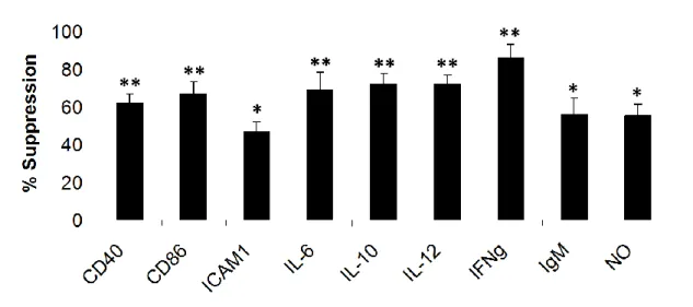
In vitro, it has been shown by many groups that mammalian telomeric suppressive ODN (A151) inhibits the production of several proinflammatory cytokines and chemokines induced by bacteria such as IL6, IL12, IFNγ, TNFα and MIP2α [79, 120-122]. As summarized in Table 1.6and Table 1.7, Suppressive ODN can significantly reduce cytokine/chemokine production and immune cell proliferation induced by CpG ODN or several other different immune activators [122]
Table 1.6. Effect of Suppressive ODN in CpG induced immune activation (Adapted from [122]). Treatment % Activation CpG ODN alone 100±2 +Mammalian DNA 27±4 +Telomeric DNA 13±3 +Non-telomeric DNA 87±5 +Control ODN 97±3
Table 1.7. Effect of Suppressive ODN on IFNγ production induced by several immune activators (Adapted from [122]).
Stimulus None Control ODN Suppressive
ODN (A151)
CpG DNA 27±5 24±6 6±2
ds RNA 8±2 10±1 2±1
Peptidoglycan 28±5 31±4 5±2
LPS 24±7 21±3 6±2
Effects of Suppressive ODN (A151) in the treatment of different autoimmune and inflammatory diseases in several animal models have been studied by us and others. In 2005, Shirota and Gursel showed that suppressive ODN treatment immediately prior to LPS challenge significantly improved survival rates in BALB/c mice injected with lethal dose of LPS [31].
In another study, effect of Suppressive ODN (A151) on the onset of systemic lupus erythematosus in NBZ/W mice has been studied. In this study, investigators injected mice twice per month starting at age 6 weeks with Suppressive ODN or Control ODN and several characteristics of the disease such as proteinuria, kidney inflammation and also survival has been followed. According to their results Suppressive ODN significantly delayed the onset of proteinuria and inflammation. Most importantly survival rates of the animals have been prolonged [123].
Rheumatoid Arthritis (RA) is the most common rheumatic disease which is characterized by destruction, disability and deformity of the joints. Effect of Suppressive ODN (A151) on RA is studied in an animal model called collagen-induced arthritis (CIA). In this study arthritis was collagen-induced by collagen administration. Authors indicated that Suppressive ODN (A151) treatment significantly reduced the incidence and severity of the arthritis and also decreased serum titers of pathogenic IgG autoantibody titers and IFNγ production by collagen reactive T cells [124]
Furthermore, effects of Suppressive ODN (A151) in Silica induced Pulmonary Inflammation have been studied. Silicosis was induced via instilling silica into the lungs of normal BALB/c mice. Silica instilling increased bronchoalveolar lavage (BAL) cellularity and production of IL12 and kerationcyte chemoattractant (KC) proteins. These effects were significantly reduced in Suppressive ODN treated groups [125].
Uveitis is an ophthalmic disorder that causes vision loss in developed countries [126, 127] and is characterized by acute, recurrent, or persistent ocular inflammation, the breakdown of the blood-ocular barrier, and infiltration of leukocytes [128]. Fujimoto et al. [129] reported that suppressive A151 ODN can inhibit ocular inflammation in two murine models, IRBP (interphotoreceptor retinoid-binding protein)-induced experimental autoimmune uveitis and adoptively transferred ocular inflammation. In 2010, we published that Suppressive ODN can reduce the symptoms associated with a very aggressive form of experimental uveitis initiated via endotoxin administration [81]. Compared to previous studies our animal model was a much more aggressive form of the disease and Suppressive ODN was still able to alleviate several disease induced parameters as described in Results section of this thesis.
Over the last few years, remarkable improvements have been achieved by researchers in Suppressive ODN field. In 2011, Klinman and colleagues showed the effects of Suppressive ODN (A151) on a type of cancer that inflammation plays a critical role called papilloma. In this study, authors used 7,12-dimethylbenz(a)anthracene (DMBA)/12-O-tetradecanoylphorbol-l3-acetate (TPA) in order to induce skin carcinogenesis. Their results indicated for the first time that Suppressive ODN blocked TPA-dependent skin hyperplasia, edema, and leukocytic infiltration. Most importantly, their results suggested that Suppressive ODN reduced papilloma formation in a dose and sequence dependent manner. Up to date, this was the first and only study suggesting that Suppressive ODN can be used in inflammation associated oncogenesis [130]
Their observations were supported by other researchers indicating that Suppressive ODNs can improve the antiproliferative effects of anticancer drugs [131]. Most recently, again Klinman and colleagues demonstrated that Suppressive ODN A151 is capable of reducing lung cancer susceptibility of mice with silicosis [132]. As mentioned before in this section of this thesis, Klinman and colleagues have already established the inhibitory role of Suppressive ODN on silicosis. In addition to their previous observations, in this study researchers investigated the role of potent carcinogens present in cigarette smoke (NNK) in development of lung cancer in mice with silicosis and protective effects on Suppressive ODNs in pathologic inflammation and cancer. In conclusion, authors reported that Suppressive ODN can inhibit pulmonary fibrosis and other inflammatory manifestations of chronic silicosis and so as lung cancer [132].
As mentioned in the beginning of this section, Suppressive ODN A151 is a sequence that contains poly G runs. Guanosines are tend to make interchain and intrachain Hoogstein Bonds and these interactions led the sequence to form higher order structures called G-tetrads. It has been shown by Gursel et al., that the suppressive activity of ODN A151 is correlated with its ability to form G-tetrads. They showed that nucleotide substitutions that inhibit G-tetrad formation abrogated the suppressive activity of these sequences [79].
Suppressive ODN (A151) is not the only sequence motif with suppressive properties. Several reports indicate that sequences other than A151 can also reduce immune responses. Sequences showing suppressive activity, used disease models, potential modes of actions and TLR dependence are given in Table 1.8
![Figure 1.2. PAMP recognition by cell-surface TLRs (Adapted from [19]) 1.4.2 Intracellular Toll-Like Receptors](https://thumb-eu.123doks.com/thumbv2/9libnet/5793221.117853/34.892.187.742.159.501/figure-pamp-recognition-surface-tlrs-adapted-intracellular-receptors.webp)
![Figure 1.3. PAMP recognition by intracellular TLRs and the role of UNC93B1 on their trafficking from ER to endolysosome (Adapted from: [19])](https://thumb-eu.123doks.com/thumbv2/9libnet/5793221.117853/38.892.203.712.142.565/figure-pamp-recognition-intracellular-tlrs-trafficking-endolysosome-adapted.webp)
![Figure 1.5. MyD88 and TRIF dependent pathways (Adapted from [15])](https://thumb-eu.123doks.com/thumbv2/9libnet/5793221.117853/41.892.181.781.421.869/figure-myd-trif-dependent-pathways-adapted.webp)