ORIGINAL PAPER
Clinicopathological studies on facial eczema outbreak
in sheep in Southwest Turkey
Ozlem Ozmen&Sima Sahinduran&
Mehmet Haligur&Metin Koray Albay
Accepted: 10 January 2008 / Published online: 23 January 2008 # Springer Science + Business Media B.V. 2008
Abstract After very hot summer, 22 sheep from 5 different flocks consisting of approximately 150–200 animals each were diagnosed with facial eczema in September 2005, in southwest Turkey. Photophobia, corneal opacity, severe ulcers of the facial skin, especially localized around the eyes and mouth, and 3% mortality were the most prominent clinical symp-toms. GGT levels of the animals were very high and varying between 261- 328 U/l. While the activities of ALT and total bilirubin were elevated and AST was normal in affected sheep. Total bilirubin level was higher than normal. Seven of the 22 sheep were euthanatized and necropsy was performed on all of these animals. Severe icterus, hepatomegaly, enlarged gallbladder, congestion of mesenteric vessels were the common necropsy findings. Histopathological changes of the liver included necrosis of the hepatocytes, cholangiohepatitis characterized by mononuclear in-flammatory cell infiltrate in the portal area and mild to severe fibrosis around bile ducts. A diagnosis of
sporidesmin toxicosis was made based on the histopa-thology of the livers, the elevation in liver enzymes, and the development of cutaneous lesions consistent with photosensitization and high spore counts in the ruminal contents. Surviving sheep were treated with procaine penicillin + dihidrostreptomycin sulfate, multivitamin complexes and flunixin meglumine. Additionally, zinc sulphate was also given at a dose of 6 gr per 100 lt drinking water for 28 days. All treated sheep recovered. Pasture spore counts were between 96,300- 267,500 spores/g grass.
Keywords Facial eczema . Sporidesmin toxicosis . Pathology . Sheep
Abbreviations
GGT Gamma Glutamyltransferase ALT Alanine Aminotransferase AST Aspartate aminotransferase
Introduction
Facial eczema is a condition of severe dermatitis in cattle, sheep and goats caused by a toxin in spores of the saprophytic fungus Pithomyces chartarum, which lives in dead vegetative material in pastures (Radostits et al. 2000). The mycotoxin that produced by this fungus is sporidesmin (Jubb et al.1993; Hansen et al. 1994). Facial eczema is an example of “secondary
DOI 10.1007/s11250-008-9132-7
O. Ozmen (*)
:
M. HaligurDepartment of Pathology, Faculty of Veterinary Medicine, University of Mehmet Akif Ersoy,
15100 Burdur, Turkey
e-mail: [email protected]
S. Sahinduran
:
M. K. AlbayDepartment of Internal Medicine, Faculty of Veterinary Medicine, University of Mehmet Akif Ersoy, 15100 Burdur, Turkey
photosensitization,” in which the skin lesions are really the results of liver damage, rather than the direct result of a plant toxin. The liver damage in facial eczema is caused by the toxin sporidesmin (Hansen et al. 1994; Radostits et al.2000). Perennial ryegrass is the grass species most associated with facial eczema. P. chartarum does not grow well in legumes (Kellerman et al. 1980; Jubb et al. 1993; Hansen et al. 1994; Radostits et al.2000).
The hepatobiliary lesions are due to the excretion and concentration of unconjugated sporidesmin in bile. Sporidesmin is also excreted in urine, and if the dose is high enough, oedema and mucosal haemor-rhage occur in the bladder. The hepatic lesion is due to irritation of mesenchymal tissues in the portal triads and around bile ducts. If the concentration of sporidesmin is high enough, the biliary epithelium undergoes necrosis, and diffusion of toxin produces irritative lesions and necrosis in the adjacent blood vessels (Jubb et al. 1993). In some cases, obliteratif cholangitis can cause occlusion of bile ducts with crystalloid material (microliths) (Coetzer et al.1983). There are considerable elevations in the liver enzymes, especially GGT, in facial eczema (Smith and Embling1991; Radostits et al.2000).
Under normal conditions, the chlorophyll in ingested forage plants is metabolized by rumen microbes into phylloerythrin, which is absorbed and transported to the liver. Healthy livers then transfer the phylloerythrin to the bile for excretion. Livers damaged by the sporidesmin toxin cannot properly metabolize phylloerythrin, which then accumulates in peripheral blood. Circulating phylloerythrin causes the photosensitization reaction in nonpigmented skin (Jubb et al.1993; Hansen et al.1994; Hargis1995).
Facial eczema is relatively common in areas of New Zealand. Facial eczema has also been observed in Australia, South Africa, and in irrigated perennial ryegrass fields in the United States (Oregon) (Smith and Embling 1991; Jubb et al. 1993; Hansen et al. 1994). There is no report about facial eczema in our country and this is the first facial eczema report in sheep in Turkey.
Materials and methods
In September 2005, after a very hot summer, 22 crossbred merino sheep from 5 different flocks
consist-ing of approximately 150–200 animals each were brought to Veterinary Teaching Hospital. Clinical signs included photophobia, corneal opacity, erythe-ma, hair loss and severe ulcers of the facial skin, especially around the eyes and mouth from the Southwest Turkey (Burdur, Isparta, Denizli). The 3.0% mortality was in animals. They were rearing in the pasture. Because of the poor prognosis, 7 of the 22 sheep were presented to Department of Pathology for euthanasia and diagnosis. Three of the animals were comatose. Necropsy was performed on all. Blood samples were taken from all of the 22 animals. MS9 blood counting equipment was used for haematological analysis of the blood drawn in EDTA tubes. GGT, AST, ALT and total bilirubin levels were analyzed in serum samples using IDEXX Vet-Test equipment and reagents. After euthanasia, all organs were removed and examined grossly. Tissue samples taken from the organs during the necropsy were fixed in 10% buffered formalin. Using standard methods, tissues were blocked in paraffin and cut to 5 μ thickness. Tissue sections were stained with Hema-toxylin-Eosin (HE) and examined microscopically. Samples were also collected for bacterial and parasi-tological examination. One drop ruminal and gut con-tents all of the necropsied animals were examined under the microscope for presence of P. chartarum spores. After diagnosis, surviving animals were treated with procaine penicilline + dihidrostreptomycin sul-phate (1 ml per 25 kg body weight, intra muscular, for five days), multivitamin complexes (5 ml/per sheep, intra muscular) and flunixin meglumine (1.1 mg/kg intravenously, for two days). Additionally, zinc sul-phate was given at a dose of 6 gr/100 lt drinking water. Zinc sulphate was added to the drinking water for 28 days of affected animals (Radostist et al.2000).
Pasture samples were collected and counted for P. chartarum spores by a modified method of di Menna (1977). Briefly, this consisted of collecting pasture from five sites in a paddock each at a distance of 10 m from the other. Samples were cut 1 cm above ground-level and examined to ensure that soil was absent. Samples from the five sites were mixed and cut to c. 4 cm lengths. Fifteen grams of the mixed sample was placed in a plastic bag (15×30 cm) and 150 ml of tap water containing Tween 20 (0.05%) was added. The sample in the bag was squeezed by hand every few seconds for 1 min to suspend the spores and 1 ml of the wash water was transferred to a 1.5 ml
micro-centrifuge tube. An aliquot of the wash water was used to determine the spore count (di Menna 1977).
Results
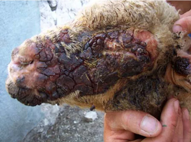
Clinical signs ranged from mild to severe. Photopho-bia and keratitis were the earliest findings. Skin lesions were most commonly seen in sun exposed areas and varied from erythema to severe ulceration with crusting around the eyes and mouth (Fig. 1). These areas were become reddened firstly, and then crusty and dark (Fig. 2). Skin lesions were often accompanied by oedema, resulting in drooping ears and puffy periocular and facial skin. Pruritus, mani-fest as scratching and rubbing was noted. Jaundice was seen at later stages. Severely affected individuals lost weight rapidly. Bilirubinuria, characterized by dark yellow urine, was noted in severely affected sheep, prior to these individuals becoming comatose. Severe icterus, hepatomegaly, enlarged gallbladder, prominent hyperaemia at the mesenterial vessels were the common necropsy findings (Fig. 3). Icterus was seen in the subcutaneous tissues, muscles, omentum, mesenterium, visceral organs. The bones were a dark yellow colour. A large gallbladder was seen in all cases. The gallbladder was oedematous and occasion-ally hemorrhagic. Bile accumulation was excessive and mucoid in nature. Liver abnormalities were seen in all cases. Some were enlarged, others were yellowish and hard. Mesenteric vessels were hyper-aemic. Gut walls were thickened and their lumens were usually empty or contained only a small amount
of ingesta. In some severely affected individuals, a small amount of fluid was noted in the abdominal cavity.
Biochemical analysis of serum samples were shown in Table 1. Leukocytosis was observed at the haematological analysis and it continued until 15th days after starting treatment. At the microscopic examination of the skin, no or a little inflammatory reaction was observed especially in acute cases although presence of severe ulcers. Subcutaneous tissue was severely oedematous and the vessels were hyperaemic. At the chronic cases secondary infections were caused slight inflammatory reaction but oedema was decreased. Histopathology of livers revealed severe necrotic changes in hepatocytes and bile ducts. Cholangiohepatitis was characterized by portal fibro-sis with bile duct proliferation, periportal
hepatocel-Fig. 1 Characteristic skin lesions in a sheep suffering from facial eczema, case 4
Fig. 2 Another acutely affected sheep, case 5, severe oedema and ulcers on the face skin
Fig. 3 Abdominal visceral organs, case 2, severe icterus, severely affected liver and swollen gallbladder
lular loss and a scant mixed periportal, cellular infiltrate (Figs.4and5). In two cases occlusion with crystalloid materials were observed. The larger intra-hepatic ducts were more severely affected. Bile stasis was common and bile pigments were seen also out of the livers. Kidneys and spleen were the most common sites of the bile pigment accumulation. Numerous piknotic cells were observed in the kidneys tubule epithelium. All of the vessels in the guts were hyperaemic and severe neutrophil and mononuclear cell infiltration was seen in the propria mucosa. Lungs were observed oedematous especially severely affect-ed sheep. No indication about viral disease was observed and no bacteria were isolated from the organs. Parasitological examination was negative. Examination of the ruminal contents was revealed 6,000–15,000 spores/ml P. chartarum spores. Most of the spores were digested and a little amount spores were intact. Pasture spore counts were changing 96,300- 267,500 spores/g grass.
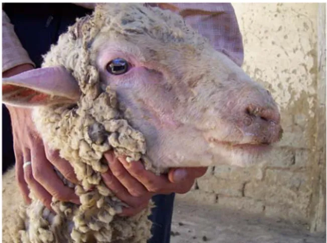
After diagnosis animals suffering from facial eczema and remaining sheep were removed from the pasture and housed in shade pens. Commercial diets were provided for them. All of the affected animals treated with procain penicillin + dihidrostreptomycin sulphate, multivitamin complexes and it was given again 15 days later and flunixin meglumine. Addi-tionally, zinc sulphate was also given 6 gr per 100 lt drinking water during the 28 days. Most animals recover from the acute phase immediately after treatment. After 15 days starting the treatment most of the skin lesions were recovered in treated sheep (Fig. 6). Liver enzyme levels were also reduced but GGT levels were still high. No animal died after starting the treatment.
Fig. 4 Liver, sheep, case 2, cholangiohepatitis characterised by
mononuclear inflammatory cell infiltration, HE, Bar=200μm
Fig. 5 Liver, sheep, case 2, necrosis at the hepatocytes and
fibrous tissue proliferation; HE, Bar=50μm
Table 1 Liver enzyme and total bilirubin levels of sheep suffering from facial eczema before and after treatment
Before treatment After treatment (15th days) Reference range (Sheep)*
GGT 261-328 U/L 86–143 U/L 20–52 U/L
AST 86–120 U/L 72-97 U/L 60–280 U/L
ALT 55-70 U/L 43-66 U/L 22–38 U/L
Total bilirubin 0.8–1.2 mg/dL 0.5-0.8 mg/dL 0.1–0.5 mg/dL *(Radostist et al. 2000)
Fig. 6 A sheep recovered from facial eczema, case 19, 15th days after starting the treatment
Discussion
Facial eczema is one of the earlied- recognized diseases caused by a mycotoxin; it was first described in sheep in New Zealand. The toxin sporidesmin principally affects the liver, and the outstanding lesion from which disease gets its name is the result of hepatotoxic photosensitization due to circulating phylloerythrin. Sporidesmin is concentrated in fungal spores, and the toxigenicity of pasture is related to the density of the spores in it and sporidesmin producing ability to the spores (Jubb et al. 1993; Jones et al. 1997; Collin et al. 1998; Radostits et al. 2000). Experimental administration of the toxin sporidesmin does produce rapid necrosis at the hepatocytes, but these are mild and nonspecific changes. If adminis-tered in suitable dosage, the toxin causes permeability alterations in many tissues and will, for example, produce corneal oedema on local application. In sheep, the face and ears are the most severely affected sites (Jubb et al.1993; Jones et al.1997; Radostits et al. 2000). In our study typical skin and liver lesions were observed. Skin lesions were secondary and they occurred because of severe liver damage.
Animals suffering from facial eczema become restless and seek shade (photophobia). Growth and milk production are dramatically reduced. Sheep may have extensive oedema of the head resulting of the facial eczema. Lesions initially include erythema and oedema, followed by blisters, exudation, necrosis, and sloughing of necrotic tissues and keratitis in some animals. Typical symptoms of facial eczema include severely ulcerated skin in non-pigmented areas of the body, particularly on the head and ears of sheep (Hargis 1995). Newly-shorn sheep are especially vulnerable. Other symptoms may include itching and rubbing of affected areas, scabs, loss of appetite, droopy and swollen ears, and swollen lips and eyelids (Jubb et al. 1993; Hargis 1995). Similar clinical findings were observed in present study. All of the sheep had extensive oedema of the head at the first stages of the illness then necrosis and sloughing of the face skin were seen. Corneal oedema and opacity were seen in 17 out of 22 sheep at the clinical examination. Most of them were unilateral but in severe cases it was bilateral. After treatment, corneal opacity was completely recovered. Corneal oedema and opacity were attributed to the photosensitization.
Blood taken for routine haematological and serum biochemical analyses confirmed an increase in liver enzymes. The increases in the serum enzyme activity in these sheep and histopathological changes in the livers of the affected animals were consistent with changes identified in sporidesmin toxicosis. Similar gross and microscopic lesions were observed in this study that as previously reported (Jubb et al. 1993; Jones et al.1997, Radostits et al.2000). Skin lesions were occurred due to severe liver damage. Based on the clinical and pathological examinations, facial eczema was diagnosed. Liver enzyme activity espe-cially GGT in serum is specific for liver damage (Jubb et al. 1993; Morris et al. 2004). There is a significant relationship between severity of liver injury and serum GGT levels (Morris et al. 2004). In this study, we also observed this correlation, and we noticed that serum GGT levels were high in sheep with severely affected livers.
The liver damage, characterized by necrosis in single centrilobular hepatocytes, is common finding. Minor to moderate portal fibroplasia and bile duct proliferation are almost always present. Cholangiohe-patitis is the outstanding hepatic lesion. Focal hepatic lesions and regenerative hyperplasia may be seen. Extrahepatic bile ducts are enlarged and oedematous, as is the wall of the gallbladder. Haemorrhages in the wall of the gallbladder and urinary bladder are common (Jubb et al. 1993; Jones et al. 1997; Radostits et al. 2000). Similar findings were also observed in our study, but the lesions were usually severe. Gross lesions were very typical and histopa-thology supported the gross findings. At the histo-pathological examination of liver, characteristic degeneration, necrosis and in some cases fibroplasia were observed.
The occurrence of facial eczema is also influenced by livestock genetics. Goats are more resistant to facial eczema than sheep, although there is large variation among breeds of goats. The ability to tolerate exposure to sporidesmin is a genetic trait with a heritability of approximately 40%, and selec-tion for resistance is possible. The field tests for this trait involves placing sheep in high-toxin pastures and after 7–10 days begin testing for elevated blood levels of the enzyme GGT and it is a symptom of liver damage (Radostits et al.2000). In this study, all of the animals were crossbred Merino sheep. This result
may be attributed that this breed has susceptibility to this disease like as the other skin disease, because of the thin skin and hair structure. Some authors reported that goats were more resistant to facial eczema than sheep (Smith et al.1987; Jubb et al.1993; Jones et al. 1997; Radostits et al. 2000). In our study we also observed similar findings because no goat suffered from this disease that was reared together with sheep with facial eczema.
Animals suffering from facial eczema should be removed from the contaminated pasture and provided with shade, cool water, and a good diet. A major goal of treatment is to reduce the phylloerythrin load on the liver. Changing pastures helps prevent consump-tion of addiconsump-tional fungus spores. Feeding hay and/or grain also reduces toxin consumption and maintains the animals until the skin lesions heal and new healthy liver tissue can grow. Feeding high levels of zinc may help prevent facial eczema (Radostits et al. 2000). In addition to these applications, we also gave the antibiotic for to prevent secondary bacterial complications of skin lesions. Anti inflammatory drugs were also found effective for treatment.
Fungal growth is favoured by warm, wet, humid weather, or with heavy dews or irrigation. Spore levels can rise quite rapidly under these conditions. The practice of intensive grazing may actually increase the incidence of facial eczema, because animals in closely-grazed pastures consume vegeta-tive matter that may be heavily contaminated with fungus spores (Kellerman et al. 1980; Jubb et al. 1993; Hansen et al. 1994; Radostits et al. 2000). Normally the southwest of Turkey has high humidity and according to the meteorological data the weather was hotter in 2005 summer than the last 150 years in southwest Turkey. The possible cause of the occur-rence of the disease was attributed the increasing spore counts in pasture, because there was no facial eczema report in Turkey before this report. Pasture P. chartarum spore counts were changing 96,300-267,500 spores/g grass and 6,000–15,000 spores/ml spores were in ruminal content. These findings were observed similar with previous reports (Smith et al. 1987). Spores count in the atmosphere is depending on the weather condition (Troutt and Levetin 2001). Smith et al. (1987) reported that total consumption of spores and high level of spores count in rumen can cause liver damage in sheep. In present study, we also
found numerous spores in rumen content. Although we couldn’t analyzed the sporidesmin levels in the P. chartarum in pasture and ruminal contend there were strong relationship between spores counts in ruminal contend and severity of clinical symptoms. The pasture also examined for new or toxic plant species but the referring veterinarian reported that there were no toxic plants or new in the paddock. This result may be attributed to P.chartarum spores possibly produce sporidesmin.
Facial eczema is a costly problem in pastoral agriculture, and has a detrimental impact on animal wellbeing. Incidence and severity of the disease can be reduced by grazing management and zinc prophy-laxis (Morris et al. 2004). In Burdur province sheep grassed in pastures during the spring, summer and autumn. Rye grass is the most common plant in the pastures. Facial eczema can also be possible threat for sheep in this region in future. At the other hand, the other organ lesions like as liver should be take in to consideration in skin problems like as facial eczema. It is possible to say that facial eczema may affect to the sheep economy in future in Turkey.
References
Coetzer, J.A., Kellerman, T.S., Sadler, W. and Bath, G.F., 1983. Photosensitivity in South Africa. V. A comparative study of pathology of the ovine hepatogenous photosensitivity diseases, facial eczema and geeldikkop (Tribulosis ovis), with special reference to their pathogenesis. Onderstepoort Journal of Veterinary Research, 50(1), 59–71.
Collin, R.G., Odriozola, E., Towers, R.N., 1998. Sporidesmin production by Pithomyces chartarum isolates from Aus-tralia, Brasil, New Zealand and Uruguay. Mycological
research, 102, 163–166.
di Menna, M. E., 1977. The wash method of counting Pithomyces chartarum spores in pasture. Proceedings of
the Ruakura Farmers’ Conference: 1–8.
Hansen, D.E., McCoy, R.D., Hedstrom, O.R., Snyder, S.P. and Ballerstedt, P.B., 1994. Photosensitization associated with exposure to Pithomyces chartarum in lambs, Journal of the American Veterinary Medical Association, 204 (10),
1668–1671.
Hargis, A.M., 1995. Thomson’s Special Pathology, (Mosby
Publishing, Missouri).
Jones, T.C., Hunt, R.D., and King, N.W., 1997. Veterinary Pathology,(Williams and Wilkins, Maryland).
Jubb, K.V.F., Kennedy, P.C., and Palmer, N., 1993. Pathology of Domestic Animals, (Academic Press. Inc., London). Kellerman, T.S., Van der Westhuizen, G.C., Coetzer, J.A.,
Bsaaon, P.A., 1980. Photosensitivity in South Africa. II The experimental production of the ovine hepatogenous photosensitivity disease geeldikkop (Tribulosis ovis) by the simultaneous ingestion of Tribulus terrestris plants and cultures of Pithomyces chartarum containing the myco-toxin sporidesmin. Onderstepoort Journal of Veterinary
Research, 47(4), 231–261.
Morris, C.A., Towers, N.R., Hohenboken, W.D., Maqbool, N., Smith, B.L., and Phua, S.H., 2004. Inheritance of resistance to facial eczema: a review of research findings from sheep and cattle in New ZealandNew Zealand
Veterinary Journal, 52(5), 205–215.
Radostits, O.M., Gay, C.C., Blood, C. and Hinchcliff, K.W., 2000. Veterinary Medicine, (W.B. Saunders Company, London). Smith, B.L. and Embling, P.P., 1991. Facial eczema in
goats-The toxicity of sporidesmin in goats and its pathology.
New Zealand Veterinary Journal, 39(1), 18–22.
Smith, B.L., Embling, P.P., and Gravett, I.M., 1987. Pithomyces chartatum spores counts in rumen contents and faeces of sheep exposed to autumn pasture at three different grazing
pressures. Journal of Applied Toxicology, 7(3), 179–184.
Troutt, C. and Levetin, E., 2001. Correlations and meteorolog-ical conditions in Tulsa, Oklahoma.International Journal