Full Terms & Conditions of access and use can be found at
https://www.tandfonline.com/action/journalInformation?journalCode=ipdp20
Fetal and Pediatric Pathology
ISSN: 1551-3815 (Print) 1551-3823 (Online) Journal homepage: https://www.tandfonline.com/loi/ipdp20
Umbilical Cord Diameter at the Junction of the
Body Wall in the Newborn. Is It a Biomarker for
Congenital Umbilical Hernia?
Nazile Ertürk
To cite this article: Nazile Ertürk (2018) Umbilical Cord Diameter at the Junction of the Body Wall in the Newborn. Is It a Biomarker for Congenital Umbilical Hernia?, Fetal and Pediatric Pathology, 37:4, 223-230, DOI: 10.1080/15513815.2018.1477886
To link to this article: https://doi.org/10.1080/15513815.2018.1477886
Published online: 27 Aug 2018.
Submit your article to this journal
Article views: 78
View related articles
View Crossmark data
Citing articles: 1 View citing articles
FETAL AND PEDIATRIC PATHOLOGY
-•--·
-·
.,.__ __
___
.._D
Taylor & FrancisUmbilical Cord Diameter at the Junction of the Body Wall
in the Newborn. Is It a Biomarker for Congenital
Umbilical Hernia?
Nazile Ert€urk
Faculty of Medicine, Department of Pediatric Surgery, Mugla Sitki Kocman University, Mugla, Turkey
ABSTRACT
Introduction: The aim is to obtain normal newborn umbilical cord diameters for use it in the evaluation of congenital umbilical hernia. Materials and methods: The umbilical cord diameter (UCD) at the abdominal wall, maternal age, birth weight, gestational age at birth, birth height, head, chest and abdominal circumferences, and the time of measurement after birth was noted. Results: Mean ± standard deviation and median (minimum–maximum) values of the UCD were 9.9 ± 1.9 mm, 10.0 (5–16 mm), respectively. There was a significance for a positive low correlation between birth height and UCD (p ¼ .039, r ¼ .143). No other birth parameter had a significant correl-ation with UCD. The gender of the newborn (p ¼ .95) and the type of delivery (p ¼ .056) did not affect UCD. Conclusion: These data may be used in determining the normality of UCD, which could be helpful in the evaluation of umbilical hernias.
ARTICLE HISTORY
Received 24 March 2018 Revised 27 April 2018 Accepted 1 May 2018
KEYWORDS
Birth weight; congenital umbilical cord hernia; umbilical cord circumference; umbilical cord diameter
Introduction
The umbilical cord may be thought as the lifeline of the fetus (1). Umbilical cord involves two arteries and one vein surrounded by Wharton’s jelly (2). The survival of the fetus requires a sufficiently functioning umbilical cord. The composition of the umbilical cord (arteries, vein, and Wharton’s jelly) changes during the gestation (3,4).
The intestines are physiologically herniated towards the proximal part of the umbilical cord in the early fetal period. That is called extracoelomic cavity (5,6). The intestines migrate back to abdominal cavity in the 10–12th weeks gestation, the umbilical ring usually closes and the extracoelomic cavity disappears. The Wharton’s jelly and the umbilical vessels form the resultant umbilical cord. On occasion, the umbilical ring does not close and intestines may remain in the extracoelomic cavity. This process is called congenital hernia of the umbilical cord (CHUC) in the postnatal period (5–9), which is estimated to occur once in 5000, with a male preponderance (3:1), and is associated with prematurity (10). In CHUC, cord clamping may cause intestinal obstruction, in that often ileum is contained within the sac (11). For this reason, the immediate evaluation of the diameter of the umbilical cord immediately after birth may have a predictive value for the diagnosis of CHUC.
CONTACTNazile Ert€urk [email protected] Mugla Sitki Kocman University, Faculty of Medicine, Department of Pediatric Surgery, Haluk Ozsoy Caddesi Mentese, Mugla 48000, Turkey.
Color versions of one or more of the figures in the article can be found online atwww.tandfonline.com/ipdp.
ß 2018 Taylor & Francis Group, LLC
2018, VOL. 37, NO. 4, 223–230
https://doi.org/10.1080/15513815.2018.1477886
C\
Taylor&
Francis~ Taylor&FrancisGroup
In this study, we aimed to assess the normal umbilical cord diameter data in the new-born and the relation between the umbilical cord diameter and some morphometric birth parameters.
Methods
Institutional Ethics Board Approval and a written informed consent were obtained before beginning the study. This prospective study included 209 term newborns. The inclusion criteria were: (1) singleton gestation, (2) normal amniotic fluid index during the pregnancy, (3) presence of three-vessel umbilical cord, (4) absence of any congenital anomaly, (5) absence of any known maternal disease, and (6) absence of any complica-tions during pregnancy.
The umbilical cord diameter was measured at its attachment to the umbilicus by a digital caliper on the first day of hospitalization by the same doctor and nurses. Maternal age, birth weight, gestational age at birth, birth height, head circumference, chest circumference, abdominal circumference and the time after birth at which umbilical cord diameter was measured were noted for each newborn. Newborns infants were divided into six 4 h groups according to the time since birth. The average value of umbilical cord diameters in each group were measured and compared with each other. ANOVA test was performed to determine a significant difference in desiccation (shrinkage) among the groups.
All statistical data were analyzed using SPSS version 15.0 (SPSS Inc., Chicago, IL). Values were expressed as mean, standard deviation and also as median (minimum–max-imum). The normality of the values were analyzed by using Shapiro–Wilk test. For the independent samples, t-test or Mann–Whitney U test was used according to the Shapiro–Wilk test result. Correlations between the variables were investigated by using Spearman correlation coefficient. Differences were considered significant at p < .05.
Results
Maternal age, birth weight, gestational age at birth, birth height, head circumference, chest circumference, abdominal circumference, umbilical cord diameter and the time of measurement after birth are shown in Table 1. All the morphological variables except birth weight had normal distribution. The 5% and 95% confidence interval limits of umbilical cord diameter were 9.8 and 10.1 mm, respectively.
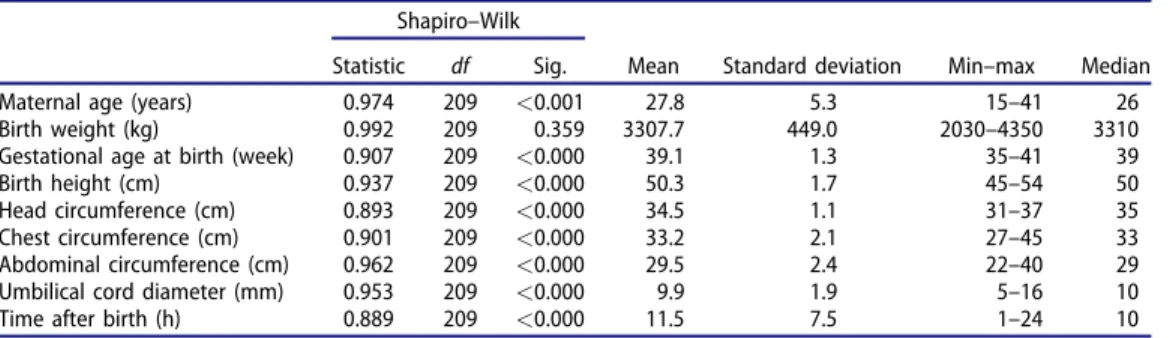
Table 1. Tests of normality and descriptive statistics. Shapiro–Wilk
Mean Standard deviation Min–max Median Statistic df Sig.
Maternal age (years) 0.974 209 <0.001 27.8 5.3 15–41 26 Birth weight (kg) 0.992 209 0.359 3307.7 449.0 2030–4350 3310 Gestational age at birth (week) 0.907 209 <0.000 39.1 1.3 35–41 39 Birth height (cm) 0.937 209 <0.000 50.3 1.7 45–54 50 Head circumference (cm) 0.893 209 <0.000 34.5 1.1 31–37 35 Chest circumference (cm) 0.901 209 <0.000 33.2 2.1 27–45 33 Abdominal circumference (cm) 0.962 209 <0.000 29.5 2.4 22–40 29 Umbilical cord diameter (mm) 0.953 209 <0.000 9.9 1.9 5–16 10 Time after birth (h) 0.889 209 <0.000 11.5 7.5 1–24 10 224 N. ERT€URK
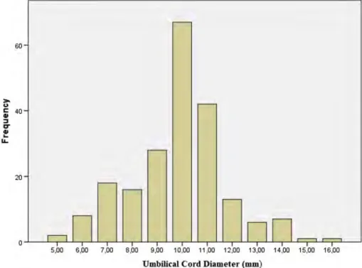
The distribution of umbilical cord diameter measurements is shown in Figure 1. The data show a normal distribution. Except birth height, none of the birth parameters had a significant correlation with the umbilical cord diameter (Table 2). There was a signifi-cance for a positive low correlation between birth height and umbilical cord diameter (p ¼ .039, r ¼ .143, Table 2). Accordingly, although there is a low correlation, the birth height and the umbilical cord thickness increase together. The gender of the newborn (p ¼ .95) and the type of delivery (p ¼ .056) did not affect umbilical cord diameter (Table 3).
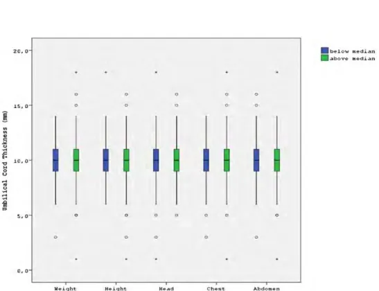
The newborns were divided into groups according to the median of the measure-ments for birth weight, chest circumference, abdominal circumference and head cir-cumference. It was found that lower and higher values of those parameters did not affect the umbilical cord diameter (Fig. 2). Findings showed no significant differ-ence in drying shrinkage among the groups during the first 24 h (p ¼ .871) (Table 4).
Within the period of our study only one baby was born with CHUC, in which the cord diameter was 21.6 mm and contained a loop of intestine, covered by a thin membrane. A primary hernia closure was required and performed.
Discussion
The aim of this study was to obtain normal newborn umbilical cord diameters for use it in the evaluation of congenital umbilical hernia. The studies related to umbilical cord are generally about the length of the cord. The length of the cord increases mainly in
Figure 1. The distribution of the umbilical cord diameter.
>
,..
60 C 40 GI ::, I:," GI...
IL 20 5,00 6,00 7,00 8,00 9,00 10,00 11,00 12,00 13,00 14,00 15,00 16,00Table 2. The correlations between umbilical cord thickness and other birth morphometric parameters. Maternal age Birth weight Gestational age at birth Birth height Head circumference Chest circumference Abdominal circumference Time after birth Umbilical cord thickness Correlation coefficient 0.071 0.085 0.034 0.143 0.023 0.049 –0.008 0.016 Significance 0.308 0.219 0.624 0.039 0.744 0.478 0.907 0.813 226 N. ERT€URK
the first and second trimesters and reaches a mean of 32 cm at 20 weeks gestation. The male and female cord length reaches to 53.75 cm and 51.75 cm at 34 weeks, respect-ively (12,13). Short umbilical cords have been associated with low Apgar scores, hypotonia, jitteriness and respiratory problems (13,14).
The morphology and components of the umbilical cord affect birth results (14–16). The length of the umbilical cord is affected by both genetic variations and fetal move-ments. Increased number of fetal movements is related to a longer cord and vice versa. The shorter umbilical cord is related with/to a less active fetus, fetal malformations, myopathic and neuropathic diseases, Down syndrome and oligohydramnios (14,16,17). Thin umbilical cord can accompany unexplained fetal death, whereas umbilical cord enlargement can be observed in association with fetal hydrops, rhesus sensitization or twin–twin transfusion (18).
The length and diameter of the umbilical cord were shown as 55–60 and 1.5–2.0 cm, respectively (14). In our study, we found umbilical cord diameter as 9.9 (±1.9 SD) mm. Supposing the umbilical cord as a circle, a 9.9 mm diameter means a circumference of
Table 3. The differences in umbilical cord diameters regarding gender and birth type. Parameters
Umbilical cord diameter (mm)
Mean (SD) (minimum–maximum) p Gender Male 9.9 ± 1.8 (5–15) .952
Female 9.8 ± 1.9 (5–16)
Birth type Cesarean section (n ¼ 31) 10.4 ± 2.3 (5–16) .056 Vaginal delivery (n ¼ 178) 9.7 ± 1.8 (5–15)
Figure 2. The differences in the umbilical cord diameter in the newborns with lower and higher birth weight, chest circumference, abdominal circumference and head circumference.
20,0 0 15, 0 0 0
!
:: " C "' ~ .c..
;l 10, 0 0 u .... ., -~ ....i
., 5,0 0 0 0 o,o W•ight K•ight 0 0 0 0 Kead 0 a 0 0 0 Ch•st 0 0 0 Abdomen•
below .median29.1 mm (19,20). After examining a newborn with incidentally diagnosed CHUC, Patel et al. (2) decided to perform a study to assess the normal umbilical cord circumference to be able to detect the newborns with umbilical cord hernia. The authors found umbil-ical cord circumference as 37.6 ± 7.3 mm and stated that none of the neonates in their study had a cord circumference of 70 mm as seen in the newborns with umbilical cord hernia. Patel et al. (2) reported a progressive increase in umbilical cord circumference with an increase in weight and chest and abdominal circumferences. The authors also stated that an unusually large cord should raise the suspicion of a hernia and the new-born should closely be examined before clamping the cord. In contrast to Patel’s study, we found no significant correlation between umbilical cord diameter and birth weight as well as the head, chest, and abdominal circumferences. Interestingly, in this study we found that the umbilical cord thickness increases with the birth height but not with body weight, in contrast to Patel’s study (2).
In the literature, there is limited data showing the umbilical cord diameter at birth in newborns. The first study belongs to Patel et al. (2) and the second study is the present study. The situation after clamping of umbilical cord during birth in the newborn; there are few studies about shrinking, drying, and detachment. After clamping, the cord begins to dry and the cord thickness gradually decreases. The period of the complete desiccation of the cord varies from the end of the first day to the fifth day; on the third day it is generally completed (19). For this reason, it is normally expected that there is a negative relationship between the time passed after birth and the thickness of the umbilical cord. In other words, as the time after birth (hour) increases, the thickness of the umbilical cord is expected to decrease. However, in our study, it was determined that there was no meaningful relationship between these two variables durıng the fırst 24 h after bırth. This indicates that significant dessication shrinkage is not evident in the first 24 h. As the umbilical cord stump detached between 3 and 45 days, the result of our study is reasonable (21).
In our study, the upper limit of the umbilical cord diameter was 16 mm with a 95% confidence interval limit. Again, supposing the umbilical cord as a circle, this corre-sponds to a circumference of 30.3 mm. In our study, the highest umbilical cord
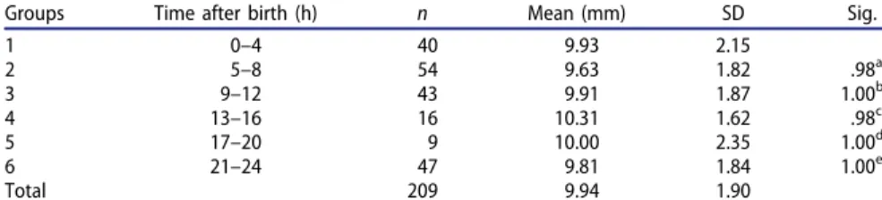
Table 4. Newborns were divided into groups of 4 h intervals, measured from the time of birth, to determine if the umbilical cord diameter decreased due to drying during the first day.
Groups Time after birth (h) n Mean (mm) SD Sig.
1 0–4 40 9.93 2.15 2 5–8 54 9.63 1.82 .98a 3 9–12 43 9.91 1.87 1.00b 4 13–16 16 10.31 1.62 .98c 5 17–20 9 10.00 2.35 1.00d 6 21–24 47 9.81 1.84 1.00e Total 209 9.94 1.90
The average value of umbilical cord diameters in each group were measured and compared with each other. Findings show that there is no significant difference in diameters among the groups. n: number of patients, mean–mean of groups diameters of umbilical cord.
The group 1 was a reference group. a p ¼ .98 vs. group 1, b p ¼ 1.00 vs. group 1, c p ¼ .98 vs. group 1, d p ¼ 1.00 vs. group 1, e p ¼ 1.00 vs. group 1. 228 N. ERT€URK
diameter measurement was 16 mm, corresponding a circumference of 48mm. According to this study, a circumference above 48 mm should raise suspicion of an umbilical cord hernia and the physicians should be careful not to cause intestinal obstructions in a newborn with CHUC. As mentioned above, Patel et al. (2) stated that the umbilical cord diameter of the newborn with CHUC in their report was 70 mm.
It was reported that CHUC emerges in the early intrauterine life and may be diag-nosed in the second trimester by ultrasonography (22,23). The umbilical cord is almost always visible by ultrasound in the first trimester (3). In the first trimester, a significant correlation was reported with gestational age and crown–rump length (24). However, Ince et al. (5) indicated that they could diagnose only six out of 15 cases of CHUC by antenatal ultrasonography. The correlation between umbilical cord parameters and fetal growth was not investigated so far (3).
In this study, we assessed normative umbilical cord diameter measurements at term. Larger studies including the umbilical cord diameters in the newborns with and without CHUC are needed to explore the value of these data in the diagnosis of CHUC.
Disclosure statement
The author declares no conflict of interests.
ORCID
Nazile Ert€urk http://orcid.org/0000-0001-9541-9741
References
1. Ente G, Penzer PH. The umbilical cord: normal parameters. J R Soc Health.1991;111(4):138–40. 2. Patel D, Dawson M, Kalyanam P, Lungus E, Weiss H, Flaherty E. Nora EG. Umbilical cord
circumference at birth. Am J Dis Child.1989;143(6):638–9.
3. Ghezzi F, Raio L, Di Naro E, Franchi M, Bruhwiler H, D’Addario V, Schneider H. First-tri-mester sonographic umbilical cord diameter and the growth of the human embryo. Ultrasound Obstet Gynecol.2001;18(4):348–51.
4. Nanaev AK, Kohnen G, Milovanov AP, Domogatsky SP, Kaufmann P. Stromal differenti-ation and architecture of the human umbilical cord. Placenta.1997;18(1):53–64.
5. Ince E, Temiz A, Ezer SS, Gezer HO, Hicsonmez A. Poorly understood and often miscate-gorized congenital umbilical cord hernia: an alternative repair method. Hernia. 2017;21(3):449–54.
6. Pal K. Congenital hernia of the umbilical cord associated with extracelomic colonic atresia and perforation of gut in a newborn. Afr J Paediatr Surg.2014;11(1):74–6.
7. Klein MD, Hertzler JH. Congenital defects of the abdominal wall. Surg Gynecol Obstet. 1981;152(6):805–8.
8. Seashore JH. Congenital abdominal wall defects. Clin Perinatol.1978;5(1):61–77.
9. Varghese AS, Vause S, Kamupira SR, Emmerson AJ. Congenital abdominal wall defects. Arch Dis Child Educ Pract Ed.2017;102(1):19.
10. Fahmi M. Umbilicus and umbilical cord. Berlin, Germany: Springer International Publishing AG;2018. p. 199.
11. Chapman-Sheath P, Wilcox D, Mok Q, Drake D. Iatrogenic ileal obstruction: a complica-tion of umbilical cord clamping. Br Med J.1996;313(7057):613–4.
12. Moessinger AC, Blanc WA, Marone PA, Polsen DC. Umbilical cord length as an index of fetal activity: experimental study and clinical implications. Pediatr Res.1982;16(2):109–12. 13. Miller ME, Jones MC, Smith DW. Tension: the basis of umbilical cord growth. J Pediatr.
1982;101(5):844.
14. Kosif R, Bayar U, Basaran M, Gezer S, Kacar D, Kiyan A. Investigation of the relationship between umbilical cord morphometry and fetal and maternal parameters. Med Bull Zeynep Kamil.2010;41(3):129–35.
15. Sornes T. Short umbilical cord as a cause of fetal distress. Acta Obstet Gynecol Scand. 1989;68(7):609–11.
16. Jaya DS, Kumar NS, Bai LS. Anthropometric indices, cord length and placental weight in newborns. Indian Pediatr.1995;32(11):1183–8.
17. Harder T, Rodekamp E, Schellong K, Dudenhausen JW, Plagemann A. Birth weight and subsequent risk of type 2 diabetes: a meta-analysis. Am J Epidemiol.2007;165(8):849–57. 18. Predanic M. Sonographic assessment of the umbilical cord. Ultrasound Rev Obstet
Gynecol.2005;5(2):105–10.
19. Billard C.M. A treatise on the discases of infants, founded on recent clinical observations and investigations in pathological anatomy, made at the Hospice des Enfans–Trouves: with a Dissertation on the Viability of the Child. Am J Med Sci.1839;25:360–2.
20. Shen Y, Hosseini AR, Wong MH, Malliaras GG. How to make Ohmic contacts to organic semiconductors. Chemphyschem.2004;5(1):16–25.
21. Novack AH, Mueller B, Ochs H. Umbilical cord separation in the normal newborn. Am J Dis Child.1988;142(2):220–3.
22. Achiron R, Soriano D, Lipitz S, Mashiach S, Goldman B, Seidman DS. Fetal midgut hernia-tion into the umbilical cord: improved definihernia-tion of ventral abdominal anomaly with the use of transvaginal sonography. Ultrasound Obstet Gynecol.1995;6(4):256–60.
23. Haas J, Achiron R, Barzilay E, Yinon Y, Bilik R, Gilboa Y. Umbilical cord hernias: prenatal diagnosis and natural history. J Ultrasound Med.2011;30(12):1629–32.
24. Hill LM, DiNofrio DM, Guzick D. Sonographic determination of first trimester umbilical cord length. J Clin Ultrasound.1994;22(7):435–8.