Ankara Üniv Vet Fak Derg, 52, 2005 131 Ankara Üniv Vet Fak Derg, 52, 131-134, 2005
Short Communication / Kısa Bilimsel Çalışma
Sebaceous gland adenoma in two dogs
Ayhan ATASEVER1, Latife BEYAZ1, Yücel ÇAM2, Meryem EREN3
1 Erciyes Üniversitesi, Veteriner Fakültesi, Patoloji Anabilim Dalı, Kayseri; 2 Erciyes Üniversitesi, Veteriner Fakültesi,
İç Hastalıkları Anabilim Dalı, Kayseri;3 Erciyes Üniversitesi, Veteriner Fakültesi, Biyokimya Anabilim Dalı, Kayseri.
Summary: In this report, biopsy materials taken from two dogs are examined histopathologically. First of all, a mass
surrounded by skin and located in the metacarpaphalangeal region of the left front leg in a 4 year old female cross bred street dog, measuring 7.5 x 6.5 x 10 cm and weighing 300 g, was fully removed at necropsy. Secondly, the smallest of the multilocational foci of a 11 year old male Terrier (skin) was with the size of a ventil whearas the largest foci being a size of a hazelnut, and a biopsy material measuring 0.1 x 0.5 x 0.2 cm was taken from these foci with an operation. However, this dog has been brought to the clinics for nutritional disorder. In both cases, sebaceous gland adenoma consisting of many mature sebaceous cells were identified in microscopical examination.
Key words: Adenoma, dog, sebaceous gland.
İki köpekte yağ bezi adenomu
Özet: Bu raporda; iki köpekten alınan biopsi materyalleri histopatolojik olarak incelendi. İlk olarak; 4 yaşlı, dişi, melez bir
so-kak köpeğinde ön sol ayağın metacarpaphalangea bölgesinde deri ile çevrilmiş 7.5x 6.5x10 cm boyutlarında ve 300g ağırlığında olan kitle nekropsi esnasında tamamen alındı. İkinci olarak; 11yaşlı, erkek, Terrier köpekte deride multilokalizasyon gösteren, mercimek-ten nohut büyüklüğüne ulaşan kitlelerden 0.1x0.5x 0.2 cm boyutlarındaki bir tanesi operasyonla biyopsi materyali olarak alındı. Bu köpek kliniğe beslenme yetersizliği nedeniyle getirilmişti. Mikroskobik olarak, her iki olguda da deride çok sayıda olgun yağ hücre-lerinden oluşan yapılar görüldü. Bu görünüm yağ bezi adenomu olarak tanımlandı.
Anahtar sözcükler: Adenom, köpek, yağ bezi..
Sebaceous gland adenomas are the most frequently encountered skin tumour in dogs (8). Sebaceous gland adenomas are reported to be observed rarely in cats and other animal species (11,14). Adenomas are most fre-quently observed in dogs between the ages 8 and 13 (1,9). Cocker spaniels are know to have predisposition for the mentioned tumour (1), whereas adenoma has been determined to be more widespread among Poodles (9). Female dogs are reported to be more sensitive (6). Pre-disposition arising from sex has not been reported (7). Sebaceous gland adenomas can be located in any region of the body (4,5). However, they are reported to be lo-cated primarily in the head in dogs (7,11,15). Sebaceous gland tumours are classified as hyperplasia, adenoma, ductal adenoma, epithelioma and carcinoma, based upon the differentiation degree of sebaceous cells (3,6,16). Macroscopical examination has revealed sebaceous gland adenomas to be formed of many lobules of irregular shape and various sizes which may reach to a diameter of 2 cm. These lobules are completely separated from the surrounding tissue (6). Alopecia, hyperpigmentation, secondary bacterial infections and ulcerous areas may be
associated with these nodular structures (7). Upon micro-scopical examination, two types of cells have been de-tected to exist in these lobules: namely, in-differentiated generative cells and mature sebaceous cells (6,10). Gen-erative cells are reported to be identical with cells located at the periphery of normal sebaceous glands (10). In regions where transformation into ductal structure is present, squamous changes and keratinization have been observed (7,15). In such cases, many connected ducts have been detected to surround the keratinised sebaceous gland duct. A great number of basal cells and widespread proliferation are observed at the periphery of sebaceous glands (6).
Infrequent reports of sebaceous gland tumours ex-cept for retrospective studies and the low number of adenoma cases encountered, constituted the aim of this study.
A 4 year old, female cross breed dog which died at the municipal boarding home for dogs at Kayseri and a male, 11 year old terrier that submitted to the Internal Diseases Clinic of Erciyes University, Faculty of Medi-cine for a medical treatment, constituted the material of
Ayhan Atasever - Latife Beyaz - Yücel Çam - Meryem Eren 132
this study. Blood samples were collected for haemato-logical and biochemical examination. Haematohaemato-logical examination included detection of erythrocyte, leucocyte and thrombocyte counts, haemoglobin concentration, haematocrit values, mean erythrocyte volume (MCV), mean haemoglobin amount (MCH), mean erythrocyte haemoglobin concentration (MCHC), erythrocyte disper-sion volume (RDV) and mean thrombocyte volume (MPV) by means of a blood count apparatus. Biochemi-cal examination included detection of serum alkaline phosphatase (ALP), aspartate aminotransferase (ALT), alanine aminotransferase (ALT) and keratin kinase (CK) activities; glucose, total cholesterol, triglyceride and urea concentrations by using commercial kits and a Shimadzu –UV 1208 model spectrophotometer. Tissue samples taken from tumour masses (measuring 7.5 x 6.5 x 10 cm and weighing 300 g ) and biopsy material (measuring 0.1 x 0.5 x 0.2 cm ) were fixed in 10% formalin and blocked in paraffin prior to preparation of 5-6 µ thick cross-sections. These cross-sections were later dyed with Hematoxylene-Eosin and examined under light micro-scope.
Clinical findings were not observed in the first case of the study, because the dog was brought to the Veteri-nary Faculty after it died at the municipal boarding home for dogs at Kayseri. The second dog was observed to be active and have a good appetite. Body temperature, pulse and respiration rates were determined as 38.9, 10/minute and 22/minute, respectively. All haematological and biochemical parameters were determined to be within normal ranges except for the mild increase in leucocyte count (18. 3 x 10³ µL).
Case 1: The tumour was located in the
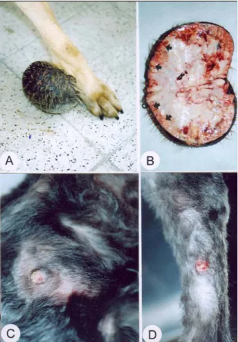
metacar-paphalangeal region of the front left leg in the 4 year old, female cross breed dog and was covered completely with hairy skin. Palpation revealed the mass to have an elastic consistency. Decubitus ulcers were observed to exist in certain areas and to be associated with hair loss. The tumoural mass was determined to measure 7.5 x 6.5 x 10 cm and weigh 300 g (Figure 1A). On the cut surface the tumour was observed to be yellowish in colour, lobular in structure and similar to gel in appearance.
Reddish-black coloured soft areas were detected es-pecially in regions located near the skin. White coloured, hard-fluctuating foci and haemorrhages were also present in these areas (Figure 1B). During necropsy, any patho-logical lesion has not seen in the other organs of the dog, except for the mass located on the left front leg.
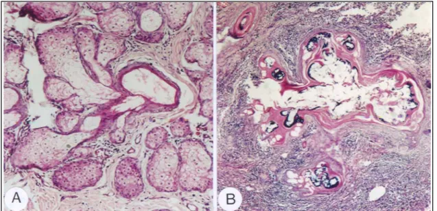
Microscopically examination revealed hyperplasia and hyperkeratinization in the epidermis. Many multi-lobular glands were detected in the dermis. In most of the glands, mature sebaceous cells were evident. Generative cells surrounded the normal lipid glands. In most regions, sebaceous glands were observed to acquire a ductal
structure (Figure 2A). In such regions, evident squamous changes and keratinization drew attention and the seba-ceous glands were observed to transform into complete ductal structure. Intensive calcification was observed in highly keratinized ducts (Figure 2B). Furthermore, ex-tensive areas of granulation tissue were located outside the sebaceous gland lobules.
Any microscopically findings have not been seen during the histopathological observation of the other organs of the dog.
Figur 1. Sebaceous gland adenoma (Şekil 1. Yağ bezi adenomu) A. Mass covered with hairy skin in the metacarpaphalangeal region (A. Metacarpaephalangea bölge-sinde kıllı deriyle örtülü kitle). B. Proliferative regions on the back with crusty surfaces (B. Kıllı deriyle örtülü kitlenin kesit yüzeyi yer yer kanamalı ve yağ dokusu görünümü ). C. Partly haemorrhagic appearance of the cross-section of the mass covered with hairy skin and the appearance of sebaceous tissue (C. Sırt bölgesinde yüzeyi kabuklanmış üremeler). D. Generalised papillomatous proliferation at the lateral of the antebrachium (D. Antebrachium’un lateralinde generalize papilomatöz yüzeyi kabuklanmış üremeler).
Case 2: A mass with elastic consistency, measuring
0.1 x 0.5 x 0.2 cm, which removed surgically at opera-tion from the neck region in a case suspected to be papilloma, constituted the material. The tumour was determined to display multilocation in the head, neck,
Ankara Üniv Vet Fak Derg, 52, 2005 133
Figur 2. Case 1. Sebaceous gland adenoma (Şekil 2. Yağ bezi adenomu). A. Sebaceous gland adenoma at the periphery of the lipid gland duct consisting of generative cells and mature sebaceous cells, HE x200. (A. Yağ bezi kanalı etrafında generatif ve olgun yağ hücrelerinden oluşan yağ bezi adenomu, HE x200.). B. Keratinisation, sqaumous differentiation (epithelium), calcification areas and intense granulation tissue in the sebaceous ducts, HE x200 (B. Yağ kanal yapılarında keratinizasyon, squamöz farklılaşma (epitel), kireçlenme sahaları ve yoğun granulasyon dokusu, HE x200).
abdomen, back and many regions of the leg. The smallest of these foci with the size of a lentil whereas the largest foci being a size of a hazelnut. Some of them were observed to be haemorrhagic on the cut surface and most of them with a crusty scare (Figure 1C,1D). On the cut surface were greyish white in colour.
Microscopical examination revealed the presence of findings observed in Case 1 except for proliferation of granulation tissue and calcification in sebaceous glands (Figure 3). Furthermore, severe haemorrhage and ulcer-ous areas were observed in the epidermis.
Figur 3. Case 2. Adenomatous structures consisting of mature lipid cells (Sebaceous gland adenoma), HE x200.) (Şekil 4. Olgun yağ hücrelerinden oluşmuş adenomatöz yapılar (Yağ bezi adenomu), HE x200).
Although, sebaceous gland adenomas have been reported as the most frequently encountered skin tumour in dogs (1, 2, 6, 12), unavailability of research carried out on these tumours in the last years except for retrospective studies, constituted the aim of this study.
Cocker spaniels have been reported to have a pre-disposition for sebaceous gland adenomas and the men-tioned tumours are reported to be frequently encountered in Poodles (15). The diagnosis of sebaceous gland adenoma in a cross breed dog and Terrier in this study was supportive of the claim that breed predisposition may be valid and also provided indicative evidence that these tumours may also be observed in cross breed dogs. Despite the age group in which sebaceous gland adeno-mas are observed has been reported to be 8-13 years (4,7), the fact that one of the animals included in this study was 4 years old, has shown that this tumour may also be observed in young dogs. Research has revealed sebaceous gland adenomas to be located in the head, thorax and body (4, 13). In one of the studies included in the afore mentioned research, sebaceous gland adenomas have been reported to be observed in the head, thorax and body at a rate of 54% (15) In another study, this tumour has been reported to be observed more frequently in the head of dogs. The data obtained in this study with regard to the location of the tumour in one of the dogs, was in accordance with the reports of other researchers, whereas, differently, the tumour was found to be located in the metacarpal region in the other dog. Previously, sebaceous gland adenomas are thought to be seen often in female purebred dogs (11,15). However, in recent years the research indicated no sex differences (7). Since the number of the cases evaluated in this study was only
Ayhan Atasever - Latife Beyaz - Yücel Çam - Meryem Eren 134
few (one male one female), drawing a conclusion would be hard on this results. Sebaceous gland adenomas are reported to consist of many lobules arising from mature sebaceous cells and generative cells according to data obtained from microscopical examination (10). Regions in which lobules are observed to transform completely into ductal structure are reported to be associated with severe keratinization and squamous changes. Similar findings were determined in this study. However, apart from the above mentioned common observation, severe calcification was also observed in the first case. Intense granulation tissue was also detected in these areas.
In conclusion, histopathological examination re-vealed the tumoural masses detected in both dogs to be sebaceous gland adenomas. Haematological and bio-chemical parameters measured in one of the dogs re-vealed only a mild increase in leucocyte count and other values to be within the normal range. Therefore, it is thought that the sebaceous gland adenoma did not exhibit a systemic effect in this animal, because there are not enough resources about this subject at the literature.
References
1. Erer H, Kıran MM (1993) : Konya’da 1985-1992 yılları
arasında köpeklerde görülen tümörler. Selçuk Üniv Vet
Fak Derg, 9, 87-89.
2. Ertürk E, Tanzer F, Bulucu M (1971) :
Patolojik-Anatomi Kürsüsü’nde 1964-1970 yılları arasında incele-nen köpek ve kedi tümörleri. Ankara Üniv Vet Fak Derg,
18, 383-386.
3. Goldschmidt MH (1984) : Sebaceous and hepatoid gland
neoplasms of dogs and cats . Am J Dermatopathol, 6,
287-293.
4. Halouzka R, Nevole M (1976) : Sebaceous gland tumors
in dogs. Vet Med, 21, 565-572.
5. Jubb KVF, Kennedy PC, Palmer N (1993) : Pathology
of Domestic Animals. Fourth ed., Academic Press, Inc. San
Diego, pp.714-715.
6. Levene A (1984) : Sebaceous gland differentiation in
tumours of the feline oral mucosa. Vet Rec, 114, 69.
7. Meuten DJ (2002) : Tumors in Domestic Animals. In: Tumors of the Skin and Soft Tissues. Fourth ed., Iowa State Press , Ames, Iowa, pp.64-67.
8. Mikulastik J, Bednar B (1989) : Sebaceous skin tumors. Ceskoslovenska Pathol, 25, 94-98.
9. Moulton JE (1990) : Tumors of the Skin and Soft Tissues. In: Tumors in Domestic Animals. Third Ed, University of California Press, Berkeley, Los Angeles, pp.64-66. 10. Nielsen SW, Cole CR (1960) : Cutaneous epithelial
neoplasms of the dog. A report of 153 cases. Am J Vet
Res, 21, 931-948.
11. Paikne DL, Kadhane DL, Purohit BL, Deshmukh MS (1971) : Pulmonary spirocercosis and sebaceous gland
adenocarcinoma in dog. Indian Vet J, 48, 22-24.
12. Pamukçu M, Ertürk E (1962) : Ankara’da köpeklerde
görülen tümör çeşitleri. Ankara Üniv Vet Fak Derg, 9, 1-9.
13. Scott DW, Anderson WI (1990) : Canine sebaceous gland
tumors. A 172 cases. Canine Pract, 15, 19-21.
14. Scott DW, Anderson WI.(1991) : Feline sebaceous gland
tumors. A nine cases. Feline Pract, 19, 16-18.
15. Strafuss AC (1976) : Sebaceous gland adenomas in dogs. JAVMA, 169, 640-642.
16. Warren S, Warvi WH (1943) : Tumors of sebaceous
glands. Am J Pathol, 19, 441-459.
Geliş tarihi: 24.06.2004 / Kabul tarihi: 25.10.2004
Yazışma adresi:
Doç. Dr. Ayhan Atasever
Erciyes Üniversitesi Veteriner Fakültesi Patoloji Anabilim Dalı
Sümer Mah. Barış Manço Cad. 38090, Kocasinan/Kayseri