Histomorphometric examination of the pineal gland in foals and adult
horses
Durmuş BOLAT
1*, Aytül KÜRÜM
2, Sadullah BAHAR
3, Siyami KARAHAN
2,4Kırıkkale University, Faculty of Veterinary Medicine, 1Department of Anatomy; 2Department of Histology and Embryology,
Kırıkkale; 3Selçuk University, Faculty of Veterinary Medicine, Department of Anatomy, Konya; 4Kırıkkale University Scientific and
Technological Laboratories, Kırıkkale, Turkey.
Summary: This study was conducted to evaluate the pineal glands of the foal and adult horses with histomorphometry. The pineal glands were sectioned at a thickness of 40 µm and stained with AgNOR for stereological analyses. The weight and volume of the pineal gland as well as the number of pinealocytes were significantly higher in the adult horses (P=0.009). However, the number of pinealocytes in per volume was similar between foals and adult horses. Such data indicate that growth in the size of the gland is related to increase in the number of pinealocytes. The pinealocyte nucleus is significantly larger in adults (P= 0.009). Such a size difference should be further investigated if it is due to an increase in the number of cells with increased DNA content. Melanin was distributed throughout the foal pineal gland whereas it was focally localized to connective tissue in adults. The different patterns in melanin distribution suggest that foals and adult horses may differ by means of melanin metabolism in the pineal gland.
Keywords: Aging, histochemistry, pinealocyte, stereology.
Glandula pinealis’in taylarda ve ergin atlarda histomorfometrik olarak incelenmesi
Özet: Bu çalışma, tayların ve yetişkin atların pineal bezlerinin histomorfometrik olarak incelenmesi amacıyla yapılmıştır. 40 μm kalınlığında alınan kesitler, stereolojik analizler için AgNOR ile boyandı. Yetişkin atlarda pineal bezin ağırlığı, hacmi, ve pinealosit sayısı anlamlı derecede yüksek bulundu (P = 0.009). Bununla birlikte, hacim başına düşen pinealosit sayısı tay ve yetişkin atlarla benzerdi. Bu veriler, pinealosit sayısındaki artışa bağlı olarak bezin büyümesine işaret etmektedir. Pinealosit çekirdeği yetişkinlerde belirgin olarak daha büyüktü (P=0.009). Bu durum DNA içeriği artmış hücrelerin sayısının artmasından kaynaklanıyorsa böyle bir boyut farklılığı daha fazla araştırılmalıdır. Melanin, tay pineal bezinin her bölgesine dağılmış bir durumda idi, ancak yetişkinlerde stromanın bağ dokusuna dağılmış vaziyette idi. Melanin dağılımındaki farklılıklar, pineal bezdeki melanin metabolizmasının taylarda ve yetişkin atlarda farklılık gösterebileceğini düşündürmektedir.
Anahtar sözcükler: Histokimya, pinealosit, stereoloji, yaşlanma.
Introduction
The pineal gland is an endocrine gland located in the midbrain and its caudal part is almost in contact with the third ventricle (6). As a neuroendocrine organ, the pineal gland is responsible for synthesis of melatonin that regulates circadian rhythm in mammals and estrus cycle in seasonal breeders, e.g. horse (23).
Aging is an ongoing process that occurs in all organ systems. Age related morphological and physiological changes occur at organ, tissue and cellular levels and consequently influence their functions accordingly. It is commonly accepted that age related changes in the central nervous system are generally characterized by a decreased number of cerebral and modified glial cells in the brain (10, 21).
As in the other mammalian species, pinealocytes and interstitial cells compose the parenchyma of the organ in
the horse pineal gland. Pinealocytes are diffusely distributed throughout the gland whereas interstitial cells are mainly localized in close proximity to perivascular spaces. The external pial capsule extends and distribute throughout the vicinity of the pineal gland, forming the stroma that contains melanin containing large cells, mononuclear phagocytes, and pericytes. In adult animals, significant amount of lipofuscin is also deposited at the confluence of connective tissue septa (6).
Although there have been investigations conducted to reveal morphology of the horse pineal gland (6), many aspects including the number of pinealocytes, pineolocytic and astrocytic areas, and aged related changes are remained to be investigated in the light of the modern stereological techniques. Therefore, we aimed to investigate the horse pineal gland using a well-developed stereology technique supported with a powerful software.
Materials and Methods
The study protocol was approved by the Ethic Committee for Animal Experiments of Selçuk University (SUVFEK 2011/015-099). The samples of the pineal gland were collected during the fall season from five purebred Arabian female foals aged between 2 months and 4 months and five adult horses (a 12-year-old male Thoroughbred weighing 450 kg, a 10-year-old male Thoroughbred weighing 420 kg, a 15-year-old male Belgian horse weighing 480 kg, a 13-year-old female Arabian horse weighing 300 kg and 5-year-old female Arabian horse weighing 340 kg). The samples were weighed in a water-filled cup on an electronic balance (16) and then fixed in 10% neutral formalin solution.
Histochemistry: The samples of the pineal gland
were dehydrated in a graded ethanol series, cleared in xylene, and then embedded in paraffin. The organ samples in paraffin blocks were cut on a rotary microtome at a thickness of 40 µm. Tissue sections from each block were sampled according to a systematic random sampling rules (11). Every 10th sections of the pineal glands were
evaluated by stereological analysis. At least 8 sections from each foals and 12 sections from each adult horses were used for stereology. The consecutive sections were stained with Crossman’s modified triple staining (8) to calculate the ratio of pinealocytic, astrocytic and connective tissue areas, as well as vessels. Another set of thick sections was stained with AgNOR (20) for calculation of the total number of pinealocytes.
A total of 24 thin sections were taken at a thickness of 5 µm from each block for hematoxyline&eosine (H&E) and histochemical staining. Thin sections were stained with Periodic Acid-Schiff (PAS) (7) and Alcian blue (pH: 2.5) (27) to locate and differentiate melanin and lipfuscin pigments. Some sections were treated with 10% hydrogen peroxide (H2O2) for 12 hours for differentiation of melanin
from lipofuscin.
Stereological analysis: The total number of
pinealocytes was counted by the “optical fractionator”. The ratios of subcomponents were calculated by the “area fraction fractionator probe” (9, 28). At the beginning of the study, we conducted a pilot study through which we determined the “optical disector height” by measuring the size of nuclei, not the size of whole pinealocytes. As we did not observe any binucleated pineolocyte, the nuclei of pinealocytes were counted in order to calculate the total number of pinealocytes. In the pilot study, the mean diameter of pinealocyte nuclei was found to be less than 10 µm, and thus, the optical disector height and the upper guard zone were set to 25 µm and 3 µm, respectively. The diameter of nucleus was calculated by employing an “unbiased counting frame” (12). The nucleus that hits on
the right upper corner of the unbiased counting frame was selected and the diameter of this nucleus was measured using the quick measurement tool of a computer-assisted stereological analysis system (Stereo Investigator V.10, MBF). The diameter of the nucleus was recorded by measuring the pole with the highest length (Figure 1A). To count pinealocytes, following formula was used: N = ∑Q- × t/h × 1/asf × 1/ssf, where “N” refers to the estimated
number of pinealocytes, “∑Q-” refers to the counted cells
in the sampled area, “t” refers to thickness, “h” refers to the disector height, “asf” refers to the area sampling fraction, and “ssf” refers to the section sampling fraction (28).
In each sampling step, a 20×20 µm unbiased counting frame was used to calculate the total number of pinealocytes. The optical disectors with a height of 25 µm were used under a high magnification of oil-immersion objective (100 ×, Plan Apo, NA 1.4). In this procedure, between 100 and 300 sampling steps were evaluated. At least 300-500 nuclei of pinealocytes were counted for per animal in order to obtain an acceptable limit of Coefficient of Error (CE) (15) (Figure 1B). The CE value for the total number of pinealocytes was calculated using the following formula: 1/√n, where “n” refers to counted particles (25, 28). The real thickness following histological procedure measured by the software was found to be 30.70 ± 0.58 µm. Estimated population of pinealocytes, which was calculated from the mean section thickness, was used to compare the total number of pinealocytes in the pineal gland. The volume of an object is equal to weight of the object suspended in water (23). Therefore, we used the weight of the gland in water as gland volume instead of calculating volume of the gland. This process also helped us to avoid tissue shrinkage that can occur after histological procedure. The “numerical density” was calculated as follows: the total number of pinealocytes was divided by the volume of the gland that gives the number of pinealocytes in a volume of 1 mm3 in this study.
The principle of “volume fraction” is an unbiased technique for estimation of volume fraction from tissue sections by using the following formula: VV (Y, ref) = AA
(Y, ref), where “Y” refers to the object of interest, “V” refers to whole volume and “v” refers to volume of the object of interest. This area fraction technique is applied by conducting a fractionator scan of the area of interest while overlaying a rectangular lattice of points. In this technique, the hitting points in the region of interest are selected and then the area and volume or the ratio of subcomponents of the tissue are estimated without any bias (11). Briefly, systematic random sampling of grids with a size of 600 × 600 µm and a rectangular lattice of points with a size of 20 × 20 µm were used to calculate
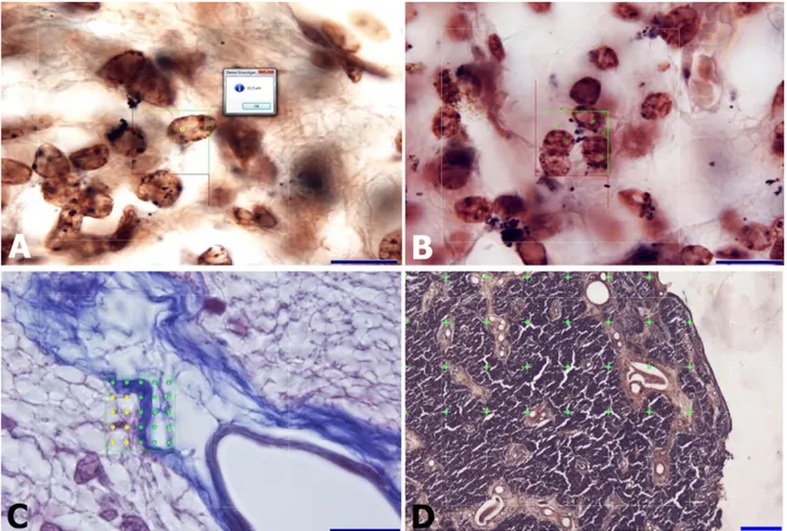
Figure 1. The stereological methods used to analyses the horse pineal gland.
A: The diameter of cell nuclei was calculated without bias using a quick measurement tool with the guide of an unbiased counting frame in foal. AgNOR staining, bar = 20µm.
B: Counting of pinealocyte nuclei using the optical fractionator method with the guide of an unbiased counting frame in foal. The area of unbiased counting frame was 400 µm2. AgNOR staining, bar = 20 µm.
C: Calculation of pinealocytic, astrocytic, connective tissue and vascular areas using the area fraction fractionator probe in foal. The green crosses indicated the connective tissue areas, the yellow crosses indicate pinealocytic areas. Crossman’s modified triple staining, bar = 20 µm.
D: Area calculation of the pineal gland section using the Cavalieri’s method in foal. The area of a single cross was 0.25 mm2. The
green crosses show the region of interest. AgNOR staining, bar = 500µm. Şekil 1. At pineal bezini analiz etmek için kullanılan stereolojik yöntemler.
A: Hücre çekirdeğinin çapı, taylarda tarafsız sayım çerçevesi kılavuzu kullanarak hızlı ölçüm aracı ile tarafsız olarak hesaplandı. AgNOR boyama, çubuk = 20μm.
B: Pinealosit çekirdeğinin, optik parçalama yöntemi ile hayvanlarda tarafsız sayım çerçevesi yardımı ile sayımı. Tarafsız sayım çerçevesinin alanı 400 μm2 idi. AgNOR boyama, çubuk = 20 μm.
C: Taylarda saha bölme aracı probu kullanılarak pinealositik, astrositik, bağ doku ve vasküler alanların hesaplanması. Yeşil renkli artılar bağ dokusu alanlarını, sarı artılar ise pinealositik alanları göstermektedir. Crossman'ın modifiye üçlü boyaması, çubuk = 20 μm. D: Cavalieri yöntemi kullanılarak pineal bezin alan hesaplamaları. Tek bir artının alanı 0.25 mm2'dir. Yeşil artılar ilgilenilen bölgeyi
göstermektedir. AgNOR boyama, çubuk = 500μm.
area and ratio of subcomponents. For every animal, at least 100 sites were visited. More than 1000 points for the pinealocytic, astrocytic and connective tissue areas were calculated using following formula:  = 1/asf × a(p) × P(Yi) (Figure 1C). Using the same formula, about 500 points were calculated for vessels. In this formula, “” refers to estimated area, “asf” refers to the area sampling fraction, “a (p)” refers to the area of a point, and “P(Yi)” refers to the point hitting the reference volume. On the
rectangular lattice, a distance between two points was set to 4 µm. The 100 × oil immersion objective (Plan apo, NA 1.4) was used during calculation. We also calculated the area of cross section to control the accuracy of area fraction fractionator calculations. (Figure 1D).
Statistical analysis: Nonparametric tests followed by
Mann-Whitney U comparison test was performed to compare the data using GraphPad Prism version 6.00 for Windows, GraphPad Software, La Jolla California USA.
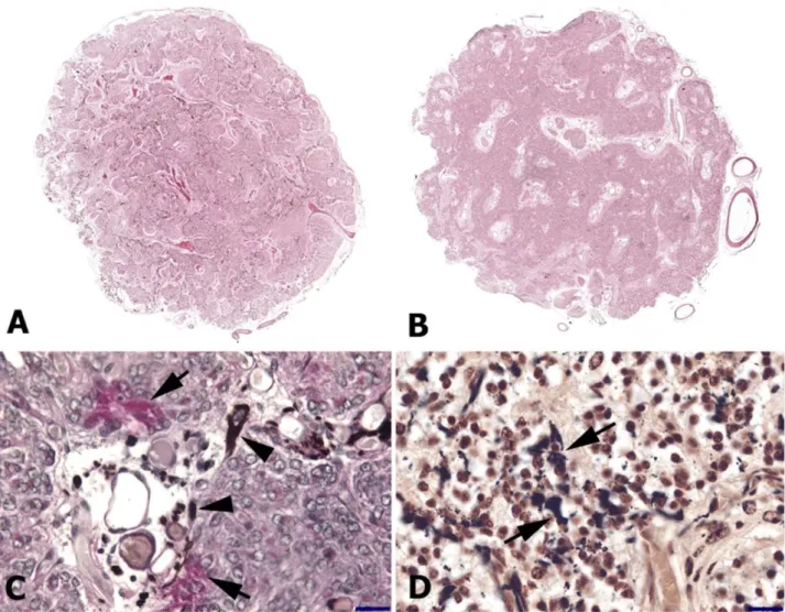
Figure 2. Histological features of the pineal gland in foals and adult horses.
A. General view of the cross sectional area of the foal pineal gland, reconstructed by virtual slice module of Stereo investigator under 20×objective. Lobules formed by the connective trabeculae are more remarkable in foals. H&E staining.
B. General view of the cross sectional area of the pineal gland in adult horses, reconstructed by virtual slice module of Stereo investigator under 20×objective. Trabecular pattern and lobules are not as conspicuous. The loose connective tissue contains large vessels. H&E staining.
C. PAS positive lipofuscin (arrow) and PAS negative melanin pigments (arrow head) can be seen in this section of the pineal gland of an adult horse. PAS staining, bar = 50 µm.
D. Melanin pigment (arrow) can also be seen in this section of the pineal gland of a foal. AgNOR staining, bar = 50 µm.
Şekil 2. Taylar ve erişkin atlarda pineal bezin histolojik özellikleri. A. Stereo investigator'ın virtual slice modülü tarafından 20 × objektif altında yeniden yapılandırılan tay pineal bezinin kesit alanının genel görünümü. Trabeküller tarafından oluşturulan lobüller, taylarda daha dikkat çekicidir. H & E boyası. B. Yetişkin atlardaki pineal bezin kesit alanının genel görünüşü, 20 × objektif altında Stereo investigator’ın virtual slice modülü ile yeniden oluşturulmuştur. Trabeküler ve lobüller belirgin değildir. Gevşek bağ dokusu büyük damarlar içerir. H & E boyası. C. Yetişkin bir atın pineal bezinin bu bölümünde PAS pozitif lipofuksin (ok) ve PAS negatif melanin pigmentleri (ok başı) görülebilir. PAS boyama, çubuk = 50 μm. D. Melanin pigmenti (ok), tayların pineal bezinin bu bölümünde de görülebilir. AgNOR boyama, çubuk = 50 μm.
Results
Histological findings: The pineal glands were
conical in shape both in foals and adult horses. The pial capsule encapsulated the gland and gave branches into the glandular parenchyma. These connective tissue extensions, also called trabeculae, divided the glandular parenchyma into several lobules. Such a lobulation was more conspicuous in foals compared to adult horses. The pineal gland connective tissue was denser in foals
compared to that of adult horses. Connective tissue areas in adults were also characterized by presence of remarkable vascularization (Figures 2A and 2B).
Pinealocytes were easily differentiated in Crossman’s modified triple staining and H&E stained sectioned. Pinealocytes had a euchromatic nucleus. Regular histological and AgNOR staining indicated that melanin granules were present in cytoplasm of pinealocytes and among connective tissue fibers (Figures
2C and 2D). Moreover, melanin granules were also present in the adventitia of the vessels. Presence of melanin granules was confirmed by H2O2 bleaching as
melanin granules disappeared after bleaching. Melanin granules gave a negative reaction with alcian blue at pH 2.5 while they became more intense in AgNOR staining. Unlike in adult horses, PAS positive granules were mainly absent in foals. Importantly, PAS positive granules became more apparent in PAS staining when applied after H2O2 bleaching. Melanin deposition was diffusely
distributed throughout the parenchyma in foals (Figure 2D). On the other hand, melanin granules were mainly deposited in the pineal connective tissue of adult horses (Figure 2C). The pineal gland did not contain brain sands, also called calcareous concentrations, either in foals or adult horses.
Stereological findings: The weight of the pineal
gland was significantly higher in the adult horses
(70.20±5.89 mg) compared to that of the foals (30.20±1.20 mg) (P = 0.009).
Table 1 presents the volume of the pineal gland, the numerical density, the total number of pinealocytes and the diameter of nuclei of pinealocytes in foals and adult horses. The volume of the pineal gland was significantly higher in adult horses (P = 0.009). Similarly, the number of pinealocytes was significantly higher in adult horses compared to foals (P = 0.009). However, there was no significant difference (P = 0.602) for the numerical density of pinealocyte in per 1 mm3 of volume between foals and
adult horses. The diameter of pinealocyte nucleus was significantly higher in adult horses (P = 0.0001).
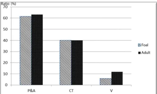
There were no significant difference between foals and adult horses for the ratios of pinealocytic, astrocytic and connective tissue areas. However, the ratio of areas occupied by the vessels was significantly higher in adult horses (P = 0.009, Figure 3).
Table 1. The volume of pineal glands, the total number of pinealocytes, the numerical density of pinealocytes and the diameter of nuclei of pinealocytes.
Tablo 1. Epifiz bezlerin hacmi, toplam pinealosit sayısı, pinealositlerin sayısal yoğunluğu ve pinealositlerin çekirdek çapı.
Parameters Foal Adult P Value
Mean ± SEM Mean ± SEM
The volume of pineal gland (mm3) 30.20 ± 1.20 70.20 ± 5.89 0.009
The total number of pinealocytes (106) 5932 ± 0.405 13332 ± 1.239 0.009
The numerical density of pinealocytes 196423 ± 341 189914 ± 289 0.602
Diameter of nuclei of pinealocytes (µm) 5.79 ± 0.08 7.89 ± 0.10 0.032
Note: the coefficient of error for the total number of pinealocytes was calculated as 0.04 and 0.05 in foal and adult horses, respectively while the coefficient of variance for the same parameter was calculated as 0.07 and 0.09 for foal and adult horses.
Not: Taylarda ve ergin atlarda toplam pinealosit sayısı için hata katsayısı sırasıyla 0.04 ve 0.05, taylarda ve ergin atlar için aynı parametrenin varyans katsayısı 0.07 ve 0.09 olarak hesaplanmıştır.
Figure 3. The ratio of subcomponents of the pineal gland calculated by the area fraction fractionator probe. P&A= Pinealocytic and astrocytic areas , CT = Connective Tissue, V = Vessels.
Şekil 3. Area fraction fractionator probu ile hesaplanan pineal bezin alt komponentlerinin oranı. P & A = Pinealositik ve astrositik bölgeler, CT = Bağ dokusu, V = Damarlar.
Discussion and Conclusion
There has been more or less bias in conventional histomorphometric methods employed in previous studies. To eliminate bias, advanced stereological methods such as the “fractionator” have been used for the calculation of the amount of tissue structures in region of interest. The “optical fractionator” is used as a counting method which is employed on thick sections for optical sectioning. The optical fractionator is very efficient for time and preparing tissue sample (17). In the present study, the optical fractionator was employed for the calculation of the number of pinealocytes in the horse pineal gland (Figure 1). The “area fraction fractionator” (9) was utilized for calculating subcomponents of the pineal gland such as pinealocytic and astrocytic area, ratio of connective tissue and vessels (Figure 3).
The pineal gland undergoes morphological and physiological changes during the aging process. As also indicated by previous studies conducted in cats (2), dogs (3), horses (5), and humans (18), the present study concluded that age related morphological changes in the horse pineal gland are not gender specific. A study conducted in dogs found that the size of the gland increases during the postnatal period until adult ages (3). In the present study, we determined the pineal gland in adult horses was at least two folds heavier than that of foals. As cited by Khavinson and Linkova (18) the increase in the weight of human pineal gland during aging was mostly related to deposition of fibrous tissue and cysts without a significant decrease in the number of pinealocytes. In contrast, the present study indicated that there was no difference between foals and adult horses for the percent volume of connective tissue in the pineal gland, suggesting that the amount of connective tissue deposition was proportional to the size of the organ. Furthermore, the number of pinealocytes in per volume was similar between foals and adult horses. However, the blood vessels in the pineal gland connective tissue are larger and occupy higher volumes in adult horses.
The human pineal gland is morphologically classified into three categories: cellular, trabecular, and alveolar (18). In the dog and cat, such a classification has not been reported (2, 3). In addition, the dog pineal gland has a limited connective tissue areas (3). In the present study, we observed some differences between foals and adult horses by means of stromal organization in the pineal gland. In foals, the stroma was characteristically composed of trabecular septa with branches that divided the gland into lobules. In adult horses, on the other hand, the trabecular septa and such branches were not conspicuous, but a loose connective tissue was discernible (Figures 2A and 2B). Redondo et al. (22) described a trabecular pattern in the pineal gland of young sheep, similar to what we observed in foals.
The total number of pinealocytes in the human pineal gland decreases by 18% by aging (18). The data we obtained in horses did not immediately support this conclusion. Although the number of pinealocytes was at least 2.5 folds higher in adult horses, there was no significant difference between foals and adult horses by means the number of pinealocytes in per mm3. However,
absence of a group of horses in very old ages was a shortcoming of the present study so that it was impossible to make a definitive conclusion. In the meantime, the present study suggested that the increase in weight and volume of the organs occurs as a result of an increase in almost every component of the gland, the number of pinealocytes, connective tissue, etc. The pineal gland undergoes some but not significant morphological changes by aging. Presence of calcareous concentrations has been reported in gerbil (26) and even in young horses (6). Increase in deposition of calcium and lipofuscin pigment is often considered as the functional state of the gland, not directly related to aging (18). In the present study, we did not observe any calcareous concentrations either in foals or adult horses. It is methodologically important to state that no decalcifying agent was used in the present during routine histological processes. Likewise, we did not come across with a reference stating that formalin fixative or any other reagents used in the routine histological process dissolve calcareous concentrations.
In the pineal gland, some pinealocytes had a nucleus with double DNA content and their frequency is higher in elderly (18). In our search of literature, we did not come across with reports on the presence of dikaryotic pinealocytes. In the present study, we did find any dikaryotic pinealocyte, either. Pinealocytes in both foals and adult horses had a euchromatic nucleus. A remarkable finding was that the diameter of the pinealocyte nucleus was significantly higher in adult horses. Such a large nucleus might indicate presence of a large number of cells with an increased DNA content (29). It is well known that the pineal gland is significantly influenced by aging process (18), and the nuclear size of pineolocytes is one of the parameters investigated (11, 13, 15). A study by Gusek and Meier (13) claims that the nuclear size of the pinealocytes is larger in younger ages although the pineolocyte cell size is similar between the younger men (30 to 40-years-old) and elderly (13). Human in 30-40 years of age is considered in mature ages (18). In our study, horses in very advanced ages were not investigated. In such advanced ages, horses might have pineoclocytes of very different sizes compared to young and mature ages. A study conducted in hamsters (15) concluded that the nuclear size and whole cell size are influenced by age and photoperiod. Rodents are active in dark and their circadian rhythm is different compared to most mammals.
It is well known that horse is a seasonal breeder and thus influenced by the day light period. The samples used in our study were collected during the autumn season. Thus, additional studies should be conducted on how day light period influences horse pinealocytes by means of nuclear and cytoplasmic sizes. Another study on young and adult mice claimed that pinealocyte nuclear size differed slightly by region (11). In our study, we did not specifically investigate the regional difference by means of the pinealocyte nuclear size; however, we observed that pinealocytes with smaller nuclei were commonly found close to the habenular commissure where the pinealocyte population was lower compared to other regions of the gland.
The light and electron microscopic studies concluded that melanin granules are present in the pineal glands of all ages (3). The horse pineal gland is also unique for the presence of large quantities of melanin granules (5). As we also found in the present study, melanin deposition occurred not only in pinealocytes but also along the connective tissue fibers in the horse pineal gland (4). Capucchio et al. (4) also claims that melanosis or deposition of melanin in the pineal gland is not related to age of the horses. In contrast, Koshy and Vettivel (19) claims that melanin deposition in human increases by aging. Al-Hüssain (1) cited that the pineal gland of the elderly does not contain melanin. In this study, we found a different pattern of melanin deposition in foals and adult horses. Melanin deposition was diffuse and widespread in foals while it was focally localized to connective tissue in adult horses. It has been proclaimed that there is a relationship between gonadal development and melanin containing cells in the pineal gland (24). In this respect, one can suggest that a higher amount of melanin content could be associated with gonadal development in horses. However, Calvo et al. (3) concluded that pigmented cells in the pineal gland are unrelated to maturation of the gonads and sexual development in dogs. Therefore, a further comparative study would be more useful.
In conclusion, the equine anatomy is often difficult to study due to scare material. Despite the small number of material used in the current study, the results revealed the differences between pineal glands of foals and adult horses in the light of modern methods.
References
1. Al-Hüssain SM (2006): The pinealocytes of the human pineal gland: A light and electron microscopic study. Folia Morphol, 65, 181-187.
2. Boya J, Calvo JL, Rancano D (1995): Structure of the pineal gland in the adult cat. J Pineal Res, 18, 112-118. 3. Calvo J, Boya J, Garcia-Maurino A, et al. (1990):
Postnatal development of the dog pineal gland. Light microscopy. Histol Histopathol, 5, 31-36.
4. Capucchio MT, Marquez M, Pregel P, et al. (2010): Parenchymal and vascular lesions in ageing equine brains: Histological and immunohistochemical studies. J Comp Pathol, 14, 61-73.
5. Cozzi B, Ferrandi B (1984): The pineal gland of the horse. Morphological and histochemical results. (With notes on the donkey and mule pineal). Basic Appl Histochem, 28, 81-90.
6. Cozzi B (1986): Cell types in the pineal gland of the horse: An ultrastructural and immunocytochemical study. Anat Rec, 216, 165-174.
7. Culling CFA, Allison RT, Barr WD (1985): Carbohydrates. 214-255. In: Culling CFA, Allison RT, Barr WD (Ed), Cellular Pathology Technique. Butterworth & Co. (Publishers) Ltd, London.
8. Denk H, Kunzele H, Plenk H, et al. (1989): Romeis Mikroskopische Technik. Urban and Schwarzenberg, München.
9. Elias HD, Hyde DM (1983): A Guide to Practical Stereology. S. Karger AG, Basel.
10. Flood DG, Coleman PD (1988): Neuron numbers and sizes in aging brain: Comparisons of human, monkey, and rodent data. Neurobiol Aging, 9, 453-463.
11. Gundersen HJ, Jensen EB, Kieu K, et al. (1999): The efficiency of systematic sampling in stereology-reconsidered. J Microsc, 193, 199-211.
12. Gundersen, HJ (1977): Notes on the estimation of the numerical density of arbitrary profiles: The edge effect. J Microsc, 111, 219-223.
13. Gusek W, Meier D (1988): Size of the cell nucleus and nucleus-cytoplasm relations in pinealocytes in humans of middle and advanced age. Z Gerontol, 21, 79-82.
14. Hayasaka K (1988): Electron-microscopic observations on pinealocytes in various regions of the pineal gland of young and adult mice. Hokkaido J Med Sci, 63, 115-129. 15. Hira Y, Sakai Y, Matsushima S (1989): Comparisons of
sizes of pinealocyte nuclei and pinealocytes in young and adult Chinese hamsters (Cricetulus griseus) under different photoperiod conditions. J Pineal Res, 7, 411-418.
16. Hughes SW (2005): Archimedes revisited: A faster, better, cheaper method of accurately measuring the volume of small objects. Phys Educ, 40, 468-474.
17. Jastrow H, Von Mach MA, Vollrath L (1997): Adaptation of the disector method to rare small organelles in TEM sections exemplified by counting synaptic bodies in the rat pineal gland. J Anat, 191, 399-405.
18. Khavinson VK, Linkova N (2012): Morphofunctional and molecular bases of pineal gland aging. Hum Physiol, 38, 101-107.
19. Koshy S, Vettivel S (2001): Melanin pigments in human pineal gland. J Anat Soc India, 50, 122-126.
20. Ploton D, Menager M, Jeannesson P, et al. (1986): Improvement in the staining and in the visualization of the argyrophilic proteins of the nucleolar organizer region at the optical level. Histochemical J, 18, 5-14.
21. Pontén J, Stein WD, Shall SA (1983): Quantitative analysis of the aging of human glial cells in culture. J Cell Physiol, 117, 342-352.
22. Redondo E, Regodon S, Masot J, et al. (2003): Postnatal development of female sheep pineal gland under natural inhibitory photoperiods: An immunocytochemical and
physiological (melatonin concentration) study. Histol Histopathol, 18, 7-17.
23. Reiter RJ (1991): Pineal gland interface between the photoperiodic environment and the endocrine system. Trends Endocrin, 2, 13-19.
24. Santamarina E (1958): Melanin pigmentation in bovine pineal gland and its possible correlation with gonadal function. Can J Biochem Physiol, 36, 227-235.
25. Schmitz C, Hof PR (2000): Recommendations for straightforward and rigorous methods of counting neurons based on a computer simulation approach. J Chem Neuroanat. 20, 93-114.
26. Swietoslawski J (1999): The age-related quantitative ultrastructural changes in pinealocytes of gerbils. Neuro Endocrinol Lett, 20, 391-396.
27. Totty BA (2002): Mucins. 163-200. In: Bancroft JD, Gamble M (Ed), Theory and Practice of Histologycal Techniques. Churchill Livingstone, Edinburg.
28. West MJ, Slomianka L, Gundersen HJ (1991): Unbiased stereological estimation of the total number of neurons in the subdivisions of the rat hippocampus using the optical fractionator. Anat Rec, 231, 482-497.
29. Winick M, Rosso P, Waterlow J (1970): Cellular growth of cerebrum, cerebellum, and brain stem in normal and marasmic children. Exp Neurol, 26, 393-400.
Geliş tarihi: 10.02.2017 / Kabul tarihi: 10.04.2017 Address for correspondence:
Doç. Dr. Durmuş BOLAT
Kırıkkale University, Faculty of Veterinary Medicine, Department of Anatomy,
Yahşihan, Kırıkkale, Turkey. e-mail: [email protected]