A Preliminary Investigation on the Presence of
Calcifying Nanoparticles in the Breast Tumor
Pınar OZKAL-BAYDIN1, Sedef J. GOCMEN2, Esra ERDEMLI3, Ibrahim G. TUNC4,
Hasan B. SENER5, Berrak GUMUSKAYA5, Asuman SUNGUROGLU1 1 Ankara University, Faculty of Medicine, Department of Medical Biology 2 Başkent University, Faculty of Medicine , Department of Medical Microbiology
3 Ankara University, Faculty of Medicine, Department of Histology
4 Dışkapı Yıldırım Beyazıt Training and Research Hospital, Department of General Surgery 5 Dışkapı Yıldırım Beyazıt Training and Research Hospital, Department of Pathology, Ankara / TURKEY
ABSTRACT
Calcium phosphate is deposited in many diseases, but the molecular basis of mineralization remains largely unknown. Biomineralizied calcifications that are formed by calcium deposits are also detected in breast mammograms. Some of the detected microcalcifications are thought to be related with malignancy. Taken together, calcifying nanoparticles (CNP) may be thought as a source of malign calcifications in breast cancers. The aim of the study is to research the presence of CNP in breast tumor tissue. With this aim, the presence of CNP was investigated by culturing 16 patients’ breast tumor tissue and from 2 pathologic tissues with transmission electron microscopy (TEM) and scanning electron microscopy (SEM). Their growth was monitored by optical density (OD) at a wavelength of 650 nm. CNP couldn’t be found in the analysed tissues. The presence of CNP in the breast tumor tissue was researched for the first time. We could not find CNP in the breast tumor tissue, but we think this research will open a new field of study for researchers.
Keywords: Breast tumor, Calcification, Calcifying nanoparticles, Scanning electron microscopy, Transmission electron microscopy
ÖZET
Meme Tümöründe Kalsifiye Edici Nanopartiküllerin Varlığına Dair Bir Ön İnceleme
Kalsiyum fosfat depolanması pek çok hastalıkta görülür, fakat mineralizasyonun moleküler nedeni büyük oranda bilinmemektedir. Kalsiyum depolanmasıyla oluşan biyomineral kalsifikasyonlar mamogramla saptanmaktadır. Saptanan mikrokalsifikasyonların bazılarının maligniteyle ilişkili olabileceği düşünülmektedir. Tüm bu bilgiler göz önüne alındığında, kalsifiye edici nanopartiküller (CNP) malign kalsifikasyonların nedeni olarak düşünülebilir. Bu çalışmanın amacı, meme tümör dokusunda CNP varlığını araştırmaktır. Bu amaçla, CNP varlığı 16 hastanın meme tümör dokusunun kültürü sonrası ve 2 patolojik dokunun geçirimli ve taramalı elektron mikroskopları (TEM ve SEM) ile incelenmesi ve 650 nm’de optik densitenin ölçülmesi ile araştırılmıştır. Analiz edilen dokularda CNP saptanamamıştır. Bu çalışmayla, CNP varlığı ilk defa meme tümör dokusunda araştırılmıştır. Meme tümör dokusunda CNP bulunamamakla birlikte bu çalışmanın araştırmacılara yeni araştırma alanları açacağı düşünülmektedir.
Anahtar Kelimeler: Meme tümörü, Kalsifikasyon, Kalsifiye edici nanopartiküller, Taramalı elektron mikroskobu, Geçirmeli elektron
INTRODUCTION
The formation of organized inorganic crystalline structures within macromolecular extracellular matri-ces is referred to as “biomineralization”. Mammalian bone and dental enamel are examples of biominerali-zation. The formation mechanisms of mineralization remain speculative and largely unknown.1
Calcifica-tion of soft tissues, termed “pathologic calcificaCalcifica-tion” is a common process in a wide variety of diseases2 like
aortic valve stenosis, atherosclerosis and psammoma bodies in ovarian cancer.3-6 Breast calcifications are
deposits of calcium that can be seen on breast mam-mograms.7 One of the most important roles of
cal-cification detection in mammography is the identifi-cation of malignancy. The other important role is in the follow up of breast cancer patients having breast conservation therapy as calcifications have been de-tected in 29-80% of recurrences. Morover, there is an increased risk of malignancy associated with an increased number of calcifications.2 Calcifications
associated with benign conditions are usually larger, fewer in number, widely dispersed, and round.7
There are two forms of microcalcification in breast disease: Type I and Type II. Type I is composed of calcium oxalate and these oxalate calcifications are generally associated with proliferating but noninva-sive diseases of the breast. On the other hand, Type II consists predominantly of calcium phosphate. Calci-um phosphate in the crystalline form of hydroxyapa-tite is usually correlated to invasive malign tumors. The mechanism that causes deposition of the bone-specific mineral hydroxyapatite has not yet been il-luminated.7
Calcifying nanoparticles (CNP) (nanobacteria, na-nons, nanobes, nanobacteria-like particles) were dis-covered as cell culture contaminants by Kajander et al in bovine and human blood in the last decade of 20th century. The name is derived from the very small size of these nanoparticles, which varies from 50 nanom-eters to 500 nanomnanom-eters. The defining characteristics of CNP is their outer envelope composed of calcium phosphate that presents on transmission electron microscope images as an electron-dense shell sur-rounding a translucent central core. They produce carbonate apatite on their cell envelope from soluble calcium and phosphorus at physiologic concentra-tions and condiconcentra-tions. However, there is still a big de-bate about whether these self-mineralizing particles are bacteria or inorganic substances with the capacity
for extensive crystallization.8-10
Discovery of CNP initiated further studies and have now been implicated in a wide array of human dis-eases associated with calcifications like: kidney stone formation1, prostatitis11, psammoma bodies in ovarian
cancer6,12, Randall’s plaque13, testicular
microlithi-asis14, calcification in placenta15, calcific aortic valve
stenosis4,16, atherosclerotic plaques5, and dental pulp
stones.17
Taken together, since microcalcifications are one of the most common abnormalities detected on screen-ing mammography for breast cancer7 and CNP has
been shown to contribute to different benign and malign calcifications11-16, CNP may also contribute
to malign calcifications in breast cancer. Thus, in the light of these investigations and results, this study fo-cuses on researching the presence of CNP in calcified breast tumor tissues for the first time.
MATERIALS AND METHODS Clinical sample collection
All enrolled patients agreed to sign written consent forms and the study was approved by Kırıkkale Uni-versity Ethics Board. Sixteen women, aged 43-74 (median 57), all having breast carcinoma (7; infiltra-tive ductal, 5; invazive ductal, 1; mucinose, 1; meta-plastic, 1; mix type-infiltrative ductal+infiltrative lob-uler-, and 1; inflamatuar carcinoma) were accepted for this study. In addition to 16 patients’ tumor tis-sues, 2 paraffin embedded breast tumor tissues are included in this study.
Cultivation of calcifying nanoparticles
Before culturing tumor tissues, we prepared cultures with DMEM supplemented with 10% fetal bovine serum (FBS) (filtered through 0.22 µm filters) to observe CNP under mammalian cell culture condi-tions (370C, 5% CO2, 95% air). These were our
posi-tive control cultures. The incubation period was 6-8 weeks.
The breast tumor tissues were divided into two parts: one of which was kept at 40C for direct analysis
with-out culturing and the other part was crushed in a ster-ile glass petri by sterster-ile surgical blades under laminair flow. It was demineralized by adding 1 M HCl, which was subsequently neutralizied with 1 M NaOH. The resultant was filtrated with a 0.22 µm membrane fil-ter and cultured in DMEM supplemented with 10%
gamma-irradiated (γ) and 0.22 µm membrane filtrat-ed FBS at 370C, under 5% CO2 atmosphere.16 Also,
cultures without tumor tissue cultured under the same conditions were used as the negative controls. After 6-8 weeks of incubation, a culture scraping was ex-ecuted in each flask, then centrifuged at 5000 x g for 45 minutes and pellets were obtained.
Transmission electron microscopy (TEM)
PBS washed pellets were fixed by immersion in 2.5% gluteraldehyde in 0.1M phosphate buffer (pH 7.4) at room temperature for 3 hrs and post – fixed in 1%
phosphate buffered osmium tetroxide for 2 hrs. Sam-ples were dehydrated by being passed through the graded ethanol series and embedded in Araldite CY-212. Ultra thin sections were contrast stained with uranyl acetate and lead citrate and examined using the Leo 906 E (80 kV) (Oberkohen-Germany) transmis-sion electron microscope.16,18
For preparing slides from pathological samples which were previously processed for standard histological examination, paraffin blocks were placed in 100% xylene for 1-2 hrs until no evidence of paraffin re-mains then rehydration in ethanols was performed. After incubation in 1% osmium tetroxide for 1 hr, they were dehydrated and embeded in Araldite CY 212 as for routine TEM sample preparation.19
Also, breast tumor tissues of 16 patients that were kept at 40C, were screened by direct x-ray to see
cal-cificated areas. From 7 patients’ calcal-cificated breast tu-mor tissue areas, slides were prepared to be observed under TEM.
Scanning electron microscopy (SEM)
Only positive control cultures were screened by SEM. Pellets were washed with PBS then subcultured in serum-free RPMI-1640 on glass cover slips for 24 hrs. The glass cover slips were washed with PBS and fixed with 2% gluteraldehyde for 30 min at room tem-perature. Fixed samples were dehydrated with abso-lute alcohol; dried in a critical point dryer and layered with gold followed by examination in SEM.
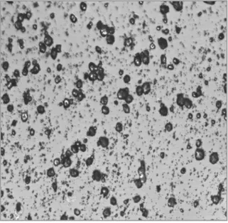
Figure 1. Scanning electron micrograph of calcifying
nano-particles.
Figure 2. Transmission electron micgraph of calcifying nano
particles (Original magnification is 100.000X).
Figure 3. Transmission electron micgraph of calcifying
Microbiologic tests
To be sure about the non-existance of classical mi-crobes in our CNP cultures, subcultures were per-formed on 200 µl pellets in a mycoplasma liquid medium, sheep-blood agar and eosin methylen blue (EMB) agar both in aerobic and anaerobic conditions. Morover, Gram and Giemsa stainings were done. Spectrophotometry
Maximum absorbances of pellets were measured by spectrophotometer.
RESULTS
Calcifying nanoparticle culture results, imag-ing by TEM and SEM: After 8 weeks of culture, we could observe CNP like particules in our positive control cultures including DMEM supplemented with 10% FBS both with SEM and TEM. SEM of the posi-tive control culture revealed coccobacillary particles with a diameter ranging between 100 and 200 nm. The particles were prokaryotic in shape. (Figure 1). TEM also showed coccoid structures wrapped by a high electron-dense, thick outer shell (Figure 2 and 3). These morphologic and distinctive features are the same as those of CNP described in previous stud-ies.1,18,20 But neither in the cultures with breast tumor
tissues nor in our negative control cultures could we observe CNP.
TEM of pathologic tissue and calcified area re-sults: From 2 pathologic breast tumor tissues and 7 patients’ breast tumor tissues we tried to sign the calcified areas and tried to prepare slides for TEM only from these areas. CNP could not be imaged from these samples.
Microbiologic test results: A subculture of pellets on sheep blood agar, mycoplasma medium and EMB agar resulted without growing. Also, they couldn’t be dyed by Gram and Giemsa.
Spectrophotometric results: Maximum absorbance of breast tumor tissue pellets was 550-560 nm. DISCUSSION
CNP appear as self-propagating calcifying macromo-lecular complexes found in bovine and human blood and blood products. There are still discussions about their nature, as living or non-living and it is certain that the discussions will continue until their genome can be accurately sequenced. Whether they are
phys-icochemical phenomenon or bacteria, they exist and cause diseases.10 The disease-causing mechanisms of
CNP include the known effects of calcium on blood vessels, blood coagulation and thrombus formation; elevation of intracellular [Ca2+] levels and its
conse-quences. They grow better in the presence of other bacteria and this could change the normal flora and promote bacterial infection. CNP adhere to foreign objects, such as implants, and could result in bio-film formation later attracting bacteria, leading to a chronic infectious problem that is often untreatable with antibiotics.21
Guneslioglu, et al.3 and Altundag, et al.7 hypohesized
a possible association between breast implant capsule contracture, malignant calcifications in breast cancer and CNP. Breast calcification is an important feature in the radiological assessment of breast lesions. Good understanding of the morphologic features and the mechanism of calcifications is crucial for health care professionals involved in breast care. When we take different calcification mechanisms into consideration, pathologic calcification can be classified as dystropfic and metastatic calcification. Dystropfic calcification may be intra- or- extracellular. While the initiation in extracellular sites begins at membrane bound vesi-cles derived from degenerating cells; the initiation in intracellular sites begins in the mitochondria of the dead or dying cells. Calcifications detected in the breast is probably derived from dystropfic calcifica-tion, not from metastatic one.2 There are two types
of calcification molecules in breast tissue. One is calcium oxalate, which is crystalline and is present mostly in benign lesions. The other is non crystalline, which is a form of calcium phosphate and is associ-ated with malign lesions.22 Osteopontin also appears
to be a crucial mediator involved in the process of calcification. This mediator was found in the histio-cytes around the lesional epithelial breast tissue, but at much lower level in tumor cells. In the scenario of high grade tumor, osteopontin expression may represent part of inflammatory response to cellular damage. In addition to construction mechanism of microcalcifications and the particles they include, the density, mass and number of calcifications are very important in the prognosis of breast lesions.2 For
these reasons, in this study we planned to investigate the presence of CNP in calcified breast tumor tissues. Ciftcioglu, et al.23 found that CNP were best
culture: One type was a positive control including DMEM with 10% FBS. This cultures’ aim was to cul-ture and image CNP. These bacteria like particles can not grow and reproduce under common conditions for microbial culture, but need media for mammalian cell culture such as Roswell Park Memorial Institute (RPMI) 1640 medium or Dulbecco modified eagle medium to grow (DMEM).10,11 FBS was used as a
supplement and source of CNP. The second type was the negative control cultures including DMEM with 10% γ FBS. If FBS was added into the culture medi-um directly, it gives false positive results. Fortunately, the destructive effects of γ ray irradiation on FBS can effectively eliminate powerful inhibitors of apatite crystal formation.24 So, we used γ FBS to avoid false
positive results and did not expecting to observe CNP in these cultures as in the previous studies.1,24,25 The
last culture was the same with our negative cultures, but we added our demineralized, neutralized and 0.22 µm pore filtered breast tumor tissues to these cultures. Morover, FBS and γ FBS were both filtered before cultivation to prevent contamination by other bacte-ria or fungi. It has also been shown that, nucleation capacity could be lost after 0.1 or o.05 µm membrane filtration.25 After 8 weeks of culture, we were able to
manage to image CNP only in our positive controls, but not in the cultures of breast tumor tissues. Also, spectrophotometric measurements would indicate the presence of CNP if we measured OD650, but our re-sults were OD550-OD560. This result made us think about the amount of calcification of our tissues. To be sure about imaging calcified breast tumor areas, we screened 16 patients breast tumor tissue that we were keeping at 40C by direct x-ray. For seven of the
16 tissues, we were able to check off calcified areas and tried to prepare slides for TEM from these calci-fied areas. Morover, we again prepared 2 slides for TEM from paraffin embedded calcified brest tumor tissues. But with the same result as our previous one, we could not observe CNP by TEM in these samples. Several methods are being used to detect CNP, like bacterioscopic, bacteriological, serological, genomic or proteomic methods, although some of these are still questinable.10 However, TEM is still a powerful
tool for detecting and studying CNP.9 If we are able to
see CNP by TEM in tumor tissue samples, we could try PCR-based detection of the universal 16S rRNA gene9,26 or immunefluorescence staining with 8D10.27
Kajander and Ciftcioglu have discovered CNP in
hu-man and cow blood first1; then they developed their experiments by researching other mammalian cell cultures. They have shown the adherence of CNP to 3T6 and other fibroblasts. CNP were bound as clus-ters on the cell surfaces. Adherence took place on cellular extensions and to the perinuclear area of the cells. They thought CNP are internalized either by receptor-mediated endocytosis or by a closely related pathway.1,20 This important experiment have shown
that, CNP have the ability to invade mammalian cells. Many mammalian malign cells have receptors for CNP adherence1 that could introduce CNP into
the tumor with subsequent calcification.20 Guo et al
9 speculated that the CNP detected in their placental tissue calcification was a novel type of nanoscale bac-teria distinct from the nanobacbac-teria isolated from kid-ney stones and human blood, as reported elsewhere. They assumed that different nanobacteria may induce calcification by receptors in different human tissues, like nanobacteria receptor mediated tumor tissue cal-cification. There might exist a special cell adhesion molecule in the outer membrane of the CNP to invade a cell. In the light of this research, the receptor-medi-ated endocytosis pathway mechanism used by CNP may not be suitable for breast cells at all.
In our study, the reason we could not observe CNP in the calcified breast tumor tissue may be a result of in-sufficient calcified areas such that the amounts of cal-cified areas were too small to isolate or they were lost in some steps. It is also said that calcification depends on the amount of calcifying nanoparticles present.9
Moreover, it might be possible for us to see CNP in the calcified breast tumor tissue, if we could have a chance to culture more breast tumor tissues.
In this study, the presence of CNP in the breast tumor tissue was researched for the first time. Isolating and culturing CNP from breast tumor tissue has not been reported before. We think that this investigation will open a new area for researchers. Therefore, we won-der if new culture parameters, tools and techniques can be used and developed in this new research area.
Acknowledgment
This work was supported by Ankara University Re-search Foundation (Project No: 2007-08-09-052 HPD). The authors would like to acknowledge the contribution of Prof. Alp Can from Ankara Univer-sity, Faculty of Medicine, Department of Histology for SEM image.
REFERENCES
1. Kajander EO, Ciftcioglu N. Nanobacteria: An alternative mechanism for pathogenic intra-and extracellular calcification and stone formation. Proc Natl Acad Sci USA 95: 8274-8279, 1998.
2. Tse GM, Tan PH, Cheung HS, et al. Intermediate to highly suspicious calcification in breast lesions: a radio-pathologic correlation. Breast Cancer Res Treat 110: 1-7, 2008. 3. Gundeslioglu O, Altundag O, Altundag K. Nanobacteria and
breast implant capsule contracture and calcification: a hy-pothesis. Aesth Plast Surg 29: 582, 2005.
4. Jelic TM, Chang H, Roque R, et al. Nanobacteria-associated calcific aortic valve stenosis. J Heart Valve Dis 16: 101-105, 2007.
5. Puskas LG, Tislavics L, Razga ZS, Torday LL, Krenacs T, Papp JGy. Detection of nanobacteria-like particles in human atherosclerotic plaques. Acta Biol Hungarica 56: 233-245, 2005.
6. Sedivy R, Battistutti WB. Nanobacteria promote crystallization of psammoma bodies in ovarian cancer. APMIS 111: 951-954, 2003.
7. Altundag K, Altundag O, Akyurek S, Atik MA. Possible asso-ciation between nanobacteria and malign microcalcifications in breast cancer. Breast J 12: 287, 2006.
8. Jelic TM, Roque R, Yasar U, et al. Calcifying nanoparticles associated encrusted urinary bladder cystitis. Int J Nanomed 3: 385-390, 2008.
9. Guo Y, Zhang D, Lu H, et al. Association between calcifying nanoparticles and placental calcification. Int J Nanomed 7: 1679-1686, 2012.
10. Kutikhin AG, Brusina EB, Yuzhalin AE. The role of calcifying nanoparticles in biology and medicine. Int J Nanomed 7: 339-350, 2012.
11. Zhou Z, Hong L, Shen X, et al. Detection of nanobacteria in-fection in type III prostatitis. Urology 71: 1091-1095, 2008. 12. Hudelist G, Singer CF, Kubista E, et al. Presence of
nanobac-teria in psammoma bodies of ovarian cancer. Evidence for pathogenic role in intrtumoral biomineralization. Histopathol-ogy 45: 633-637, 2004.
13. Ciftcioglu N, Vejdani K, Lee O, et al. Association between Randall’s plaque and calcifying nanoparticles. Int J Nanomed 3: 105-115, 2008.
14. Zhang Q, Lu G, Shen X, et al. Nanobacteria may be linked to testicular microlithiasis in infertility. J Androl 31: 121-125, 2010.
15. Lu H, Guo Y, Liu S, Zhang D. Nanobacteria may be linked to calcification in placenta. Ultrastruct Pathol 36: 160-165, 2012.
16. Bratos-Perez MA, Sanchez PL, deCruz SG, et al. Association between self-replicating calcifying nanoparticles and aortic stenosis. A possible link to valve calcification. Eur Heart J 29: 371-376, 2008.
17. Yang F, Zeng J, Zhang W, Sun X, Ling J. Evaluation of the interaction between calcifying nanoparticles and human den-tal pulp cells: a preliminary investigation. IInt J Nanomed 6: 13-18, 2011.
18. Khullar M, Sharma SK, Singh SK, et al. Morphological and im-munological characteristics of nanobacteria from renal stones of a North Indian population. Urol Res 32: 130-35, 2004. 19. Dykstra MJ. A Manual of Applied Techniques for Biological
Electron Microscopy. New York and London, Plenum Press, 1993: 75-77.
20. Ciftcioglu N, Kajender EO. Interaction of nanobacteria with cultured mammalian cells. Pathophysiology 4: 259-270, 1998.
21. Kajander EO. Nanobacteria-propagating calcifying nanoparti-cles. Lett Appl Microbiol 42: 549-552, 2006.
22. Gonzalez JE, Caldwell RG, Valaitis J. Calcium oxalate crstals in the breast. Pathology and significance. Am J Surg Pathol 15: 586-591, 1991.
23. Ciftcioglu N, Bjorklund M, Kuorikoski K, et al. Nanobacteria. An infectious cause for kidney stone formation. Kidney Int 56: 1893-1898, 1999.
24. Wen Y, Li Y, Yang Z, et al. Detection of nanobacteria in serum, bile and gallbladder mucosa of patients with cholecystolithi-asis. Chin Med J 118: 421-424, 2005.
25. Cisar JO, Xu D, Thompson J, Swaim W, Hu L, Kopecko DJ. An alternative interpretation of nanobacteria-induced biomin-eralization. PNAS 97: 11511-11515, 2000.
26. Raoult D, Drancourt M, Azza S, et al. Nanobacteria are miner-alo fetuin complexes. Plos Pathogens 4: e41, 2008. 27. Hu R, Zhao Y, Sun Y, et al. Detection of nanobacteria-like
material from calcified cardiac valves with rheumatic heart dis-ease. Cardiovasc Pathol 19: 286-292, 2010.
Correspondence
Dr. Pınar ÖZKAL BAYDIN Ankara Üniversitesi Kök Hücre Enstitüsü
Ceyhun Atıf Kansu Cad. No: 169 Cevizlidere, Balgat
ANKARA / TURKEY Tel: (+90.312) 595 71 16