Abstract.
Background/Aim: To compare
simultaneous-integrated boost (SIB) versus sequential-boost (SB) delivered
in the context of whole-breast irradiation (WBI) via
volumetric-modulated arc therapy (VMAT) and helical-tomotherapy (HT).
Materials and Methods: Planning target-volume (PTV)
dosimetric parameters and organs at risk (OAR) were analyzed
for SB plan (50 Gy plus 16 Gy boost) and SIB plan (50.4 Gy
WBI and 64.4 Gy tumor bed boost) in VMAT and HT
techniques. Results: Conformity and homogeneity for
target-volume doses were better in HT plans compared to VMAT
plans. There were no significant differences in ipsilateral lung
doses between VMAT and HT plans for SB/SIB techniques,
except for a significantly higher lung V5 value with VMAT-SB,
and lung V13 value with HT-SIB technique. HT provided a
statistically significant decrease in contralateral lung mean V5.
Conclusion: The SIB technique showed better target-volume
dose distribution in both HT and VMAT plans, and better
sparing heart in HT compared to the SB technique.
Breast cancer (BC) is the most common cancer and is a
major cause of cancer-related death in women (1). Currently,
adjuvant radiotherapy (RT), especially whole-breast
irradiation (WBI) after breast-conserving surgery (BCS), is
accepted as standard-of-care for early BC patients as this
multimodal treatment increases local control and improves
overall survival in early BC (2, 3). Additional tumor-bed
boost after WBI has also been shown to reduce local
recurrence (4); however, a long-term follow-up study found
that inhomogeneous radiation in tumor-bed boosts, and use
of photon beam boosts led to poorer cosmetic outcomes in
patients (5). Therefore, in the past few decades, a lot of
attention has focused on the development of new techniques
to achieve a homogeneous dose distribution, better local
control, and improve cosmetic outcomes for WBI after BCS.
Breast RT techniques range from conventional tangential
field RT, field-in-field conformal RT (CRT), intensity
modulated RT (IMRT), volumetric arc therapy (VMAT) to
helical tomotherapy (6-11). For tumor-bed boost, relatively
simple electron or hybrid techniques (i.e., a combination of
different techniques for WBI and boosts), have been used.
Recent dosimetric studies have demonstrated that multi-field
3D-CRT, IMRT, or VMAT are superior to electron therapy
with respect to the target-volume coverage and sparing
normal tissues (12-15). Moreover, simultaneous-integrated
boost (SIB), which is a technique wherein a boost is delivered
to the tumor bed concurrently with WBI, has been reported
to be technically and dosimetrically feasible (11-13, 15-17).
The rationale underlying the SIB technique is to escalate
radiation doses in the area at highest risk without prolonging
treatment duration. This dose escalation provides improved
patient comfort while shortening the duration of RT, and also
exploits the higher sensitivity of breast tumor cells to larger
single doses. However, more advanced techniques are
required to increase the uniformity of the radiation dose to
improve patients’ cosmetic outcomes and limit the doses to
organs at risk (OAR) to reduce late radiation toxicities.
Recently, it has been demonstrated that SIB, in combination
with inversely planned IMRT, improves tumor-bed dose
homogeneity and decreases normal tissue doses, when
compared to 3D-CRT with sequential boost (12, 15, 18-20).
Although the dosimetric advantages of SIB technique have
been previously evaluated (11, 12), there is no strict standard
for the irradiation technique used for the SIB.
Very little data are currently available on the dosimetric
analysis of VMAT and HT in the context of WBI and SIB
(11, 12). Furthermore, no study to date has compared the SIB
This article is freely accessible online.Correspondence to: Cem Onal, MD, Prof., Baskent University Faculty of Medicine, Adana Dr. Turgut Noyan Research and Treatment Center, Department of Radiation Oncology, 01120 Adana, Turkey. Tel: +90 3223444444/1304, Fax: +90 3223444445, e-mail: [email protected]
Key Words: Breast cancer, radiotherapy, helical tomotherapy, volumetric-modulated arc therapy, simultaneous integrated boost.
Dosimetric Comparison of Sequential Versus
Simultaneous-integrated Boost in Early-stage Breast Cancer
Patients Treated With Breast-conserving Surgery
CEM ONAL, ESMA EFE, OZAN C. GULER and BERNA A. YILDIRIM
technique in the context of VMAT and HT technologies.
Therefore, we performed dosimetric comparisons of VMAT
and HT for WBI with either an SB or a SIB in early-stage
BC patients who had undergone BCS.
Patients and Methods
Patients. Thirty patients who received RT following BCS for early BC were enrolled for this study. Patients had stage I or II (TIN0M0, T2N0M0) disease according to the 2009 7th edition of the American Joint Committee on Cancer (AJCC)/Union for International Cancer Control (UICC) staging system. In this study 15 left-sided and 15 right-sided tumors were evaluated. We included a diverse patient population having big or small breast volume, large or small tumor size, and deeply or superficially located tumors.
Delineation of target-volumes and organs at risk. All patients had undergone a 2.5-mm slice thickness, free-breathing computed tomography (CT) scan in the supine position on 10˚–15˚ angle breast-tilting board with both the arms elevated for treatment planning purposes.
The delineation of the target-volume and normal tissue was finally approved by two experienced radiation oncologists. Clinical target-volume (CTV) for whole breast was delineated according to European Society for Radiotherapy and Oncology guidelines (21). The CTV included all breast parenchyma seen on planning CT. The planning target-volume for the entire breast (PTVbreast) was created by 5-mm
expansion of CTV in all directions around the tumor bed except for the skin. The anterior border of PTVbreastwas trimmed from the skin
by 2 mm. The lumpectomy cavity or tumor bed was delineated according to the metal clips placed during BCS or the post-operative residual seroma. Additionally, pre-operative mammography and/or MRI images were used for better defining the PTVboost which
included the primary tumor site with a 7-mm margin in all directions within the breast tissue. The pectoralis muscles were excluded from the CTVboostunless clinically warranted by tumor pathology. The PTVboost was created with 5 mm expansion of PTVboost in all
directions. The PTVbreast was generated by subtracting PTVboostfrom
whole-breast volume.
In addition to the ipsilateral lung, the contralateral breast and lung, and heart were delineated on the CT image of each slice. Heart was delineated from pulmonary trunk to its most distant extent near the diaphragm, excluding pericardial fat tissue.
Treatment plans. For each patient four different plans, –VMAT-SB, VMAT-SIB, HT-SB, and HT-SIB– were generated using same CT images and the volumes delineated. For the SB technique, dose prescribed for the whole breast was 50 Gy delivered in 25 fractions, followed by a sequential boost to a total dose of 16 Gy and 2 Gy per fraction in eight fractions. For SIB technique, WBI with a total dose of 50.4 Gy, 1.8 Gy per fraction, with an integrated boost of 2.3 Gy per fraction (for a total dose of 64.4 Gy in 28 fractions) was delivered to the tumor bed. Both plans aimed to cover 95% of the target-volumes (PTVbreastand PTVboost) with 95% of the prescribed
dose. Furthermore, efforts were made to reduce the treatment volume receiving >107% of prescribed dose to <1%.
VMAT plans were calculated with the Monaco treatment planning system version 5.0 (CMS; Elekta, Crawley, UK) using the Monte Carlo algorithm. All treatment plans were delivered with 6
MV energy Axesse linear accelerator (Elekta AB, Stockholm, Sweden) with 5-mm multileaf collimator (MLC) thickness. SB plans comprised two different plans that were optimized separately, one for whole breast receiving 50 Gy, and other for tumor-bed boost receiving 16 Gy. In VMAT, double ipsilateral partial arcs with a maximum individual length of 180˚ starting from the mid-sternum, were implemented. An additional arc was generated for the tumor bed in the SB technique. The SIB technique consisted of single optimization, with the breast receiving the prescribed 50.4 Gy dose, and the tumor bed receiving 64.4 Gy with two different daily doses of 1.8 Gy for PTVbreast and 2.3 Gy for PTVboost. For SIB plan, the
isocenter was placed on the center of PTVboost, and two arcs were
generated for whole breast and boost volumes.
HT plans were generated using a Hi-Art Tomotherapy system (TomoTherapy Inc., Madison, USA), which is a helical fan-beam IMRT system equipped with inverse planning software and a 6-MV photon beam. The HT plans were made for TomoEdge Dynamic Jaws system of the TomoHDA series. A collimator aperture of 2.5 cm, pitch of 0.287, and modulation factor of 2.5 were used. Dose calculations were performed using the fine-dose calculation grid (3 mm in the craniocaudal direction over a 256×256 matrix in the axial plane from the original CT scan). An inverse planning optimization was used to ensure that both PTVbreast and PTVboost receive 95% of
the prescribed doses. During planning, contralateral breast, hemi-body, and the posterior part of the ipsilateral side of the body were blocked.
For OARs, the dose constraints were prescribed, but an effort was made to deliver as low a dose as possible. The dose constraints prescribed for OARs were: (a) <10% of the heart volume may receive >30 Gy, (b) <20% of the ipsilateral lung may receive >20 Gy, and (c) the mean dose to the contralateral breast and lung should be limited to <5 Gy.
Plan evaluation. For all patients, cumulative dose-volume histograms and dosimetric parameters were calculated and compared for the PTVbreast, PTVboost, and OARs, for both SB
and SIB plans. The D2 and D98, the minimal doses to 2% and 98% of the target-volumes, respectively, were used as surrogates for maximum and minimum doses. The volume of target-volumes receiving 95% (V95) and 107% (V107) of prescribed dose was calculated. Target homogeneity and conformal index values (HI and CI, respectively) were compared. The HI was calculated as HI=[(D2–D98)/D50], where a greater HI value indicates poorer uniformity of the dose distribution. The CI was calculated as (VTref/VT) × (VTref/Vref), where VTref: target-volume covered by isodose; VT: target-target-volume; Vref: total volume covered by 95% of isodose. The value of CI ranges from 0-1, with a value closer to 1 indicating better conformity of the dose to the PTV.
Doses to OARs, including ipsilateral lung and heart, were analyzed. The heart doses were analyzed for entire group, and also evaluated separately for left- and right-sided tumor beds. Additionally, the doses of contralateral breast and lung were assessed. The parameters that were compared included Dmean and V5, V13, V20, and V30 for ipsilateral lung, Dmean, V10, V20, and
V30 for heart, Dmeanand V5 for contralateral breast and lung.
Statistical analysis. Statistical analyses were performed using SPSS 20.0 software (SPSS, Chicago, USA). Wilcoxon’s matched-pairs test was applied to determine the significance of differences between
volumes and doses in the VMAT and HT approaches for both SB and SIB plans. Results are presented as mean±standard deviation unless otherwise specified. All p-values reported are two-sided, and p<0.05 was considered statistically significant.
Results
Target-volume doses. The mean PTV
breastand PTV
boostwere
986 cm
3(range=328-1268 cm
3) and 68 cm
3(range=17-125
cm
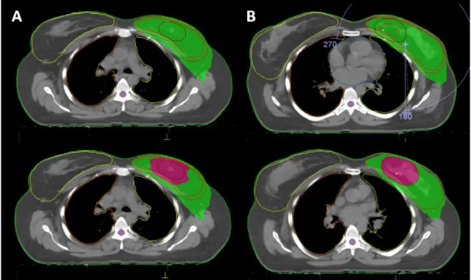
3), respectively. Figures 1 and 2 show representative axial
sections depicting the PTV
breastand PTV
breastdose
distributions for VMAT and HT approaches of SB and SIB
plans within a representative patient, respectively. The
dosimetric parameters for target-volumes are summarized in
Table I. All plans met the criteria for PTV coverage.
VMAT versus HT plans. The maximum doses (D2) for
PTV
breastand PTV
boostwere significantly higher in
VMAT-SB plan compared to HT-VMAT-SB plan. The minimum dose
PTV
breast(D98) was higher in HT-SB plan compared to
VMAT-SB plan; however, the minimum dose for tumor bed
was higher in VMAT-SB plan compared to HT-SB plan.
Although no PTV
breastand PTV
boostreceived more than
107% of the prescribed dose in the HT plan, mean V107 for
PTV
breastand PTV
boostwere 2.31% and 1.34%, respectively,
with the VMAT-SB plan. The CIs for PTV
breastand PTV
boostwere significantly higher, and HIs were significantly lower
in HT-SB compared to VMAT-SB plans.
The D2 values for PTV
breastand PTV
boostwere significantly
higher in VMAT-SIB compared to HT-SIB. Although the V95
was higher in HT-SIB plans compared to VMAT-SIB plans, it
was still within planned limits. No PTV
breastand PTV
boostreceived more than 107% of the prescribed dose in both plans.
The dose homogeneity and conformity of whole breast and
tumor bed were significantly better with the HT-SIB approach
than with the VMAT-SIB approach.
SIB versus SB technique. With the HT-SIB technology, the
D2, D98, and V95 values for whole breast were significantly
lower compared to the HT-SB technique. The dose
homogeneity and conformity for tumor volumes were similar
for SB/SIB plans except for the HI of PTV
breast, which was
higher with SB technique compared to the SIB technique
(0.36 vs. 0.32; p<0.001).
The D2, D98, and V107 values for PTV
breastand PTV
boostwith VMAT technology were significantly higher with the
SB technique compared to the SIB technique. Although the
PTV
breastV95 was higher in SB technique, the V95 of
PTV
boostwas found to be higher in SIB technique with
VMAT plan. For whole breast, conformity was better with
the SIB technique whereas homogeneity was better with the
SB technique. There were no significant differences in CI
and HI for tumor bed between SIB and SB techniques in
VMAT plans.
OAR doses. The average dosimetric data of OARs for SB
and SIB techniques using VMAT and HT plans are presented
in Table II. There were no significant differences in
ipsilateral lung doses between VMAT and HT approaches
with SB and SIB techniques, except for significantly higher
(a) lung V5 value in VMAT-SB, and (b) lung V13 value in
HT-SIB. Also, there were no significant differences in
ipsilateral lung and heart doses between SB and SIB
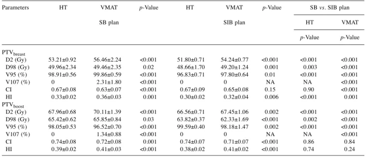
techniques for both VMAT and HT technologies. The mean
heart doses for all patients with right-sided tumors were
significantly lower compared to left-sided tumors both in
VMAT (2.69±0.94 Gy vs. 3.94±1.44 Gy; p<0.001) and HT
plan (2.25±0.87 Gy vs. 4.28±1.13 Gy; p<0.001) (Figure 3).
Similarly, mean heart doses were significantly lower in
right-sided breast and left-right-sided breast in SB and SIB techniques
with VMAT and HT plans (Table III). For right-sided tumor,
the heart doses were significantly higher in SIB technique
compared to SB technique with VMAT plan (2.80±0.88 Gy
vs. 2.57±1.01 Gy; p=0.01). However, with HT plan, SIB
plan was more advantageous in heart doses compared to SB
plan (2.13±0.69 Gy vs. 2.37±1.03 Gy; p=0.002). For
left-sided tumor, although no significant difference in heart doses
was found between SB and SIB plans with VMAT, the mean
heart doses were significantly higher in SB plan compared
to those in SIB plan (4.79±0.73 Gy vs. 3.78±1.24; p<0.001)
with HT technique.
HT provided statistically significant decreases in
contralateral lung mean V5 (44% relative decrease with SB
technique and 60% relative decrease with SIB technique).
Additionally, a 68% relative decrease in contralateral breast
mean V5 was observed with the SB technique. A 42%
relative decrease was observed with the HT-SIB technique
in mean contralateral breast doses compared to the
HT-VMAT plan. Contralateral lung V5 was significantly lower
with HT-SIB compared to the VMAT-SIB plan, and
contralateral mean lung dose was lower with HT-SIB
compared to VMAT-SB technique. Contralateral mean breast
dose and V5 were significantly lower with the VMAT-SIB
technique compared to the VMAT-SB technique.
Discussion
In the present study, we compared the target-volume
coverage and dose to OARs when HT and VMAT
technologies were employed in conjunction with SB and SIB
techniques after BCS in early-stage BC patients. Our results
showed that HT had better target-volume coverage compared
to VMAT. Although there were no significant differences in
dose ipsilateral lung between the two technologies, HT
provided lower V5 values to contralateral breast and lung.
Furthermore, SIB technique offered better target-volume
dose distribution with both HT and VMAT approaches,
compared to the SB technique. The mean heart doses were
Figure 1. Dose distribution demonstrating 95% prescribed dose of whole-breast irradiation (green area) and tumor-bed boost (magenta area) of sequential boost (A) and simultaneous-integrated boost techniques generated with VMAT plan.
Figure 2. Dose distribution demonstrating 95% prescribed dose of whole-breast irradiation (green area) and tumor-bed boost (magenta area) of sequential boost (A) and simultaneous-integrated boost techniques generated with HT plan.
significantly lower for both left- and right-sided tumors using
SIB technique compared to SB technique with HT plans.
Additionally, the VMAT-SIB technique was advantageous in
contralateral breast and lung doses over VMAT-SB
technique.
With image-based RT, the PTV
boostcan be delineated on
planning CT, and the dosimetric analysis of any tumor-bed
boost plan can be easily performed. The tumor-bed boost can
be delivered either sequentially after WBI or with SIB
technique. Previous dosimetric studies demonstrated
feasibility of SIB with 3D-CRT, IMRT, and VMAT (7, 11,
12, 15-17). Furthermore, the dosimetric advantages of SIB
over SB have been previously demonstrated (14, 15, 22).
Compared to the most widely used sequential electron boost,
Table I. Target-volume doses according to treatment plans and irradiation techniques.Parameters HT VMAT p-Value HT VMAT p-Value SB vs. SIB plan SB plan SIB plan HT VMAT
p-Value p-Value PTVbreast D2 (Gy) 53.21±0.92 56.46±2.24 <0.001 51.80±0.71 54.24±0.77 <0.001 <0.001 <0.001 D98 (Gy) 49.96±2.34 49.46±2.35 0.02 48.66±1.70 49.20±1.24 0.001 0.003 <0.001 V95 (%) 98.91±0.56 99.86±0.59 <0.001 96.83±0.71 97.80±0.64 0.01 <0.001 <0.001 V107 (%) 0 2.31±1.80 <0.001 0 0 NA NA <0.001 CI 0.67±0.08 0.63±0.07 <0.001 0.67±0.09 0.65±0.08 0.15 0.90 <0.001 HI 0.33±0.02 0.36±0.03 0.001 0.30±0.02 0.32±0.04 0.006 <0.001 0.001 PTVboost D2 (Gy) 67.96±0.68 70.11±1.39 <0.001 66.56±0.71 67.45±1.06 0.002 <0.001 <0.001 D98 (Gy) 65.42±0.62 65.85±0.84 0.03 63.82±0.37 62.33±1.69 <0.001 0.002 <0.001 V95 (%) 98.05±0.53 96.52±0.70 <0.001 99.59±0.40 98.18±1.47 0.002 <0.001 <0.001 V107 (%) 0 1.34±0.88 <0.001 0 0 NA NA <0.001 CI 0.74±0.08 0.72±0.08 0.001 0.74±0.07 0.71±0.07 <0.001 0.86 0.84 HI 0.39±0.02 0.41±0.03 <0.001 0.38±0.02 0.41±0.02 <0.001 0.74 0.24
Table II. Organs at risk doses according to treatment plans and irradiation technique.
Parameters HT VMAT p-Value HT VMAT p-Value SB vs. SIB plan SB plan SIB plan HT VMAT p-Value p-Value Ipsilateral lung V5 43.73±7.25 53.67±7.93 0.02 41.23±5.76 43.29±3.96 0.28 0.09 0.13 V13 17.39±3.68 18.36±3.23 0.42 16.57±2.28 14.81±3.08 0.03 0.32 0.84 V20 10.99±2.73 11.42±2.53 0.53 10.66±1.98 9.42±2.57 0.08 0.14 0.15 V30 6.45±2.19 6.56±2.21 0.83 5.82±1.72 5.21±2.10 0.26 0.19 0.07 Dmean 8.66±1.43 9.16±1.08 0.27 8.11±0.73 7.83±0.92 0.23 0.05 0.44 Heart V10 14.56±7.27 17.22±8.46 0.42 14.26±8.59 13.63±10.23 0.79 0.95 0.50 V20 2.30±0.42 2.89±1.79 0.39 2.47±1.44 2.06±1.91 0.50 0.09 0.71 V30 1.35±0.15 1.56±0.71 0.08 1.14±0.55 1.11±1.06 0.94 0.22 0.50 Mean 3.58±1.51 3.24±1.39 0.21 2.96±1.29 3.38±1.35 0.02 0.002 0.64 Contralateral lung V5 1.98±0.73 3.52±1.40 0.03 1.61±1.06 4.04±3.22 0.009 0.003 0.04 Dmean 2.47±0.58 2.29±0.46 0.29 2.40±0.79 2.10±0.70 0.11 0.14 0.03 Contralateral breast V5 1.48±1.19 4.61±3.55 0.03 1.33±0.46 1.82±0.77 0.64 0.18 0.02 Dmean 2.35±1.15 2.75±1.40 0.31 1.83±1.57 3.14±1.44 0.02 0.88 0.01
SIB technique with IMRT seems to have the advantages of
better skin sparing and boost volume conformity especially
for deeper boost volumes (23, 24). In the present study, our
dose distribution analysis showed more conformity and less
dose spilling to the ipsilateral breast tissue outside the boost
volume with the SIB technique compared to SB technique.
This can be technically explained by the boost volume being
surrounded by the PTV
breastfor the SIB technique. With the
SB technique, the surrounding breast tissue has to be spared
during boost irradiation in order to reduce cumulative breast
doses. However, with the SIB technique, whole breast and
tumor bed are simultaneously irradiated with different
prescribed doses. Additionally, for the SB technique, field
apertures must be several millimeters larger than the
PTV
boostvolume to account for the penumbra, which causes
higher doses to surrounding breast tissue. However, for the
SIB technique no extra margin around the PTV
boostis
required to obtain target coverage, so the dose conformity is
higher. In the current study, similar to previous studies, we
found that SIB technique provides better in target-volume
dose distribution compared to SB with both HT and VMAT
plans. Furthermore, HT has shown superiority with absence
of V107 values in SB plan, and significantly lower HI
compared to VMAT in SB and SIB plans, which potentially
reduces the risk of breast fibrosis.
There is no standard technique for SIB in WBI after BCS,
because of conflicting results due to different RT techniques,
treatment planning systems, and dose constraints for OARs
used in these studies. Maier et al. (25) have evaluated IMRT,
VMAT, and tangential arc VMAC using Monaco treatment
planning for right-sided BC patients using SIB technique. The
authors found that best target coverage and homogeneity was
observed with VMAT, and lowest doses to the contralateral
lung and breast were observed with tangential arc VMAT. Hijal
et al. (11) have analyzed the dosimetric data of HT and
3D-CRT with SIB technique in early-stage BC patients. The
authors found that both HT and 3D-CRT provided adequate
dose coverage and low heart doses; however, HT reduced
higher doses within breast and decreased ipsilateral lung doses.
Figure 3. Box-plot graphics demonstrating the heart doses using sequential boost and simultaneous-integrated boost techniques with (A) VMAT plan and (B) HT plan for right- and left-sided breast irradiation.Table III. The heart doses according to left- and right-sided breast irradiation techniques.
Heart dose (mean±SD) (Gy) Technique Right breast Left breast p-Value SB technique VMAT 2.57±1.01 3.91±1.43 0.006 HT 2.37±1.03 4.79±0.73 <0.001 SIB technique VMAT 2.80±0.88 3.96±1.51 0.02 HT 2.13±0.69 3.78±1.24 <0.001
Michalski et al. (12) have compared HT, IMRT, and 3D-CRT
for left-sided breast tumors with SIB technique, and found that
HT offered superior target-volume doses and lower doses to
ipsilateral lung and heart, compared to 3D-CRT and IMRT. In
dosimetric studies with more conformal techniques, VMAT
(16, 25) and HT (11, 12) provide better target-volume coverage
and lower lung and heart doses. In the current study comparing
VMAT and HT technologies with the SIB technique, we found
that HT had better target-volume coverage and provided lower
doses to contralateral breast and lung compared to VMAT.
With the improved efficacy of BC therapies, the likelihood
of long-term survival of patients has increased substantially.
Due to the technical characteristics of IMRT, VMAT, and HT
plans, radiation scattering to the surrounding normal organs
is slightly higher than with conventional techniques (26).
Therefore, radiation toxicities during long-term follow-up are
the subject of increasing attention. Henson et al. (6) have
found that the BC patients experienced 1.2 times the number
of cardiac deaths compared to the general population.
Although one study that conducted long-term follow-up in
35,000 patients undergoing RT found that the incidence of
cardiovascular disease increased remarkably after RT (18),
there were no increase in cardiovascular mortality in patients
who underwent RT after 1980, which may be due to
development/utilization of more conformal RT techniques
(27). VMAT may lead to more radiation being scattered to
organs due to its technical characteristics, and dosimetric
studies have demonstrated that tangential field IMRT
decreases ipsilateral V5 and V10 compared to multi-field
IMRT and VMAT (26, 28). Although ipsilateral lung volumes
receiving low-dose radiation were higher with the VMAT
technology (suggesting that VMAT may be associated with a
higher possibility of radiation-induced lung cancer), the
results of long-term follow-up are still lacking. Caudrelier et
al. (29) have evaluated 30 patients for long-term cardiac and
pulmonary effects from mammaria interna radiotherapy using
the IMRT technique. The authors concluded that V25Gy
should be included in the evaluation of dose distributions
besides mean heart dose with regard to long-term cardiac
effects with IMRT. In the presented study, the V30Gy value
was below the recommended limit of 5%. Furthermore, we
found that SIB technique with HT is advantageous in heart
doses for both left- and right-sided tumors. Additionally, the
dose levels of lung and heart in these dosimetric studies were
still within limits of the ‘Quantitative Analysis of Normal
Tissue Effects in Clinic’ (QUANTEC) guidelines (30, 31).
According to QUANTEC guidelines, heart V25 <10% was
associated with less than 1% probability of cardiac mortality
15 years after RT (30), and the limits for lung V20 and mean
lung dose were set as <30–35% and <20–23 Gy, respectively
(31). However further studies evaluating the long-term
cardiac and pulmonary effects of VMAT and HT technologies
deployed with the SIB technique are warranted.
Another important feature of VMAT and HT is the
substantial contralateral breast and lung doses that raises
concerns about secondary malignancies especially for
early-stage BC patients. Although the V5 values of breast and lung
were very low with both techniques, HT is advantageous over
VMAT in sparing contralateral lung and breast, and VMAT is
unsafe in terms of secondary BC, especially in young women.
This study possesses some limitations. First, our study
demonstrates the dosimetric advantages of SIB technique
with HT plan, however, we did not analyze
radiation-associated late toxicities and long-term cosmetic outcomes
of this technique. Second, we could not compare the
treatment time and monitor units between VMAT and HT,
since both systems are technically different. Lastly, we did
not assess the inaccuracies in the setup for these complicated
technologies. However, in order to improve accuracy in these
complex techniques, a strike immobilization and image
guidance with daily cone beam CT or breath-hold techniques
are required. Notwithstanding these limitations, our
dosimetric study demonstrated the applicability of SIB
technique with HT plan.
Conclusion
In conclusion, our study suggests that HT may be more
suitable for the SIB technique for WBI after BCS than
VMAT as it offers appropriate target coverage and acceptable
doses to OARs, especially the contralateral lung and breast.
Furthermore, the SIB technique provided a better
target-volume dose distribution in both HT and VMAT plans,
compared to the SB technique. The most important finding
is that SIB technique with HT plan is better in sparing heart
for both right- and left-sided tumors. Additional clinical
studies with larger sample sizes are needed to assess the
long-term feasibility and efficacy of SIB using HT.
Conflicts of Interest
The Authors declare no conflicts of interest regarding this study.
Authors’ Contributions
Study conception and design: CO, OCG. Provision of study materials or patients: OCG, EE, BAY. Collection and assembly of data: EE, BAY. Data analysis and interpretation: CO, EE. Manuscript writing: CO, OCG. Final approval of manuscript: CO, EE, OCG, BAY. All authors read and approved the final manuscript.
References
1 Ghoncheh M, Pournamdar Z and Salehiniya H: Incidence and mortality and epidemiology of breast cancer in the world. Asian Pac J Cancer Prev 17(S3): 43-46, 2016. PMID: 27165206. DOI: 10.7314/apjcp.2016.17.s3.43
2 Clarke M, Collins R, Darby S, Davies C, Elphinstone P, Evans V, Godwin J, Gray R, Hicks C, James S, MacKinnon E, McGale P, McHugh T, Peto R, Taylor C, Wang Y and Early Breast Cancer Trialists’ Collaborative G: Effects of radiotherapy and of differences in the extent of surgery for early breast cancer on local recurrence and 15-year survival: An overview of the randomised trials. Lancet 366(9503): 2087-2106, 2005. PMID: 16360786. DOI: 10.1016/S0140-6736(05)67887-7
3 Fisher B, Anderson S, Bryant J, Margolese RG, Deutsch M, Fisher ER, Jeong JH and Wolmark N: Twenty-year follow-up of a randomized trial comparing total mastectomy, lumpectomy, and lumpectomy plus irradiation for the treatment of invasive breast cancer. N Engl J Med 347(16): 1233-1241, 2002. PMID: 12393820. DOI: 10.1056/NEJMoa022152.
4 Bartelink H, Horiot JC, Poortmans PM, Struikmans H, Van den Bogaert W, Fourquet A, Jager JJ, Hoogenraad WJ, Oei SB, Warlam-Rodenhuis CC, Pierart M and Collette L: Impact of a higher radiation dose on local control and survival in breast-conserving therapy of early breast cancer: 10-year results of the randomized boost versus no boost EORTC 22881-10882 trial. J Clin Oncol 25(22): 3259-3265, 2007. PMID: 17577015. DOI: 10.1200/JCO.2007.11.4991.
5 Immink JM, Putter H, Bartelink H, Cardoso JS, Cardoso MJ, van der Hulst-Vijgen MH, Noordijk EM, Poortmans PM, Rodenhuis CC and Struikmans H: Long-term cosmetic changes after breast-conserving treatment of patients with stage I-II breast cancer and included in the EORTC ‘boost versus no boost’ trial. Ann Oncol 23(10): 2591-2598, 2012. PMID: 22499858. DOI: 10.1093/annonc/mds066.
6 Henson KE, Reulen RC, Winter DL, Bright CJ, Fidler MM, Frobisher C, Guha J, Wong KF, Kelly J, Edgar AB, McCabe MG, Whelan J, Cutter DJ, Darby SC and Hawkins MM: Cardiac mortality among 200 000 five-year survivors of cancer diagnosed at 15 to 39 years of age: The teenage and young adult cancer survivor study. Circulation 134(20): 1519-1531, 2016. PMID: 27821538. DOI: 10.1161/circulationaha.116.022514 7 Zhao H, He M, Cheng G, Han D, Wu N, Shi D, Zhao Z and Jin
J: A comparative dosimetric study of left sided breast cancer after breast-conserving surgery treated with VMAT and IMRT. Radiat Oncol 10(231), 2015. PMID: 26577189. DOI: 10.1186/ s13014-015-0531-4.
8 Koivumaki T, Heikkila J, Vaananen A, Koskela K, Sillanmaki S and Seppala J: Flattening filter free technique in breath-hold treatments of left-sided breast cancer: The effect on beam-on time and dose distributions. Radiother Oncol 118(1): 194-198, 2016. PMID: 26709069. DOI: 10.1016/j.radonc.2015.11.032. 9 Onal C, Oymak E, Kotek A, Efe E and Arslan G: Correlation of
conventional and conformal plan parameters for predicting radiation pneumonitis in patients treated with breast cancer. J Breast Cancer 15(3): 320-328, 2012. PMID: 26709069. DOI: 10.1016/j.radonc.2015.11.032.
10 Onal C, Sonmez A, Arslan G, Oymak E, Kotek A, Efe E, Sonmez S and Dolek Y: Dosimetric comparison of the field-in-field technique and tangential wedged beams for breast irradiation. Jpn J Radiol 30(3): 218-226, 2012. PMID: 221838 29. DOI: 10.1007/s11604-011-0034-7.
11 Hijal T, Fournier-Bidoz N, Castro-Pena P, Kirova YM, Zefkili S, Bollet MA, Dendale R, Campana F and Fourquet A: Simultaneous integrated boost in breast conserving treatment of breast cancer: A dosimetric comparison of helical tomotherapy
and three-dimensional conformal radiotherapy. Radiother Oncol 94(3): 300-306, 2010. PMID: 20171752. DOI: 10.1016/j.radonc. 2009.12.043.
12 Michalski A, Atyeo J, Cox J, Rinks M, Morgia M and Lamoury G: A dosimetric comparison of 3D-CRT, IMRT, and static tomotherapy with an SIB for large and small breast volumes. Med Dosim 39(2): 163-168, 2014. PMID: 24393498. DOI: 10.1016/j.meddos.2013.12.003.
13 Son SH, Choi KH and Kim SW: Dosimetric comparison of simultaneous integrated boost with whole-breast irradiation for early breast cancer. PLoS One 12(3): e0173552, 2017. PMID: 28273157. DOI: 10.1371/journal.pone.0173552.
14 van der Laan HP, Dolsma WV, Maduro JH, Korevaar EW, Hollander M and Langendijk JA: Three-dimensional conformal simultaneously integrated boost technique for breast-conserving radiotherapy. Int J Radiat Oncol Biol Phys 68(4): 1018-1023, 2007. PMID: 17379440. DOI: 10.1016/j.ijrobp.2007.01.037. 15 Van Parijs H, Reynders T, Heuninckx K, Verellen D, Storme G
and De Ridder M: Breast conserving treatment for breast cancer: Dosimetric comparison of sequential versus simultaneous integrated photon boost. Biomed Res Int 2014: 827475, 2014. PMID: 25162031. DOI: 10.1155/2014/827475.
16 Aly MM, Glatting G, Jahnke L, Wenz F and Abo-Madyan Y: Comparison of breast simultaneous integrated boost (SIB) radiotherapy techniques. Radiat Oncol 10: 139, 2015. PMID: 26156086. DOI: 10.1186/s13014-015-0452-2
17 Wu S, Lai Y, He Z, Zhou Y, Chen S, Dai M, Zhou J, Lin Q and Chi F: Dosimetric comparison of the simultaneous integrated boost in whole-breast irradiation after breast-conserving surgery: IMRT, IMRT plus an electron boost and VMAT. PLoS One 10(3): e0120811, 2015. PMID: 25781183. DOI: 10.1371/journal. pone.0120811.
18 Taylor CW, Bronnum D, Darby SC, Gagliardi G, Hall P, Jensen MB, McGale P, Nisbet A and Ewertz M: Cardiac dose estimates from Danish and Swedish breast cancer radiotherapy during 1977-2001. Radiother Oncol 100(2): 176-183, 2011. PMID: 25781183. DOI: 10.1371/journal.pone.0120811.
19 Hurkmans CW, Meijer GJ, van Vliet-Vroegindeweij C, van der Sangen MJ and Cassee J: High-dose simultaneously integrated breast boost using intensity-modulated radiotherapy and inverse optimization. Int J Radiat Oncol Biol Phys 66(3): 923-930, 2006. PMID: 16904837. DOI: 10.1016/j.ijrobp.2006.05.041.
20 Singla R, King S, Albuquerque K, Creech S and Dogan N: Simultaneous-integrated boost intensity-modulated radiation therapy (SIB-IMRT) in the treatment of early-stage left-sided breast carcinoma. Med Dosim 31(3): 190-196, 2006. PMID: 16905449. DOI: 10.1016/j.meddos.2005.11.001.
21 Offersen BV, Boersma LJ, Kirkove C, Hol S, Aznar MC, Sola AB, Kirova YM, Pignol JP, Remouchamps V, Verhoeven K, Weltens C, Arenas M, Gabrys D, Kopek N, Krause M, Lundstedt D, Marinko T, Montero A, Yarnold J and Poortmans P: ESTRO consensus guideline on target volume delineation for elective radiation therapy of early stage breast cancer, version 1.1. Radiother Oncol 118(1): 205-208, 2016. PMID: 26791404. DOI: 10.1016/j.radonc.2015.12.027.
22 Van Parijs H, Reynders T, Heuninckx K, Verellen D, Storme G and De Ridder M: Breast conserving treatment for breast cancer: Dosimetric comparison of different non-invasive techniques for additional boost delivery. Radiat Oncol 9: 36, 2014. PMID: 24467916. DOI: 10.1186/1748-717X-9-36.
23 Alford SL, Prassas GN, Vogelesang CR, Leggett HJ and Hamilton CS: Adjuvant breast radiotherapy using a simultaneous integrated boost: Clinical and dosimetric perspectives. J Med Imaging Radiat Oncol 57(2): 222-229, 2013. PMID: 23551785. DOI: 10.1111/j.1754-9485.2012.02473.x.
24 Peters S, Schiefer H and Plasswilm L: A treatment planning study comparing Elekta VMAT and fixed field IMRT using the varian treatment planning system Eclipse. Radiat Oncol 9: 153, 2014. PMID: 25011529. DOI: 10.1186/1748-717X-9-153. 25 Maier J, Knott B, Maerz M, Loeschel R, Koelbl O and Dobler
B: Simultaneous integrated boost (SIB) radiation therapy of right sided breast cancer with and without flattening filter – a treatment planning study. Radiat Oncol 11(1): 111, 2016. PMID: 27577561. DOI: 10.1186/s13014-016-0687-6
26 Otto K: Volumetric modulated arc therapy: IMRT in a single gantry arc. Med Phys 35(1): 310-317, 2008. PMID: 18293586. DOI: 10.1118/1.2818738.
27 Patt DA, Goodwin JS, Kuo YF, Freeman JL, Zhang DD, Buchholz TA, Hortobagyi GN and Giordano SH: Cardiac morbidity of adjuvant radiotherapy for breast cancer. J Clin Oncol 23(30): 7475-7482, 2005. PMID: 16157933. DOI: 10.1200/JCO.2005.13.755.
28 Abo-Madyan Y, Aziz MH, Aly MM, Schneider F, Sperk E, Clausen S, Giordano FA, Herskind C, Steil V, Wenz F and Glatting G: Second cancer risk after 3D-CRT, IMRT and VMAT for breast cancer. Radiother Oncol 110(3): 471-476, 2014. PMID: 24444525. DOI: 10.1016/j.radonc.2013.12.002.
29 Caudrelier JM, Meng J, Esche B, Grimard L, Ruddy T and Amjadi K: IMRT sparing of normal tissues in locoregional treatment of breast cancer. Radiat Oncol 9: 161, 2014. PMID: 25052720. DOI: 10.1186/1748-717X-9-161.
30 Gagliardi G, Constine LS, Moiseenko V, Correa C, Pierce LJ, Allen AM and Marks LB: Radiation dose-volume effects in the heart. Int J Radiat Oncol Biol Phys 76(3 Suppl): S77-85, 2010. PMID: 20171522. DOI: 10.1016/j.ijrobp.2009.04.093.
31 Marks LB, Bentzen SM, Deasy JO, Kong FM, Bradley JD, Vogelius IS, El Naqa I, Hubbs JL, Lebesque JV, Timmerman RD, Martel MK and Jackson A: Radiation dose-volume effects in the lung. Int J Radiat Oncol Biol Phys 76(3 Suppl): S70-76, 2010. PMID: 20171521. DOI: 10.1016/j.ijrobp.2009.06.091.