Orak Hücreli Anemi Tespiti
Sickle Cell Anemia Detection
Batuhan ALBAYRAK, Muazzez Buket DARICI,
Furkan KİRACI, Arif Selçuk ÖĞRENCİ, Atilla
ÖZMEN,
Electrical and Electronics Engineering Kadir Has University
Istanbul, Turkey [email protected], [email protected], [email protected], [email protected], [email protected] Kerem ERTEZ School of Medicine Medipol University Istanbul, Turkey [email protected]
Özetçe— Anemi, kırmızı kan hücrelerinin bazı fonksiyonel dezavantajları nedeniyle oksijen taşıma kapasitesindeki düşüşlere verilen yaygın bir isimdir. Patoloji Laborantları mikroskop camı üzerine dokuyu yerleştirir ve Anemi hastalığını teşhis etmeye çalışırlar.Bu süreçler uzun sürmektedir ve dikkat dağılmasına neden olmaktadır. Bu nedenle, Laborantların yanlış teşhis etmesine sebep olmaktadır. Bu çalışma %91.11 kesinlik, %92.9 hassasiyet ve %79.05 hatırlanma oranıyla görüntü işleme algortmaları kullanarak kan dokusunda sağlıklı hücreli çıkararak ve sadece orak hücrelere sahip olarak teşhis süresini kısaltır ve teşhisin hata oranını en aza indirir.
Anahtar Kelimeler __ Görüntü İşleme, Dairesel Hough
Dönüşümü(DHD), Orak Hücreli Anemi.
Abstract— Anemia is a common name given to falls in oxygen transport capacity due to some of the functional disadvantages of red blood cells. Pathology Laboratorians put the tissue on the microscope glass and try to diagnose Anemia disease. Processes have been taken for a long time and it has been caused to distract. Therefore it has been caused to misdiagnose the Laboratorian. This work shortens the diagnostic period of the disease and to minimizes error probability of this diagnosis by extracting healthy cells and just having sickle cells on the blood tissue using Image Processing Algorithms with an accuracy of 91.11 %, precision of 92.9 %, recall of 79.05 % for Sickle Cell Anemia.
Keywords — Image Processing, Circular Hough Transform(CHT), Sickle Cell Anemia.
978-1-5386-6852-8/18/$31.00 ©2018 IEEE”
I. INTRODUCTION
The technologies used in medicine are constantly envolvingin order to make the work force most efficient in daily life, time gains and obtaining the most accurate results easily. In this case, biomedical applications are one of the most used fields of image processing techniques in medicine. For example, determination of chest cavity by segmentation on echocardiographic images [13],enhancement of vessel images [12], examination of contraction mechanisms of heart muscles [10,11], Obtaining two-dimensional image in Computed Tomography image processing and applying image enhancement techniques on the resulting image could be counted as examples of image processing usage in medicine.
Almost everyone knows that there has been some sort of suspected disease detection through blood counts. These diseases include types of anemia like sickle cell anemia, thalassemia, sideroblastic anemia, etc. In the tradition of this process, the person who counts blood cells in a specific volume by looking at the blood sample under the microscope and calculates the amount that can be present in a larger volume. There are many different methods being used in tissue examination or blood sample examination however this traditional method is the most commonly used method in this area. The uniformity of this process and the need to constantly working on the microscope increases unwanted errors. Proposed algorithm is in the form of an integration of medicine and engineering that has emerged with the aim of exhibiting an innovative approach both in engineering and medicine. The proposed algorithm aims to improve the diseased cell counting technique under the analogous microscopic observation and to automate this technique by using engineering methods to detect diseases in tissues and / or blood cells in pathology and / or hematology in the field of medicine. In this regard, a method has been proposed that will help employees in the health sector to reduce both time saving and human error in detecting diseases. The blood cells to be counted in the images are separated from
the background image by various image processing and shape recognition methods and then separated cell images are counted. In the second part of the work, the literature survey is mentioned, in the third part methodology is explained, in the fourth part the results are shown and in the last part the conclusion are mentioned.
II. LITERATURESURVEY
In [2] authors have used median filter, feature extraction, K-means segmentation, edge detection to count, to classify and recognize the cells which are blood cells, white blood cells and platelets. They have used fractal dimensions method to detect sickle cell. In [3] red and white blood cell segmentation is observed based on L*a*b* color space, image filtering, color segmentation in L*a*b*, Hough Transform, ROI (Region of Interest), Canny Edge Detection to guarantee it. In [3] the accuracy is 64% for white blood cells and 81% for red blood cells. In [4] authors have focused on RBC cluster separation for use in sickle cell disease with efficiency 99% for artificial images, 98% for real images and 99.35% for synthetic images. In article [4] the authors have used concave point detection method with 87% efficiency, K-curvature, K-slope, circumference adjustment method and parameters such as elliptical coefficient etc. In [5] authors have been focused on comparison of two methods in sickle cell detection. It proposes that Hough Transform (HT) is more efficient than Watershed segmentation (WS). It [5] compares two methods with true detection numbers and elapsed time in seconds. In [1] authors have studied classification of abnormal cells in Sickle Cell Anemia and Thalassemia diseases by using metric values, feature extraction, morphological operation, KNN classifier with 80.6% accuracy and 87.6% recall percentages..
Based on these studies, this paper presents an algorithm that can successfully detect sickle cell shape for Sickle Cell Anemia by cooperation of Otsu’s Method, Hough Transform, Noise Reduction, Thresholding Method, Morphological Filters for Sickle Cell Anemia This algorithm resulted with 91.40% accuracy for Sickle Cell Anemia detection.
III. METHODOLOGY
A. Sickle Cell Anemia Detection
In sickle cell anemia, red blood cells are shaped like sickles or crescent [9]. For this reason, in this work, sickle cells shaped like sickles or crescent have been detected for diagnosing the disease by using Image Processing Algorithms.
Figure 1. Examples of sickle cell shapes seen in blood tissue
1) Methodology of Sickle Cell Anemia Detection a) Thresholding Method
The main aim of using Thresholding is to have grayscale image from RGB and binary image from grayscale image. The used method that converts grayscale image to binary is Otsu’s Method.
Gray-thresh calculates the image threshold using the Otsu Method. The main purpose of the Otsu Threshold Method is to iterate over all possible threshold values and calculate the spreading of the pixels falling in the foreground or background. The aim is to find the threshold value where the sum of foreground and background spreads is at its minimum [6].
Figure 2. A 6-level greyscale image and its histogram [7]. Based on threshold level which is found by Gray-thresh Method, gray image was converted to binary image. Then some filters were applied to reduce noise in binary image.
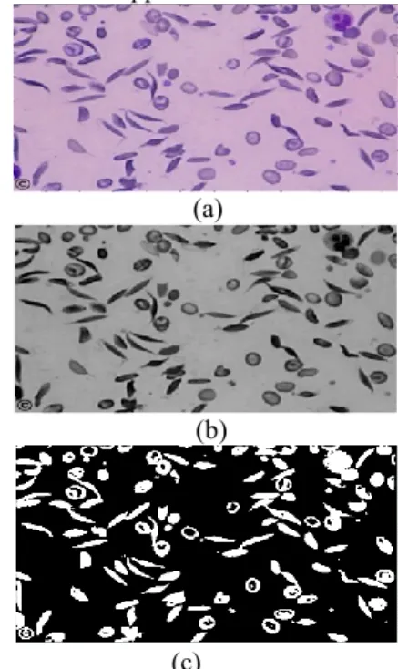
(a)
(b)
(c)
Figure 3. Result of Thresholding Method. (a) RGB image (b)Grayscale image(c) Binary image
b) Circular Hough Transform Method
Circular Hough Transformation (CHT) converts a difficult global detection problem in image space into a more easily solved local peak detection problem in a parameter space [8].
The algorithm for CHT can be summarized to [14]: Find edges
For each edge point draw a circle with center in the edge point with radius r and increment all coordinates that the perimeter of the circle passes through in the accumulator.
Figure 4. A CHT from the x,y-space (left) to the parameter space (right), this example is for a constant radius [15].
Find one or several maximum numbers in the accumulator
Map the found parameters (r, a, b) corresponding to the maximum numbers.
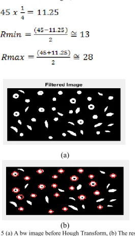
According to zoom values of blood tissue images, an interval for radius of healthy cells has been set for each images have different zoom values to determine circular cells. For example, in image which has blood cells which have 45x45 pixel value, radius values for HT were calculated
as shown
below:
For 100x zoom images;
(1) (2) (3)
(a) (b)
Figure 5 (a) A bw image before Hough Transform, (b) The result of Hough Transform
c) Removing of Objects Detected from Hough Transform
After succeeded taking the blood cells outside of the sickle into the circle by HT, it was aimed to eliminate the
whiteness in this circles and have only the sickle cells in the image. For this reason, ‘for loop’ has been created. In for loop these steps have been used:
Create a matrix which indicates centers and radius of circles
Create b and a values for all circle values which show coordinates of x axis and y axis of circles respectively. (For this, found radius values after CHT for each circular cells were used.)
Create a radius equation which is known circle’s center and two points.
Eliminate circles that are less than or equal to the circle radius of the radius that are found by the radius equation.
Figure 6. Image after removing circles
d) Connected Component Analysis and Removing Remaining Objects
After HT and deletion process, some objects like sickle cells remained in the picture and this caused the objects to be identified as a sickle cell. To solve this problem, Connected Component Analysis Algorithm (CCAA) was used to find connected components and remove them in binary image. For this, areas of all sickle cells in each image which have different zoom values were identified. So, the aim is to keep sickle cells and eliminate all other objects after CCAM. For 100x zoom images, 450 value is used based on sizes of cells in this zoom value image. The purpose of using this number is to keep objects which their areas are between 450 and maximum area value of cells, eliminate all except these objects. After that, only sickle cells stayed in binary image. This method has been used for all other images which have different zoom values.
Figure 7. Image after removing remained objects
e) Morphological Operations
Morphological operations are used in image processing to enhance the image to increase the performance of the algorithm. There are some different purposes for using morphological filters such as reduction of noises, combination of seperated objects and seperation of connected components. These filters have been used according to need throughout the study.
IV. RESULTS
A. Results of Diagnosis of Sickle Cell Anemia
It could not been obtained a lot of Sickle Cell Anemia data because of some ethical rules applied in hospitals or universities. Therefore, we had to use 8 data which are found from Hematology Atlas which is available for everyone.Here every number represents blood cell on Table 1. For example , 185 is the number of sickle cells which are detected by human eyes and software system for the 8 processed data. According to these 8 data,our results are shown below;
Precision = 185 185+14 = 92.90% (4) Recall = 185 185+49 = 79.05% (5) Accuracy = 185+14+49+467185+467 = 91.11% (6)
These percentages are based on ratios of number of sickle cells and healthy cells in the processed blood tissue which are given below in Table 1. It shows that total number of sickle cells and healthy cells mentioned previously in Section 4 according to 8 data.
TABLE 1. THE NUMBER OF SICKLE CELLS AND HEALTHYCELLS BASED ON CONFUSION MATRIX
V. CONCLUSION
The main aim of this work is to be shorten the diagnosis period of Sickle Cell Anemia and to minimize error probability of this diagnosis. Thus in this work sickle cells
shaped like sickles or crescent have been detected for diagnosing the disease by using Image Processing Algorithms.
For diagnosis of Sickle Cell Anemia, red blood cells are examined and sickle cell detection is performed by using Image Processing Algorithms which are Thresholding Method to have grayscale image from RGB and binary image from grayscale image, Hough Transform Method to detect circular erythrocyte and taking the blood cells’ outside of the sickle into the circle, Noise Reduction Filters and Morphological Filters are used to enhance images and count the remain cells in the image respectively. For better results and diversity, this study would be worked by using real and more qualified images.
VI. REFERENCES
[1] V. Sharma, A. Rathore, G. Vyas, “Detection Of Sickle Cell Anaemia And Thalassaemia Causing Abnormalities İn Thin Smear Of Human Blood Sample Using İmage Processing,” Department of Electronics & Communication Engineering, Amity University , U.P, Noida-201303. [2] M. Sahu, A. K. Biswas, K. Uma, “Detection of Sickle Cell Anemia in
Red Blood Cell,” International Journal of Engineering and Applied Sciences (IJEAS) ISSN: 2394-3661, Volume-2, Issue-3, March 2015. [3] N. H. Mahmood, M.A.A. Razak, ‘‘Blood cells extraction using color
based segmentation technique,’’ ResearchGate, Article,January 2013. [4] M. G. Hidalgo, F. A. G. Pe˜na, S. H. Garc´ıa, A. J.-i-Cap´o, P. D. M.
Fern´andez, ‘‘Red Blood Cell Cluster Separation From Digital Images for Use in Sickle Cell Disease,” IEEE Journal Of Biomedıcal And Health Informatıcs, vol. 19, no. 4, July 2015.
[5] M.A.Fadhel, A.J. Humaidi, S. R.Oleiwi, ‘‘Image Processing-Based Diagnosis of Sickle cell Anemia in Erythrocytes,” Annual Conference on New Trends in Information & Communications Technology Applications-(NTICT'2017) 7 - 9 March 2017.
[6] I. Koc , O. K. Baykan, I. Babaoglu, ‘‘Gri Kurt Optimizasyon Algoritmasına Dayanan Çok Seviyeli İmge Eşik Seçimi,” Journal of Polytechnic,2017.
[7] Dr. A. Greensted, “Otsu Thresholding” 17 June 2010.[Online]. [8] J.Illıngworth, J. Kıttler, “A Survey Of The Hough Transform, Computer
Vision, Graphics, And Image Processing,” pp.87-116, 1988. [9] R. E. Miller, “ Sickle Cell Disease,” July 2018. [Online]..
[10] T.Sato, T. Vfatanabe, H. Honjo, Y. Naito, I. Rodama, J. Toyama, “Microcomputer_Based Image Processing System for Measuring Sarcomere Motion of Single Cardiac Cells,” IEEE Transactions on Biomedical Engineering, Kasım 1988 Vol. 35 No.5 pg. 397_400 [11] B.W, Steadman, K.B. Moore, K.W. Spi'.zer, J.H.B. Bridge "A Video
System for Measuring Motion in Contracting Heart Cells" IEEE Transactions on Biomedical Engineering, Kasım 1988, Vol. 35, No.4, pp. 264-272.
[12] S. Chaudhuri, S. Chatterjee, N. Katz, M. Nelson, M. Goldbaum, “Detection of Blood Vessels in Retinal Images Using Two-Dimentional Matched Filters,” IEEE Transactions on Medical Imaging, Eylül 1989, Vol.8, No.3 pg. 263-269.
[13] J. W. Klingler, C.L. Vaughan, T.D. Fraker,L.T.Andrws “Segmentation of Echocardiographic Images Using Mathematical Morphology,” IEEE Transactions on Biomedical Engineering, Kasım 1988, Vol.35, No.ll, pp. 925-934.
[14] B. S. Morse. Class Lecture 15, Topic : “Segmentation (edge based, hough transform),” Brigham Young University, February 23, 2000.
[15] S. J. K. Pedersen. Class Lecture, Topic: “Circular Hough Transform,” Aalborg University, November 2007.