©2015 by Quintessence Publishing Co Inc.
Computed Tomography and Intraoperative Findings
in Sinus Augmentation
Gokhan Gurler, DDS, PhD1/Cagri Delilbasi, DDS, PhD2
Purpose: To investigate whether the presence of bony septum, thickness of sinus membrane (schneiderian
membrane), and residual alveolar bone height affects membrane perforation and the duration of sinus augmentation. Materials and Methods: Preoperative cone beam computed tomography (CBCT) images obtained from patients undergoing lateral sinus augmentation were evaluated for the presence of bony septum, thickness of sinus membrane, and residual alveolar bone height. During the operation, membrane perforation and duration of surgery were noted. The Student t test was used to compare descriptive statistics (mean, standard error) and quantitative variables between groups. The Fisher exact χ² test was used to compare
the qualitative data, and Pearson correlation test was used to evaluate the correlation between data. P < .05 was considered significant. Results: Data from 57 patients were evaluated. Membrane perforation occurred in 14 patients included in the study and in 8 patients with sinus septum. A significant relationship was found between the presence of septum and membrane perforation during sinus augmentation (P = .014). However, the relationship among other CBCT and intraoperative findings was not significant. Conclusion: Presence of septum in the maxillary sinus increases the risk of membrane perforation, but does not extend the duration of the sinus augmentation. Int J Oral MaxIllOfac IMplants 2015;30:1244–1248. doi: 10.11607/jomi.3797
Key words: CBCT, perforation, sinus augmentation, sinus membrane
E
ndo-osseous implants are widely used in dental practice to restore tooth loss. Because of reduced bone height and low bone density, it is challenging to insert implants in the maxillary posterior region.1,2 Si-nus floor elevation is a predictable and safe procedure to overcome this obstacle.1 Sinus floor augmentation is the preferred technique to handle this problem, par-ticularly when residual alveolar bone height is less than 5 mm. However, anatomical and pathologic variations of maxillary sinus may complicate the surgical proce-dure; therefore, the maxillary sinus must be evaluated before sinus floor augmentation to decrease intraop-erative and postopintraop-erative complications.3,4Panoramic radiographs were extensively used for this purpose until the introduction of cone beam com-puted tomography (CBCT). CBCT was first described by Mozzo et al5 and has become a popular technique in dental practice. Compared with panoramic radio-graphs and conventional computed tomography (CT), CBCT images present the accurate location of anatom-ical structures without distortion and provide informa-tion about bone dimensions and morphology with the advantage of lower radiation than CT, but slightly higher dose than panoramic radiography.6–8
The assessment of anatomical characteristics of the sinus cavity before sinus augmentation is important in surgical treatment planning. CBCT is increasingly be-ing used in dental practice and has been drawbe-ing more attention when used for the assessment of incidental findings and anatomical variations.
The most common complication encountered in sinus floor augmentation is perforation of the sinus membrane. The membrane is usually perforated dur-ing opendur-ing of the bony window, but it can also occur during elevation of the membrane, especially when there are some irregularities or bony septum in the sinus cavity, thin sinus membrane, and low residual al-veolar bone height.6,9–12
The objective of this study was to investigate whether the presence of bony septum, thickness of 1Assistant Professor, Istanbul Medipol University School of
Dentistry, Department of Oral and Maxillofacial Surgery, Unkapani-Istanbul, Turkey.
2Professor, Istanbul Medipol University School of Dentistry, Department of Oral and Maxillofacial Surgery, Unkapani-Istanbul, Turkey.
Correspondence to: Dr Gokhan Gurler, DDS, PhD, Istanbul Medipol University School of Dentistry, Department of Oral and Maxillofacial Surgery, Atatürk Bulvarı No:27, 34083 Unkapani-Istanbul. Fax: +90 212 531 75 55. Email: [email protected]
The International Journal of Oral & Maxillofacial Implants 1245
sinus membrane (Schneiderian membrane), and re-sidual alveolar bone height affects membrane perfora-tion and duraperfora-tion of sinus augmentaperfora-tion.
MATERIALS AND METHODS
This study evaluated maxillary sinus CBCT scans of patients who were undergoing sinus floor augmenta-tion at Istanbul Medipol University School of Dentistry, Department of Oral and Maxillofacial Surgery (Unka-pani-Istanbul, Turkey). The patients who were in good general health status and had no symptoms of max-illary sinus diseases were included in the study. How-ever, patients having any sinus pathology and who smoked more than 10 cigarettes per day were treated, but excluded from the study. Only one side was ran-domly included (by flipping a coin) in the study even if the patient needed bilateral surgery. Two surgeons with similar experience in sinus augmentation per-formed the procedures. The flap design for sinus floor augmentation in our clinic includes horizontal incision on the alveolar crest and a mesial vertical releasing incision. The scans were taken using i-CAT Next Gen-eration cone beam computed tomography (Imaging Sciences International). Tomography settings were as follows: 120 kV, 5 mA, and a field of view of 16 × 8 cm. Images were analyzed using i-CAT software in axial, coronal, and sagittal planes with 2-mm slices.
CBCT images from 57 patients ( 27 men and 30 women; age range, 22–78 years; mean age ± standard deviation [SD], 49.6 ± 1.14 years) were evaluated. The images were assessed by one of the authors (G.G.). For calibration and evaluation of intraobserver reliability, 10 randomly selected images were measured twice on two different days, resulting in a mean difference of 0.21 mm per image (range, 0.1–0.35 mm). For further study, each measurement was repeated and the mean value was calculated. When the difference between two values was 0.2 mm or more, a third measurement was taken.1 This study was approved by the local ethi-cal committee of Istanbul Medipol University, and in-formed consent was obtained from all patients. The following parameters were assessed and measured.
Thickness of Sinus Membrane
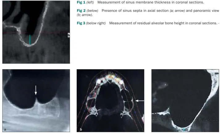
Thickness of the sinus membrane was measured on coronal images. The deepest point of the sinus inferior wall was used as a reference point. At the reference point, membrane lining and residual alveolar bone height were measured. Mucosal thickness was mea-sured with the digital ruler of i-CAT software in milli-meters (Fig 1). Sinus membrane thickness of more than 3 mm was classified as pathologic according to the cri-teria of White and Pharoah.13
Membrane Perforation
Opening of lateral bony window and elevation of the maxillary sinus membrane was carried out with a piezosurgery device (EMS Piezon Master Surgery, EMS Electro Medical Systems) and sinus elevators. Mem-brane perforation during surgery was noted but the size of the perforation was not measured.
Presence of Bony Septum
Bony septum in the maxillary sinus (Underwood sep-tum) was defined as bony walls partitioning the sinus generally in a vertical direction.14 Axial images were examined for the presence of bony septum (Fig 2).
Height of Residual Alveolar Bone
Residual alveolar bone height was measured starting from the reference point at the inferior border of the maxillary sinus extending to the alveolar crest in sag-gital sections of CBCT. The highest measurement was recorded (Fig 3).
Duration of Surgery
The length of time from the beginning of the incision to the completion of suturing was defined as duration of surgery.
Statistical Analysis
Statistical analysis was carried out using Statistical Package for Social Sciences software for Windows 15.0 (SPSS). The Student t test was used to compare descrip-tive statistics (mean, standard deviation) and quantita-tive variables between groups, and the Fisher exact χ² test was used to compare qualitative data between the groups. Pearson correlation test was used to evaluate the correlation between data. P < .05 was considered significant.
RESULTS
Sinus membrane perforation occurred in 14 patients, for a prevalence of 24.5%. Similarly, maxillary sinus sep-tum was also observed in 14 patients, for a prevalence of 24.5%. Membrane perforation occurred in 8 cases with septum (57.1%). Collagen membrane was used to cover the membrane perforations. The relation-ship between the presence of septum and membrane perforation was significant (P = .014). The mean (± SD) duration of surgery in the presence of sinus septum was 34.18 ± 8.27 minutes and without the presence of sinus septum was 30.94 ± 9.08 minutes. There was no significant effect of septum presence on the duration of surgery (P = .3). The mean sinus membrane thick-ness was 3.96 ± 2.01 mm in all patients. Membrane perforations occurred when the mean membrane
© 2015 BY QUINTESSENCE PUBLISHING CO, INC. PRINTING OF THIS DOCUMENT IS RESTRICTED TO PERSONAL USE ONLY. NO PART MAY BE REPRODUCED OR TRANSMITTED IN ANY FORM WITHOUT WRITTEN PERMISSION FROM THE PUBLISHER.
Fig 1 (left) Measurement of sinus membrane thickness in coronal sections. Fig 2 (below) Presence of sinus septa in axial section (a; arrow) and panoramic view (b; arrow).
Fig 3 (below right) Measurement of residual alveolar bone height in coronal sections.
-thickness was 3.64 ± 2.19 mm. Mean -thickness of non-perforated membrane was 4.34 ± 2.71 mm. There was no significant relationship between the membrane thickness and occurrence of perforation (P = .44). The mean thickness of sinus membrane with and with-out septum was 3.78 ± 2.19 mm and 4.26 ± 2.73 mm, respectively. The presence of sinus septum did not have a significant effect on the membrane thickness (P = .66). The mean residual alveolar bone height was 4.04 ± 1.96 mm in all patients. Membrane perforations occurred when the mean residual alveolar bone height was 3.18 ± 1.27 mm. Mean bone height in nonperfo-rated membrane was 4.32 ± 2.01 mm. The relationship between the residual alveolar bone height and occur-rence of perforation was not significant (P = .08). There was also no correlation between sinus membrane thickness and duration of surgery (r = –0.163; P = .28) as well as between residual alveolar bone height and duration of surgery (r = –0.14; P = .33). None of the pa-tients showed signs of postoperative sinus infection.
DISCUSSION
Maxillary sinus floor augmentation is a surgical proce-dure that increases the bone volume at the posterior
maxilla making it possible to place dental implants. The procedure is usually predictable with satisfactory results; however, as with any other surgical procedure, this procedure has some complications.3,14,15 One of the most common complications during surgery is perforation of the sinus membrane, with an incidence of 7% to 35%.11,16 The integrity of sinus membrane is essential for graft healing without the risk of migra-tion into the antral cavity, prevenmigra-tion of postoperative sinusitis, and success of the implant therapy.10,14 Ana-tomical variations, pathologic conditions in the sinus, as well as iatrogenic factors may increase the risk of perforation. In the present study, a perforation rate of 24.5% was noticed, all of which occurred during the el-evation of the membrane.
The sinus membrane, characterized by a perioste-um overlaid with a thin layer of pseudociliated strati-fied epithelium, plays an important role in protecting the sinus cavity. If mucus production and thus clear-ance of the mucosa is hindered, the risk for sinus infection increases.1,9,12,17 The thickness of sinus mem-brane was studied in several studies. In a postmortem study, Tos and Mogensen18 found the mean thickness of the membrane to range from 0.3 to 0.8 mm, but it may increase 10- to 15-fold in the presence of inflam-mation. Janner et al1 reported a high prevalence of
The International Journal of Oral & Maxillofacial Implants 1247
mucosal pathologies (55%) in a study carried out on 68 CBCT images. This high result can be attributed to acceptance of 2-mm thickness as a threshold value to define a pathologic membrane. Cagici et al19 con-sidered 2 mm as a threshold for pathologic swelling because mucosa could be seen only at a thickness of 2 mm or more. White and Pharoah13 considered a mu-cosal thickening of more than 3 mm to be pathologic. In the present study, 33 (57.8%) of 57 sinuses revealed a thickness of more than 3 mm in asymptomatic pa-tients. The clinical significance of the membrane thick-ness in sinus floor augmentation is still controversial.1 It is stated that perforations are most likely to occur in thin membranes, thus membranes less than 1.5 mm in thickness are more susceptible to perforation.12,14 The results of this study demonstrated that the rate of membrane perforation increased as the thickness of the membrane decreased but the difference was not significant. In addition, no significant relationship was found between membrane thickness and other parameters. Nevertheless, in clinical conditions with evidence of sinus pathology or when sinus drainage is impaired, which may risk success of the treatment, it is advisable that an ear, nose, and throat specialist be consulted.20
Antral septum was defined as a pointed bone struc-ture originating from the maxillary sinus wall. It is generally seen in an incomplete form, but rarely it can divide the maxillary sinus cavity into compartments (complete form).21–23 In previous studies, the preva-lence of antral septum was reported as 16% to 33%, with a mean height of 8 mm.14 It is seen vertically in an anteroposterior direction and is higher at the level of the medial wall. Multiple septum formation is rare.14 Septum was found in 24.5% of the maxillary sinuses in the present study. It was reported that in about half of the CBCT examinations in which an antral septum was revealed, the risk of membrane perforation dur-ing maxillary sinus lift surgery may be increased.22,23 Whether the increased possibility of perforation de-pends on the thinning of the membrane or the difficul-ty of membrane elevation in the presence of septum is debatable. In their study conducted on CTs, Cakur et al24 found that sinus septum may be the reason for membrane thinness. The present study had a similar result but the difference is not significant.
The authors encountered 57% membrane perfora-tion in sinuses with bony septum, which is statistically significant. In the presence of bony septum, the dura-tion of surgery also extends, albeit not significantly, possibly because of the increased attention given by the surgeon during membrane elevation or the extra time spent in the repair of the membrane perforation.
Maxillary sinus membrane perforation is the most commonly encountered complication of sinus
augmentation. The membrane is usually perforated during the opening of the bony window, but it can also occur during elevation of the membrane, especially when it is thin or in the presence of bony septum in the sinus cavity.9,10,12,25 Preoperative detection of any antral septum is very important for preventing unex-pected membrane perforations. Treatment options depend on the size of the perforation. The general trend is to close the tear with a resorbable collagen membrane10,14 as performed in the present study. This result was similar to that of Kasabah et al26 who also found no relation between mucosa perforation, mu-cosa thickening, and cystlike lesion in the sinus.
Residual alveolar bone height is the key factor in determining the level of bone osteotomy for lateral window opening. Alveolar bone height less than 5 mm needs a lateral sinus lift approach. Ardekian et al12 re-ported that maxillary sinus membrane perforation oc-curred more frequently with a small residual alveolar bone height. However, the present study showed no relationship between alveolar bone height and mem-brane perforation. The authors assumed that the resid-ual alveolar bone height may affect the duration of the surgery, because parallel to bone height increase, the lateral window osteotomy must be performed distant to the alveolar crest; thus the need for an extensive flap reflection led to the flap design used in the pres-ent study.
CBCT is a novel three-dimensional imaging modal-ity preferred for planning paranasal surgery. Because the anatomical structure of both the posterior maxilla and antrum is complex, advanced radiologic evalu-ation before lateral sinus lifting is suggested. CBCT is preferred over conventional CT because of the lower dose of radiation to the patient.7,8,20,27,28 The use of CBCT increases the surgeon’s confidence and makes it possible to predict complications in sinus surgery. Bac-iut et al29 compared panoramic radiography and CBCT for the evaluation of maxillary sinus before sinus lifting procedure and implant placement. They recommend-ed that CBCT be performrecommend-ed before sinus floor augmen-tation to aid in the accuracy of the surgical technique.
CONCLUSIONS
In this study, membrane perforation occurred in 8 (57.1%) of 14 patients with septum. Presence of septum in the maxillary sinus may increase the risk of membrane perforation during sinus augmenta-tion, but does not significantly extend the duration of the surgical procedure. Further studies are needed to reveal the relationship between anatomical varia-tions of the maxillary sinus and outcomes of sinus augmentation.
© 2015 BY QUINTESSENCE PUBLISHING CO, INC. PRINTING OF THIS DOCUMENT IS RESTRICTED TO PERSONAL USE ONLY. NO PART MAY BE REPRODUCED OR TRANSMITTED IN ANY FORM WITHOUT WRITTEN PERMISSION FROM THE PUBLISHER.
ACKNOWLEDGMENTS
The authors reported no conflicts of interest related to this study.
REFERENCES
1. Janner SF, Caversaccio MD, Dubach P, et al. Characteristics and dimensions of the Schneiderian membrane: A radiographic analysis using cone beam computed tomography in patients referred for dental implant surgery in the posterior maxilla. Clin Oral Implants Res 2011;22:1446–1453.
2. Jemt T, Lekholm U. Implant treatment in edentulous maxillae: A 5-year follow-up report on patients with different degrees of jaw resorption. Int J Oral Maxillofac Implants 1995;10:303–311. 3. Esposito M, Grusovin MG, Rees J, et al. Effectiveness of sinus lift
pro-cedures for dental implant rehabilitation: A Cochrane systematic review. Eur J Oral Implantol 2010;3:7–26.
4. Zijderveld SA, van den Bergh JP, Schulten EA, ten Bruggenkate CM. Anatomical and surgical findings and complications in 100 consecutive maxillary sinus floor elevation procedures. J Oral Maxil-lofac Surg 2008;66:1426–1438.
5. Mozzo P, Procacci C, Tacconi A, Matini PT, Andreis IA. A new volu-metric CT machine for dental imaging based on the cone beam technique: Preliminary results. Eur Radiol 1998;8:1558–1564. 6. Lana JP, Carneiro PM, Machado Vde C, et al. Anatomic
varia-tions and lesions of the maxillary sinus detected in cone beam computed tomography for dental implants. Clin Oral Implants Res 2012;23:1398–1403.
7. Ritter L, Lutz J, Neugebauer J, et al. Prevalence of pathologic findings in the maxillary sinus in cone-beam computerized tomography. Oral Surg Oral Med Oral Pathol Oral Radiol Endod 2011;111:634–640.
8. Suomalainen A, Kiljunen T, Käser Y, Peltola, J, Kortesniemi M. Dosimetry and image quality of four dental cone beam computed tomography scanners compared with multislice computed tomog-raphy scanners. Dentomaxillofac Radiol 2009;38:367–378. 9. Pikos MA. Maxillary sinus membrane repair: Update on technique
for large and complete perforations. Implant Dent 2008;17:24–31. 10. Ding X, Zhu XH, Wang HM, Zhang XH. Effect of sinus membrane
perforation on the survival of implants placed in combina-tion with osteotome sinus floor elevacombina-tion. J Craniofac Surg 2013;24:e102–e104.
11. Valassis JM, Fugazzotto PA. A classification system for sinus mem-brane perforations during augmentation with option to repair. J Periodontol 1999;70:692–699.
12. Ardekian L, Oved-Peleg E, Mactei EE, Peled M. The clinical signifi-cance of sinus membrane perforation during augmentation of the maxillary sinus. J Oral Maxillofac Surg 2006;64:277–282.
13. White SC, Pharoah MJ. Oral Radiology –Principles and Interpreta-tion, 5th Edition. St. Louis, MO: Mosby, 2004.
14. Meleo D, Mangione F, Corbi S, Pacifici L. Management of the Schnei-derian membrane perforation during the maxillary sinus elevation procedure: A case report. Ann Stomatol (Roma) 2012;3:24–30. 15. Testori T, Weinstein RL, Taschieri S, Del Fabbro M. Risk factor
analysis following maxillary sinus augmentation: A retrospective multicenter study. Int J Oral Maxillofac Implants 2012;27:1170–1176. 16. Tang ZH, Wu MJ, Xu WH. Implants placed simultaneously with max-illary sinus floor augmentations in the presence of antral pseudo-cysts: A case report. Int J Oral Maxillofac Surg 2011;40:998–1001. 17. Daud Galli SK, Lebowitz RA, Giacchi RJ, Glickman R, Jacobs JB.
Chronic sinusitis complicating sinus lift surgery. Am J Rhinol 2001;15:181–186.
18. Tos M, Mogensen C. Mucus production in the nasal sinuses. Acta Otolaryngol Suppl 1979;360:131–134.
19. Cagici CA, Yilmazer C, Hurcan C, Ozer C, Ozer F. Appropriate interslice gap for creening coronal paranasal sinus tomography for mucosal thickening. Eur Arch Otorhinolaryngol 2009;266:519–525. 20. Schneider AC, Bragger U, Sendi P, et al. Characteristics and dimen-sions of the sinus membrane in patients referred for single-implant treatment in the posterior maxilla: A cone beam computed tomo-graphic analysis. Int J Oral Maxillofac Implants 2013:28:587–596. 21. Ella B, Noble Rda C, Lauverjat Y, et al. Septa within the sinus:
Effect on elevation of the sinus floor. Br J Oral Maxillofac Surg 2008;46:464–467.
22. Orhan K, Seker BK, Aksoy S, et al. Cone beam CT evaluation of maxillary sinus septa prevalence, height, location and morphology in children and an adult population. Med Princ Pract 2013;22:47–53. 23. Koymen R, Gocmen-Mas N, Karacayli U, et al. Anatomic
evalu-ation of maxillary sinus septa: Surgery and radiology. Clin Anat 2009;22:563–570.
24. Cakur B, Sümbüllü MA, Durna D. Relationship among Schneiderian membrane, Underwood’s septa, and the maxillary sinus inferior border. Clin Implant Dent Relat Res 2013;15:83–87.
25. Beaumont C, Zafiropoulos GG, Rohmann K, Tatakis DN. Prevalence of maxillary sinus disease and abnormalities in patients scheduled for sinus lift procedures. J Periodontol 2005;76:461–467.
26. Kasabah S, Krug J, Simůnek A, Lecaro MC. Can we predict maxil-lary sinus mucosa perforation? Acta Medica (Hradec Kralove) 2003;46:19–23.
27. Ziegler CM, Woerttche R, Brief J, Hassfeld S. Clinical indications for digital volume tomography in oral and maxillofacial surgery. Dentomaxillofac Radiol 2002;31:126–130.
28. Koong B. Cone beam imaging: Is this the ultimate imaging modal-ity? Clin Oral Implants Res 2010;11:1201–1208.
29. Baciut M, Hedesiu M, Bran S, et al. Pre- and postoperative assess-ment of sinus grafting procedures using cone-beam computed tomography compared with panoramic radiographs. Clin Oral Implants Res 2013;24:512–516.