Images in Clinical Neurology / Klinik Görünüm
DO I:10.4274/tnd.2018.97957 Turk J Neurol 2019;25:39-40
Giant Carotid Aneurysm Causing Acute Ischemic Stroke
Akut İskemik İnmeye Neden Olan Dev Karotis Anevrizması
Fettah Eren1, Şerefnur Öztürk2 1University of Health Sciences, Konya Training and Research Hospital, Clinic of Neurology, Konya, Turkey 2Selcuk University Faculty of Medicine, Department of Neurology, Konya, Turkey
39
Dear Editor,
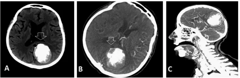
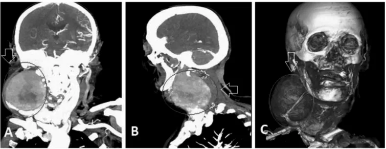
An 81-year-old female patient presented with acute-onset hemiparesis on her left side. She had diabetes mellitus for 10 years and hypertension for 8 years. In a neurologic examination, hemiparesis (2/5) and hemihypoesthesia were noted on her left side. Left Babinski sign was positive. Neck movements were restricted due to aneurysm in the right cervical area. A thrombotic aneurysm (9x9.7 cm) in the right cervical area and calcified
meningioma in the left parietal-occipital lobes were noted in brain computed tomography (CT) and CT angiography (Figures 1, 2). Partial ischemic injury in the middle cerebral artery distribution was seen in neuroimaging. Low-molecular-weight heparin (0.6 mL BID), acetyl salicylic acid (300 mg/day), amlodipine (10 mg/ day), metformin (2000 mg/day) were started. The aneurysm was partially resected. She was discharged on the 3rd day after surgery
with hemiparesis (2/5) and hemihypoesthesia. However, her neck movements were within normal limits.
Ad dress for Cor res pon den ce/Ya z›fl ma Ad re si: Fettah Eren MD, University of Health Sciences, Konya Training and Research Hospital,
Clinic of Neurology, Konya, Turkey
Phone: +90 505 860 41 46 E-mail: [email protected] ORCID ID: orcid.org/0000-0001-6834-0827
Re cei ved/Ge lifl Ta ri hi: 18.04.2018 Ac cep ted/Ka bul Ta ri hi: 02.09.2018 ©Copyright 2019 by Turkish Neurological Society
Turkish Journal of Neurology published by Galenos Publishing House.
Keywords: Thrombotic aneurysm, carotid artery, ischemic stroke Anahtar Kelimeler: Trombotik anevrizma, karotis arter, iskemik inme
Figure 1. A) Brain tomography (before intravenous gadolinium injection). B) Brain tomography (after intravenous gadolinium injection). C) Brain tomography angiography: Calcified meningioma in the left parietal-occipital lobes
40
Giant aneurysms differ from small diameter aneurysms in terms of both clinical symptoms and treatments. The majority of patients present with facial pain, ophthalmoparesis or hypoesthesia with features of mass effect. Subarachnoid hemorrhage (SAH) (50-70%) and ischemic stroke (4%) are seen in patients with giant carotid aneurysms. The most common cause of mortality in these patients is SAH (1,2,3).
Ethics
Informed Consent: Consent form was filled out by all
participants.
Peer-review: Internally peer-reviewed. Authorship Contributions
Surgical and Medical Practices: F.E., Ş.Ö., Concept: F.E., Ş.Ö., Design: F.E., Ş.Ö., Data Collection or Processing: F.E., Ş.Ö.,
Analysis or Interpretation: F.E., Ş.Ö., Literature Search: F.E., Ş.Ö., Writing: F.E., Ş.Ö.
Conflict of Interest: No conflict of interest was declared by
the authors.
Financial Disclosure: The authors declared that this study
received no financial support.
References
1. Beckerman WE, Bernik TJ, Xing S, Dardik H. Symptomatic giant carotid artery aneurysm. J Vasc Surg Cases Innov Tech 2017;3:203-204.
2. Ono H, Inoue T, Tanishima T, et al. High-flow bypass with radial artery graft followed by internal carotid artery ligation for large or giant aneurysms of cavernous or cervical portion: clinical results and cognitive performance. Neurosurg Rev 2018;41:655-665.
3. El-Sabrout R, Cooley DA. Extracranial carotid artery aneurysms: Texas Heart Institute experience. J Vasc Surg 2000;31:702-712.
Turk J Neurol 2019;25:39-40 Eren and Öztürk; Giant Carotid Aneurysm Causing Acute Ischemic Stroke